

Post-Treatment of Micro-Needling with a Dexpanthenol-Containing Ointment Accelerates Epidermal Wound Healing in Human 3D Skin Models
- PMID: 37337567
- PMCID: PMC10276988
- DOI: 10.2147/CCID.S409310
Post-Treatment of Micro-Needling with a Dexpanthenol-Containing Ointment Accelerates Epidermal Wound Healing in Human 3D Skin Models
Abstract
Purpose: In vitro study on the molecular effects of post-treatment after micro-needling applications with a dexpanthenol-containing ointment (DCO) using 3D skin models.
Patients and methods: In this in vitro study, full-thickness human 3D skin models were treated with a micro-needling device according to its clinical application. For post-treatment, some of the models were additionally treated with a dexpanthenol-containing ointment (DCO). Histological samples were taken at 0, 24 and 48 hours. Gene expression analysis was performed after 24 hours.
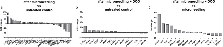
Results: Histological examination showed that DCO post-treated 3D skin models revealed a completed wound closure 24 hours after the micro-needling procedure. In contrast, DCO-untreated models still clearly exhibited the micro-needling lesions after the same period of time. After 48 hours, all models revealed a completed wound healing. In skin models that received micro-needling but no post-treatment with DCO, microarray analysis identified an upregulation of proinflammatory cytokines and chemokines and a downregulation of skin barrier and differentiation markers. In contrast, post-treatment with DCO leads to accelerated wound healing without affecting the initial inflammatory response caused by micro-needling, which leads to the subsequent collagen expression. This data was supported by qRT-PCR analyses.
Conclusion: Post-treatment with DCO accelerates epidermal wound healing after micro-needling of 3D skin models without impairing the immunostimulatory properties of micro-needling. These findings can help to optimise the aftercare routine after micro-needling procedures and to shorten the downtime for the patient after treatment.
Keywords: aftercare; cosmetic procedures; human organotypic skin equivalents; in vitro models; molecular effects; skin needling.
© 2023 Weßollek et al.
Conflict of interest statement
The authors report no conflicts of interest in this work.
Figures


References
LinkOut - more resources
Full Text Sources
