. 2023 Aug;13(8):1394-1404.
doi: 10.1002/2211-5463.13657. Epub 2023 Jul 7.

Cryo-EM structures of tau filaments from SH-SY5Y cells seeded with brain extracts from cases of Alzheimer's disease and corticobasal degeneration

Affiliations

Cryo-EM structures of tau filaments from SH-SY5Y cells seeded with brain extracts from cases of Alzheimer's disease and corticobasal degeneration

Airi Tarutani et al. FEBS Open Bio. 2023 Aug.

Abstract

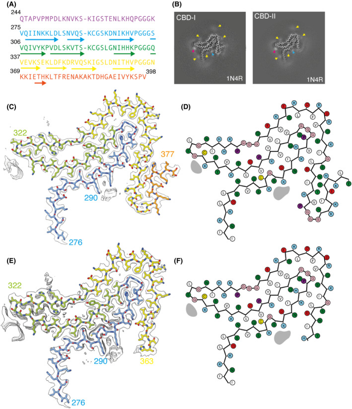
The formation of amyloid filaments through templated seeding is believed to underlie the propagation of pathology in most human neurodegenerative diseases. A widely used model system to study this process is to seed amyloid filament formation in cultured cells using human brain extracts. Here, we report the electron cryo-microscopy structures of tau filaments from undifferentiated seeded SH-SY5Y cells that transiently expressed N-terminally HA-tagged 1N3R or 1N4R human tau, using brain extracts from individuals with Alzheimer's disease or corticobasal degeneration. Although the resulting filament structures differed from those of the brain seeds, some degrees of structural templating were observed. Studying templated seeding in cultured cells, and determining the structures of the resulting filaments, can thus provide insights into the cellular aspects underlying neurodegenerative diseases.

Keywords: Alzheimer's disease; SH-SY5Y; amyloid; corticobasal degeneration; electron cryo-microscopy; tau.


Conflict of interest statement

The authors declare no conflict of interest.

Figures

Fig. 1
Fig. 1
Tau structure in SH‐SY5Y cells seeded with filaments from AD. (A) Amino acid sequence of tau residues 244–398 coloured by repeat: R1 is purple; R2 is blue; R3 is green; R4 is yellow; the C‐terminal domain is orange. (B) XY‐cross‐section of the reconstructed density with a projected thickness of approximately 1 β‐rung (4.75 Å). Extra densities are indicated with yellow arrows. (C) Reconstructed density (in transparent white) and atomic model (with the same colours as in panel A). (D) Schematic of the residues in the structure. Positively charged residues are shown in blue; negatively charged residues in red; polar residues in green; apolar residues in white; cysteines in yellow; glycines in pink and prolines in purple.
Fig. 2
Fig. 2
Comparison of the seeded structure and the structure of the seeds from AD. (A) Overlay of main‐chain traces of the seeded structure (grey) and the PHF (blue) and SF (green) from AD, aligned on the tip of the C‐shaped fold (residues 336–358). (B–F) Detailed views of the 332PGGGQ336 motif with the same colours as in panel A. Intra‐protofilament hydrogen bonds are shown with blue dashed lines and inter‐protofilament hydrogen bonds with yellow and green dashed lines. Panels (B) and (C) are top views of the comparison between the seeded structure and the PHF and SF, respectively. Panels (D–F) are side views of the seeded structure, the PHF and the SF, respectively. (G) Overlay of main‐chain traces of the seeded structure, the PHF and the SF, aligned on the ends of the C‐shaped fold (residues 358–376). (H–J) Detailed views of the C‐terminal part of the ordered cores of the seeded structure, the PHF and the SF, respectively.
Fig. 3
Fig. 3
Tau structure in SH‐SY5Y cells seeded with filaments from CBD. (A) Amino acid sequence of tau residues 244–398. (B) XY‐cross‐section of the reconstructed densities of seeded filament types 1 and 2 with a projected thickness of approximately 1 β‐rung (4.75 Å). Extra densities are indicated with yellow, pink and blue arrows. (C) Reconstructed density and atomic model for type 1 filaments. (D) Schematic of the residues in type 1 filaments. (E) Reconstructed density and atomic model for type 2 filaments. (F) Schematic of the residues in type 2 filaments. Colours are as in Fig. 1.
Fig. 4
Fig. 4
Comparison of the seeded structure and the structure of the seeds from CBD. (A) Overlay of main‐chain traces of the seeded structures (type 1 in red; type 2 in orange) and the type II filaments from corticobasal degeneration (CBD in purple) aligned on residues 283–362. (B) Detailed view of residues 356–377 in the type 1 seeded structure. (C) Detailed view of residues 356–380 in the CBD Type II filament. (D, E) Top and side views of the extra density in the type 1 seeded structure. (F, G) Top and side views of the extra density in the type 2 seeded structure. (H, I) Top and side views of the extra density in the CBD type II filament.

References

    1. Goedert M, Eisenberg DS and Crowther RA (2017) Propagation of tau aggregates and neurodegeneration. Annu Rev Neurosci 40, 189–210. - PubMed
    1. Goedert M, Spillantini MG, Jakes R, Rutherford D and Crowther RA (1989) Multiple isoforms of human microtubule‐associated protein tau: sequences and localization in neurofibrillary tangles of Alzheimer's disease. Neuron 3, 519–526. - PubMed
    1. Fitzpatrick AWP, Falcon B, He S, Murzin AG, Murshudov G, Garringer HJ, Crowther RA, Ghetti B, Goedert M and Scheres SHW (2017) Cryo‐EM structures of tau filaments from Alzheimer's disease brain. Nature 547, 185–190. - PMC - PubMed
    1. Falcon B, Zivanov J, Zhang W, Murzin AG, Garringer HJ, Vidal R, Crowther RA, Newell KL, Ghetti B, Goedert M et al. (2019) Novel tau fold in chronic traumatic encephalopathy encloses hydrophobic molecules. Nature 568, 420–423. - PMC - PubMed
    1. Falcon B, Zhang W, Murzin AG, Murshudov G, Garringer HJ, Vidal R, Crowther RA, Ghetti B, Scheres SHW and Goedert M (2018) Structures of filaments from Pick's disease reveal a novel tau protein fold. Nature 561, 137–140. - PMC - PubMed

Publication types