. 2023 Jul 1;30(Pt 4):780-787.
doi: 10.1107/S1600577523004228. Epub 2023 Jun 20.

Translocation and fate of nanospheres in pheochromocytoma cells following exposure to synchrotron-sourced terahertz radiation

Affiliations

Translocation and fate of nanospheres in pheochromocytoma cells following exposure to synchrotron-sourced terahertz radiation

Palalle G Tharushi Perera et al. J Synchrotron Radiat. .

Abstract

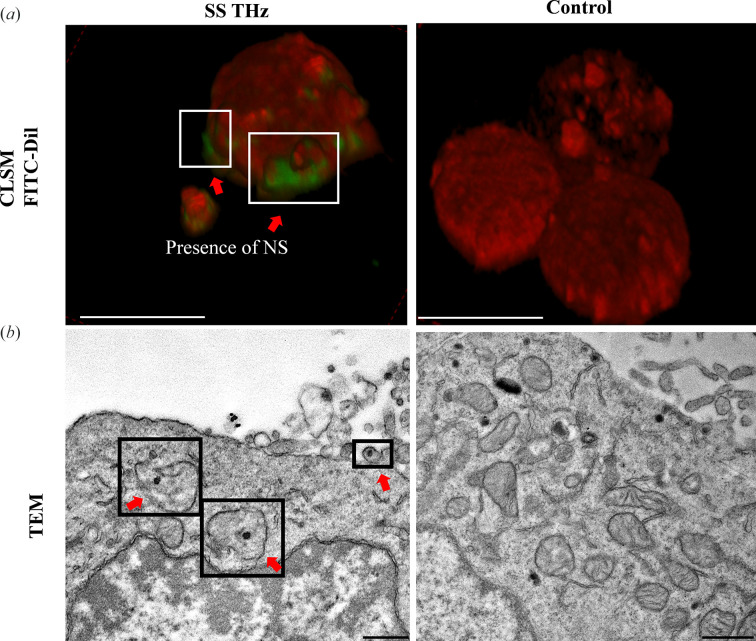
The routes by which foreign objects enter cells is well studied; however, their fate following uptake has not been explored extensively. Following exposure to synchrotron-sourced (SS) terahertz (THz) radiation, reversible membrane permeability has been demonstrated in eukaryotic cells by the uptake of nanospheres; nonetheless, cellular localization of the nanospheres remained unclear. This study utilized silica core-shell gold nanospheres (AuSi NS) of diameter 50 ± 5 nm to investigate the fate of nanospheres inside pheochromocytoma (PC 12) cells following SS THz exposure. Fluorescence microscopy was used to confirm nanosphere internalization following 10 min of SS THz exposure in the range 0.5-20 THz. Transmission electron microscopy followed by scanning transmission electron microscopy energy-dispersive spectroscopic (STEM-EDS) analysis was used to confirm the presence of AuSi NS in the cytoplasm or membrane, as single NS or in clusters (22% and 52%, respectively), with the remainder (26%) sequestered in vacuoles. Cellular uptake of NS in response to SS THz radiation could have suitable applications in a vast number of biomedical applications, regenerative medicine, vaccines, cancer therapy, gene and drug delivery.

Keywords: PC 12 neuronal cells; electromagnetic fields (EMFs); fate of nanospheres; membrane permeability; synchrotron-sourced THz radiation.


Figures

Figure 1
Figure 1
Overview of the experimental design and the technical setup at the Australian Synchrotron Far-IR/THz beamline. (a) Schematic representation of AuSi NS localization in PC 12. The SS THz exposed PC 12 samples were analysed for AuSi NPs following a 10 min exposure. (b) BEP at the Australian Synchrotron Far-IR/THz beamline, with the inset showing the beam axis in red; the beam is present within a 19 mm diameter (blue arrow) from the inner edge to the beam axis. The PC 12 cell suspension was placed on a polyethyl­ene window (red arrow) using an O-ring in the presence of the beam. (c) The spectrum of the intensity of the Australian Synchrotron THz beamline as shown by the resident Si bolometer; intensity is presented in arbitrary units, with a logarithmic scale for the THz frequencies (Vilagosh et al., 2022 ▸). (d) Depth-dependent absorption pattern of the Australian Synchrotron THz beam. The two variables share an exponential relationship where the absorption of the beam decreases as the depth of the sample increases.
Figure 2
Figure 2
SS THz induced NS uptake by PC 12 cells confirmed by CLSM and TEM. (a) Confocal micrographs displaying FITC NS uptake. The presence of NS near the membrane (white insets) is confirmed by the presence of the green signal (red arrows). Scale bars: 10 µm. (b) TEM micrographs displaying AuSi NS uptake (red arrows). NS appear to be localized inside the cytoplasm and closer to the cell membrane. Scale bars: 0.5 µm.
Figure 3
Figure 3
Localization of silica core-shell Au NS in PC 12 following exposure to SS THz radiation. (a) STEM micrographs illustrating SS THz irradiated PC 12 cells. NS are seen internalized in vacuoles (yellow arrows) inside the cytoplasm. (b) EDS spectra representative of the line data 1 in the electron microscopy image (a) confirming the presence of elemental Au (black arrows), thus confirming the presence of AuSi NS localized inside vacuoles in the cellular cytoplasm. (c) Quantification of the localization of AuSi NS. The majority of the NS are present in the cytoplasm and are sequestered in vacuoles. (d) STEM micrograph of the unexposed PC 12 cell sample and (e) representative EDS spectra. No peaks were detected for Au, line data 11, confirming the absence of Au inside the vacuoles that were observed inside the control cells.
Figure 4
Figure 4
PC 12 cells viability and morphology following exposure to SS THz radiation. (a) Optical micrographs (insets) of PC 12 cells exposed to SS THz radiation and the control. Scale bars: 5 µm. CLSM micrographs of viable PC 12 cells are shown in green – the cells were stained and imaged after exposure to SS THz; the untreated control is seen on the right. (b) Quantification of viable PC 12 cells. No statistically significant differences were observed among the two experimental groups (p = 0.28). (c) SEM micrographs depicting PC 12 cell morphology. Blebs were present in the outer membrane of the PC 12 cells in response to SS THz radiation, highlighted in the black insets by red arrows. (d) Quantification of the diameter d as shown in the insets (normal cell followed by blebbing). The average size of the blebs was 1.08 ± 0.49 µm.

Similar articles

Cited by

References

    1. Bohdanowicz, M. & Grinstein, S. (2013). Physiol. Rev. 93, 69–106. - PubMed
    1. Bolhassani, A., Khavari, A. & Oraf, Z. (2014). Application of Nanotechnology in Drug Delivery, ch. 11, edited by A. D. Sezer. InTech Open.
    1. Charras, G. T. A. (2008). J. Microsc. 231, 466–478. - PubMed
    1. Chu, Z., Zhang, S., Zhang, B., Zhang, C., Fang, C. Y., Rehor, I., Cigler, P., Chang, H. C., Lin, G., Liu, R. & Li, Q. (2014). Sci. Rep. 4, 4495. - PMC - PubMed
    1. Cocker, T. L., Jelic, V., Hillenbrand, R. & Hegmann, F. A. (2021). Nat. Photon. 15, 558–569.