. 2023 Jul 27;97(7):e0085821.
doi: 10.1128/jvi.00858-21. Epub 2023 Jun 20.

Enhanced Virus Translation Enables miR-122-Independent Hepatitis C Virus Propagation

Affiliations

Enhanced Virus Translation Enables miR-122-Independent Hepatitis C Virus Propagation

Mamata Panigrahi et al. J Virol. .

Abstract

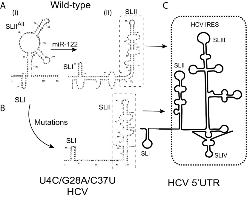
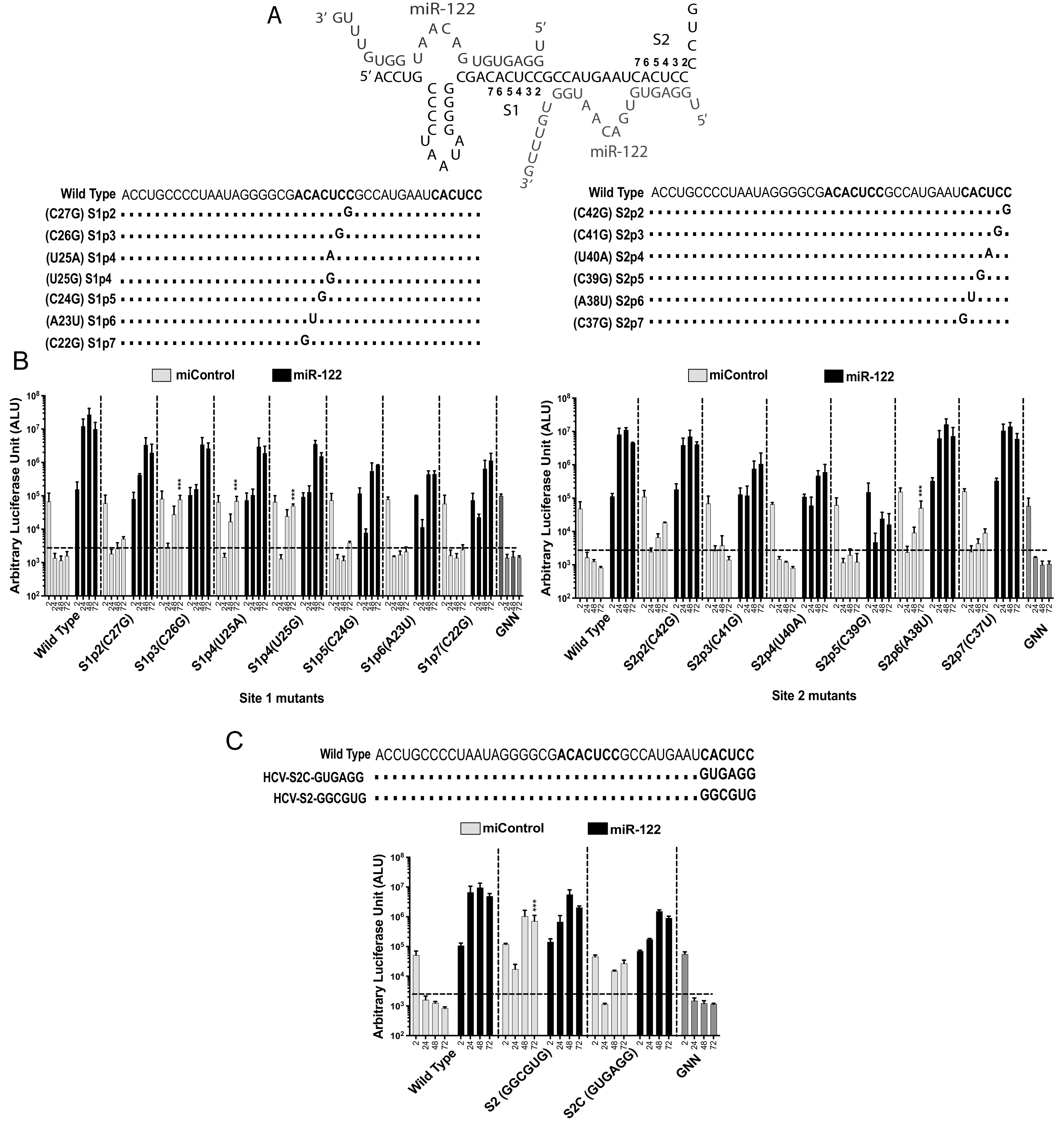
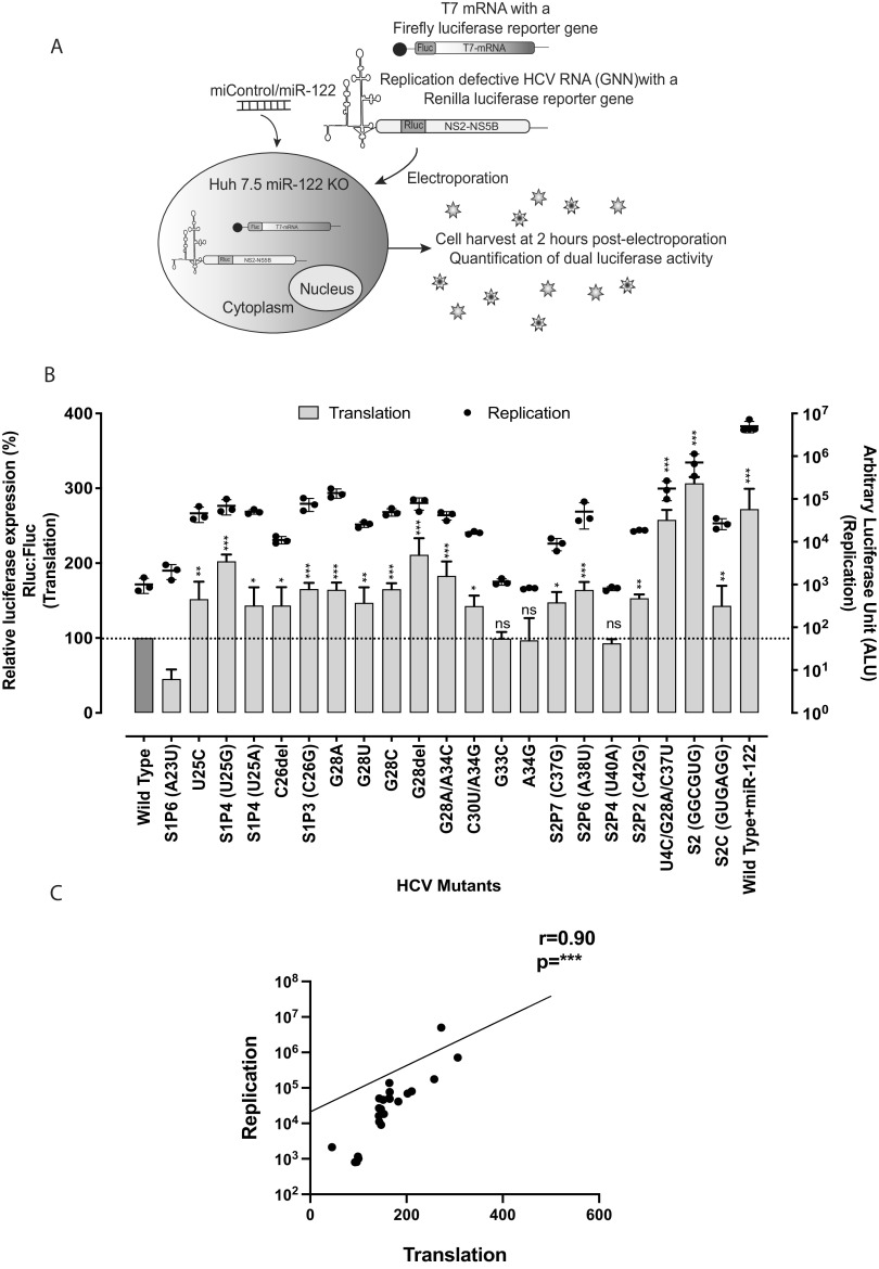
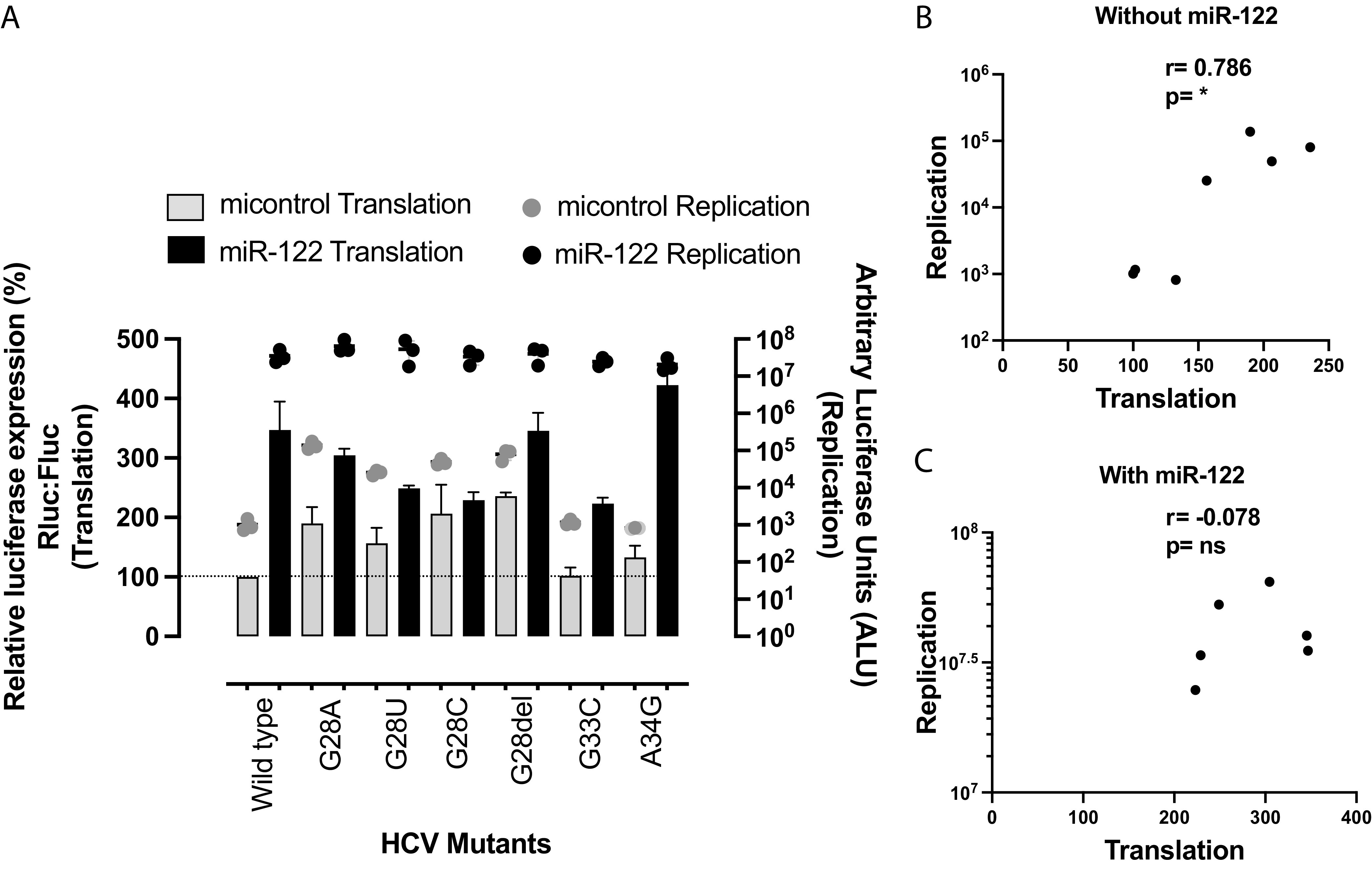
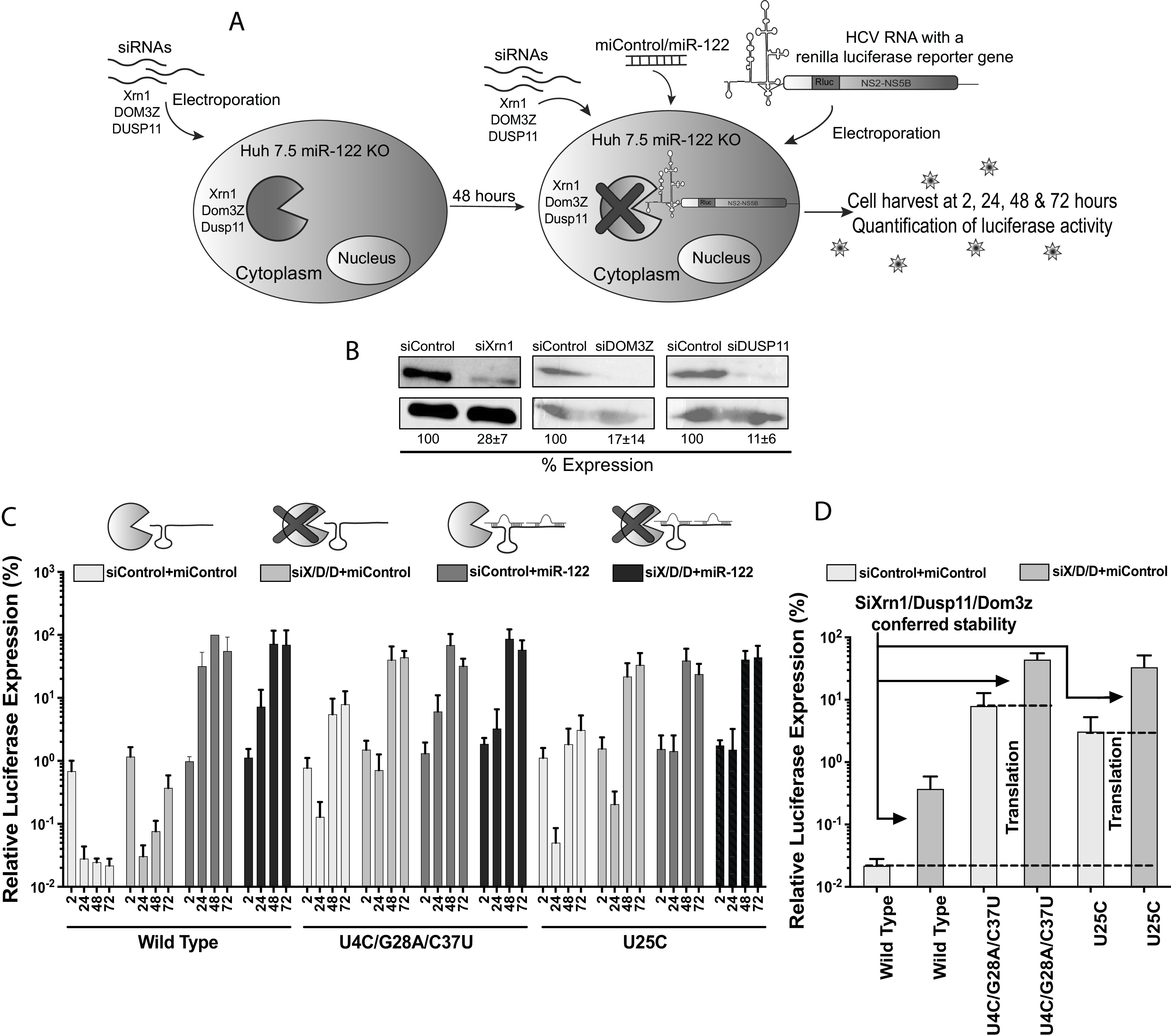
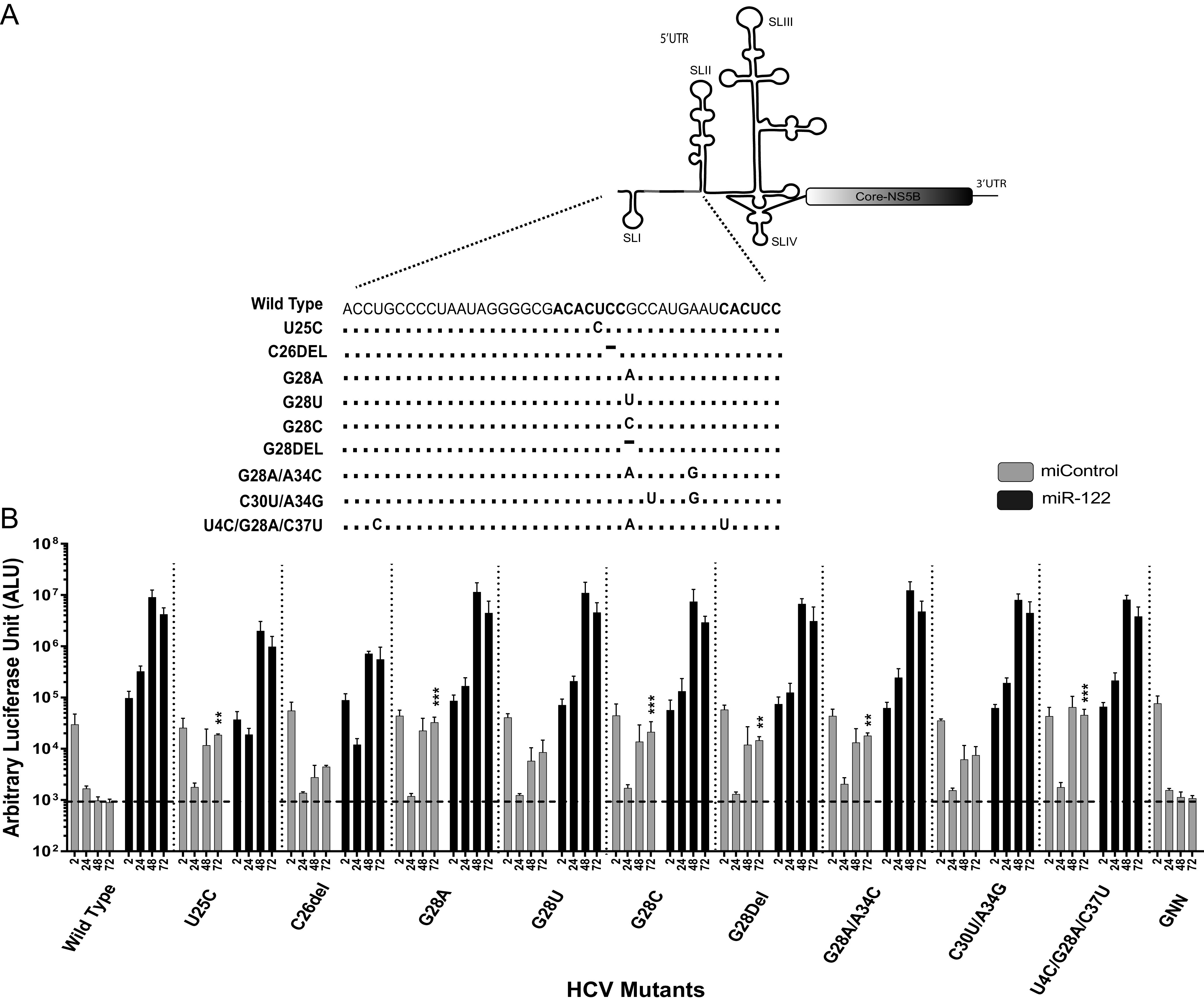
The 5' untranslated region (UTR) of the hepatitis C virus (HCV) genome forms RNA structures that regulate virus replication and translation. The region contains an internal ribosomal entry site (IRES) and a 5'-terminal region. Binding of the liver-specific microRNA (miRNA) miR-122 to two binding sites in the 5'-terminal region regulates viral replication, translation, and genome stability and is essential for efficient virus replication, but its precise mechanism of action is still unresolved. A current hypothesis is that miR-122 binding stimulates viral translation by facilitating the viral 5' UTR to form the translationally active HCV IRES RNA structure. While miR-122 is essential for detectable replication of wild-type HCV genomes in cell culture, several viral variants with 5' UTR mutations exhibit low-level replication in the absence of miR-122. We show that HCV mutants capable of replicating independently of miR-122 display an enhanced translation phenotype that correlates with their ability to replicate independently of miR-122. Further, we provide evidence that translation regulation is the major role for miR-122 and show that miR-122-independent HCV replication can be rescued to miR-122-dependent levels by the combined impacts of 5' UTR mutations that stimulate translation and by stabilizing the viral genome by knockdown of host exonucleases and phosphatases that degrade the genome. Finally, we show that HCV mutants capable of replicating independently of miR-122 also replicate independently of other microRNAs generated by the canonical miRNA synthesis pathway. Thus, we provide a model suggesting that translation stimulation and genome stabilization are the primary roles for miR-122 in promoting HCV. IMPORTANCE The unusual and essential role of miR-122 in promoting HCV propagation is incompletely understood. To better understand its role, we have analyzed HCV mutants capable of replicating independently of miR-122. Our data show that the ability of viruses to replicate independently of miR-122 correlates with enhanced virus translation but that genome stabilization is required to restore efficient HCV replication. This suggests that viruses must gain both abilities to escape the need for miR-122 and impacts the possibility that HCV can evolve to replicate outside the liver.

Keywords: 5′ UTR; 5′ untranslated region; HCV; genome stability; hepatitis C virus; miR-122; miR-122-independent replication; translation; viral translation.


Conflict of interest statement

The authors declare no conflict of interest.

Figures

FIG 1
FIG 1
Hypothetical model for miR-122 and 5′ UTR mutation-induced RNA structure modifications leading to IRES activation. (A) Structure prediction of J6/JFH-1 p7Rluc2a HCV (i) without and (ii) with miR-122. Without miR-122, the first 117 nucleotides of HCV 5′ UTR are predicted to form a noncanonical structure with SLI and an altered SLII (SLIIAlt), whereas miR-122 annealing to the viral 5′ UTR is predicted to form the canonical SLII structure. (B) The 5′ UTR of a mutant HCV (U4C/G28A/C37U) capable of miR-122-independent replication is also predicted to form the canonical SLII structure in the absence of miR-122. (C) Structure of the active HCV IRES including SLII.
FIG 2
FIG 2
Some mutations to nucleotides within miR-122 binding sites 1 and 2 allow viral propagation in the absence of miR-122. (A) HCV 5′ UTR and the location and name of mutant viruses with sequence changes within miR-122 binding sites 1 and 2. “S” stands for miR-122 binding site (1 or 2) and “p” stands for the position in the binding site (1 to 7). (B) Replication of site 1 and site 2 mutant HCV J6/JFH-1 (p7Rluc2a) RNA in Huh 7.5 miR-122 knockout cells with a control miRNA (miControl) or with miR-122. Renilla luciferase was assessed at 2 h, 24 h, 48 h, and 72 h postelectroporation as an indicator of viral propagation. The dark gray and the light gray bars represent miR-122-dependent and -independent replication, respectively. (C) Replication of J6/JFH-1 (p7Rluc2a) HCV-S2-GGCGUG, and HCV-S2C-GUGAGG mutant RNA in Huh 7.5 miR-122 knockout cells with a control miRNA (miControl) or with miR-122. Renilla luciferase was assessed at 2 h, 24 h, 48 h, and 72 h postelectroporation as an indicator of viral propagation. The dark gray and the light gray bars represent miR-122-dependent and -independent replication, respectively. miR-122-independent replication of HCV mutant RNA was compared to that of the wild-type HCV RNA, with miControl as a control. All data are presented as the averages of three or more independent experiments. Error bars indicate the standard deviations of the means, and asterisks indicate significant differences. The significance was determined by using one-way analysis of variance (ANOVA) (***, P < 0.001).
FIG 3
FIG 3
Structure prediction models of HCV J6/JFH-1 (p7Rluc2a) wild type and miR-122 binding site 1 and site 2 mutants. The sequences from nucleotides 1 to 117 of wild-type HCV and these mutants were analyzed using the “RNA structure” online tool, and the 4 lowest free-energy structure models are presented. These structures are prediction models and have not been experimentally validated.
FIG 4
FIG 4
HCV mutants replicating independently of miR-122 have higher translation efficiency than wild-type HCV, and miR-122-independent translation potency correlates with miR-122-independent replication efficiency. (A) Illustration of the translation assay method. Replication-defective Renilla reporter J6/JFH-1 (p7Rluc2a) HCV RNA and a firefly reporter mRNA were coelectroporated into Huh 7.5 miR-122 knockout cells. Cells were harvested 2 h postelectroporation, and dual-luciferase activity was measured. (B) Comparison of translation and replication efficiencies of HCV mutants and wild-type virus in Huh 7.5 miR-122 knockout cells. The left y axis and bars represent HCV RNA translation, whereas the right y axis and the dots represent HCV RNA replication. For replication of mutant and wild-type HCV, viral RNA was electroporated into Huh 7.5 miR-122 knockout cells and cells were harvested 72 h postelectroporation. Renilla luciferase was measured to analyze viral propagation. (C) Correlation of HCV RNA translation and replication was analyzed using nonparametric Spearman correlation. All data are presented as the averages of three or more independent experiments. Error bars indicate the standard deviations of the means, and asterisks indicate significant differences. The significance was determined by using one-way ANOVA for translation efficiency and t-distribution for correlation (*, P < 0.033; **, P < 0.002; ***, P < 0.001).
FIG 5
FIG 5
Translation and replication assays with and without miR-122: G28 mutants. Shown is a comparison of translation and replication efficiencies of G28 HCV mutants and wild-type virus in Huh 7.5 miR-122 knockout cells. The left y axis and bars represent HCV RNA translation, whereas the right y axis and the dots represent HCV RNA replication. Light gray represents viral translation/replication without miR-122, and dark gray represents viral translation/replication with miR-122. (B) Correlation of G28 HCV RNA translation and replication independently of miR-122 was analyzed using nonparametric Spearman correlation. (C) Correlation of G28 HCV RNA translation and replication in the presence of miR-122 was analyzed using nonparametric Spearman correlation. All data are presented as the averages of three or more independent experiments. Error bars indicate the standard deviations of the means, and asterisks indicate significant differences. The significance was determined by using t-distribution. ns, not significant.
FIG 6
FIG 6
Viral genome stabilization rescues miR-122-independent replication of 5′ UTR mutants to wild-type HCV levels. (A) Graphical representation of siRNA-induced XRN1, DUSP11, and DOM3Z knockdown and HCV replication analysis in Huh 7.5 miR-122 knockout cells. Huh 7.5 miR-122 knockout cells were electroporated with XRN1-, DUSP11-, and DOM3Z-specific siRNAs, and 48 h after the first electroporation, viral RNAs along with siRNAs and miRNAs were coelectroporated into the preknockdown Huh 7.5 miR-122 knockout cells. Cells were harvested at 2 h, 24 h, 48 h, and 72 h after the second electroporation, and luciferase activity was measured as an indicator of viral propagation. (B) Western blot showing knockdown efficiency with antibodies against Xrn1, DOM3Z, and DUSP11. Percent knockdown ± standard deviation relative to siControl is shown. (C) Replication of J6/JFH-1 (p7Rluc2a) wild-type HCV, U4C/G28A/C37U HCV, and U25C HCV RNA in Huh 7.5 miR-122 knockout cells. The solid bars with different shades of gray represent different knockdown conditions. (C) The siXRN1-, siDUSP11-, and siDOM3Z-conferred stability was determined for wild-type, U4C/G28A/C37U, and U25C HCV RNA by comparing replication without knockdown (siControl) versus replication with knockdown (siS/D/D). (D) The data shown in panel C were used to determine the contribution of stability to miR-122-independent replication by comparing replication of wild-type HCV RNA in miR-122 knockout cells with (siX/D/D) and without (siControl) knockdown of XRN1, DUSP11, and DOM3Z. Similarly, the contribution of mutation-induced translation to miR-122-independent replication was determined by comparing replication of stabilized wild-type HCV RNA (WT siX/D/D) with those of stabilized mutant RNAs (U4C/G28A/C37U siX/D/D) and (U25C siX/D/D).
FIG 7
FIG 7
Replication of HCV mutants in Huh 7.5 Drosha knockout cells. (A) Reported mutations on the extreme 5′ UTR of the viral genome that promote virus replication independently of miR-122. The number indicates the position of the nucleotide with the mutation. (B) Transient replication of HCV mutants in Huh 7.5 Drosha knockout cells electroporated with J6/JFH-1 p7Rluc2a HCV RNA with a control miRNA (gray bars) or miR-122 (black bars). Luciferase activity of cells harvested after 72 h postelectroporation was measured, and relative luciferase activity was plotted with respect to the replication-defective mutant (GNN). All data shown are the averages of three or more independent experiments. Error bars indicate the standard deviations of the means, and asterisks indicate significant differences, as determined by one-way ANOVA (**, P < 0.002; ***, P < 0.001).

Similar articles

Cited by

References

    1. The Polaris Observatory HCV Collaborators. 2017. Global prevalence and genotype distribution of hepatitis C virus infection in 2015: a modelling study. Lancet Gastroenterol Hepatol 2:161–176. doi:10.1016/S2468-1253(16)30181-9. - DOI - PubMed
    1. Hoofnagle JH. 2002. Course and outcome of hepatitis C. Hepatology 36:S21–S29. doi:10.1053/jhep.2002.36227. - DOI - PubMed
    1. Perz JF, Armstrong GL, Farrington LA, Hutin YJF, Bell BP. 2006. The contributions of hepatitis B virus and hepatitis C virus infections to cirrhosis and primary liver cancer worldwide. J Hepatol 45:529–538. doi:10.1016/j.jhep.2006.05.013. - DOI - PubMed
    1. Lukavsky PJ. 2009. Structure and function of HCV IRES domains. Virus Res 139:166–171. doi:10.1016/j.virusres.2008.06.004. - DOI - PMC - PubMed
    1. Fraser CS, Doudna JA. 2007. Structural and mechanistic insights into hepatitis C viral translation initiation. Nat Rev Microbiol 5:29–38. doi:10.1038/nrmicro1558. - DOI - PubMed

Publication types

Grants and funding