. 2023 Sep 4;222(9):e202208047.
doi: 10.1083/jcb.202208047. Epub 2023 Jun 20.

Polydom/SVEP1 binds to Tie1 and promotes migration of lymphatic endothelial cells

Affiliations

Polydom/SVEP1 binds to Tie1 and promotes migration of lymphatic endothelial cells

Ryoko Sato-Nishiuchi et al. J Cell Biol. .

Abstract

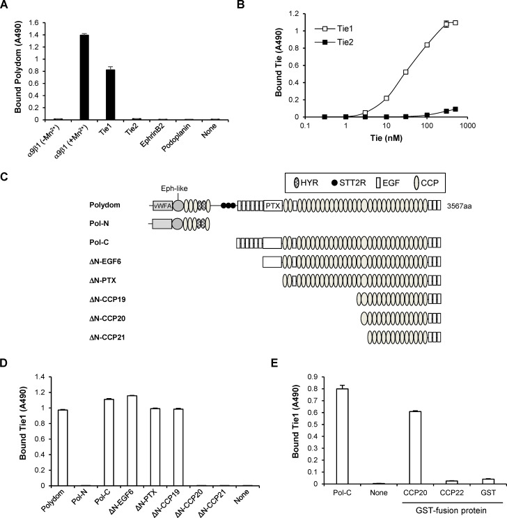
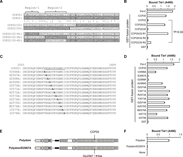
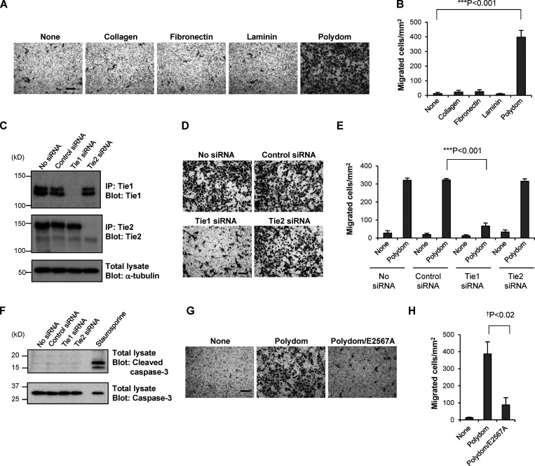
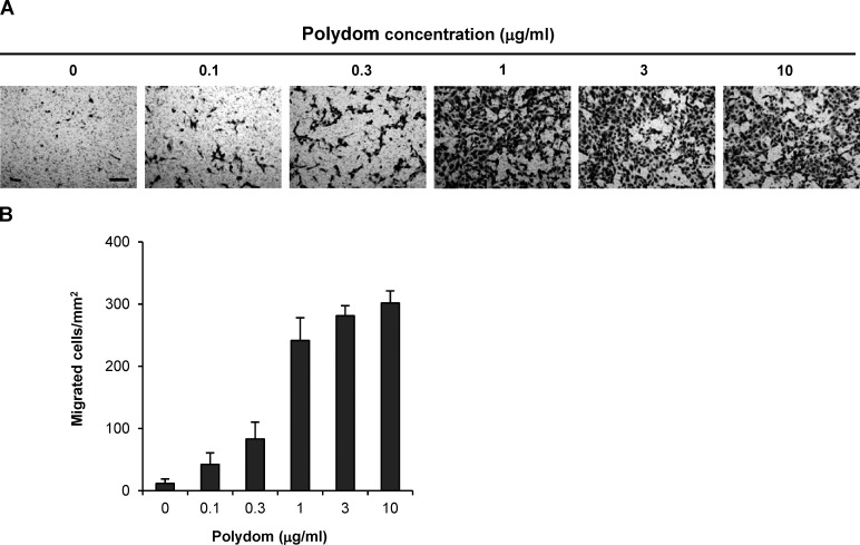
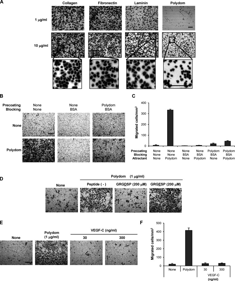
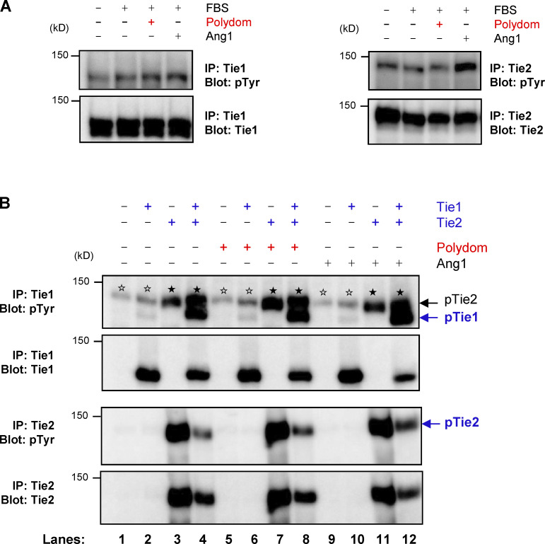
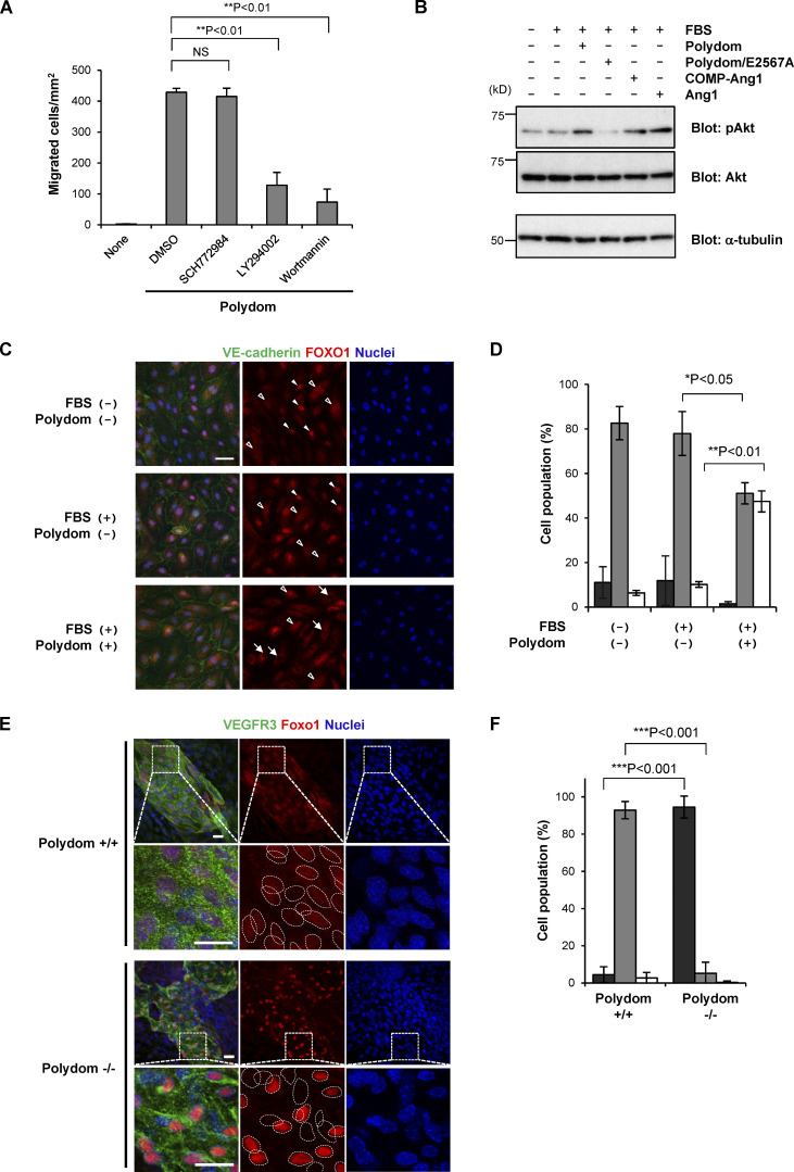
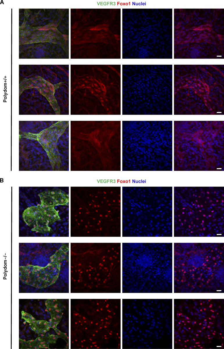
Polydom is an extracellular matrix protein involved in lymphatic vessel development. Polydom-deficient mice die immediately after birth due to defects in lymphatic vessel remodeling, but the mechanism involved is poorly understood. Here, we report that Polydom directly binds to Tie1, an orphan receptor in the Angiopoietin-Tie axis, and facilitates migration of lymphatic endothelial cells (LECs) in a Tie1-dependent manner. Polydom-induced LEC migration is diminished by PI3K inhibitors but not by an ERK inhibitor, suggesting that the PI3K/Akt signaling pathway is involved in Polydom-induced LEC migration. In line with this possibility, Akt phosphorylation in LECs is enhanced by Polydom although no significant Tie1 phosphorylation is induced by Polydom. LECs also exhibited nuclear exclusion of Foxo1, a signaling event downstream of Akt activation, which was impaired in Polydom-deficient mice. These findings indicate that Polydom is a physiological ligand for Tie1 and participates in lymphatic vessel development through activation of the PI3K/Akt pathway.


Conflict of interest statement

Disclosures: K. Sekiguchi reported personal fees from Matrixome, Inc., “other” from Nippi, Inc., grants from Mandom, Inc., and grants from Kao Corporation outside the submitted work. No other disclosures were reported.

Figures

Figure 1.
Figure 1.
Polydom binds to Tie1 via CCP20. (A) Recombinant integrin α9β1, Tie1, Tie2, EphrinB2, and Podoplanin were coated on microtiter plates at 5 μg/ml and then incubated with full-length Polydom (5 μg/ml). Polydom was allowed to bind to integrin α9β1 in the absence or presence of 1 mM MnCl2. The data represent means ± SD of triplicate determinations. (B) Titration curves of Tie1 and Tie2 binding to Polydom. Tie1 (open squares) or Tie2 (closed squares) at the indicated concentrations was allowed to bind to microtiter plates coated with full-length Polydom (10 nM). The data represent means ± SD of triplicate determinations. (C) Schematic representation of Polydom and its recombinant fragments. Polydom consists of multiple domains including von Willebrand factor type A (vWFA), Ephrin 2-like (Eph-like), hyaline (HYR), similar to thyroglobulin type 2 repeat (STT2R), EGF, and pentraxin (PTX) domains, and an array of CCP domains. (D) Polydom and its fragments were coated at 2 μg/ml on microtiter plates and incubated with Tie1 (2 μg/ml). The data represent means ± SD of triplicate determinations. (E) GST fusion proteins of CCP20 and CCP22 were coated at 2 μg/ml on microtiter plates and incubated with Tie1 (2 μg/ml). Pol-C and GST alone were included as positive and negative controls, respectively. The data represent means ± SD of triplicate determinations.
Figure 2.
Figure 2.
Glu2567 and Gly2568 in CCP20 are required for Tie1 binding. (A) Alignment of the amino acid sequences of CCP20 (white bars) and CCP22 (gray bars) by ClustalW. The asterisks denote identical amino acids, while the colons and periods denote strongly similar and weakly similar amino acids, respectively. The N-terminal half of CCP20/CCP22 is subdivided into Region-1 and Region-2. (B) Tie1 (2 μg/ml) was allowed to bind to microtiter plates coated with GST fusion proteins containing CCP20, CCP22, and their chimeras or with Pol-C or GST alone coated at 2 μg/ml. The data represent means ± SD of triplicate determinations. P < 0.02 (n = 3). (C and D) A series of alanine scanning mutants of CCP20/Region-2 (C; underlined) were expressed as GST fusion proteins and assessed for their binding activities toward Tie1 (D). The data represent means ± SD of triplicate determinations. The substituted alanine (A) and glycine (G) residues are shown in bold. (E and F) Full-length Polydom and its Glu2567Ala mutant (Polydom/E2567A; E) were assessed for their binding activities toward Tie1 (F). The data represent means ± SD of triplicate determinations.
Figure 3.
Figure 3.
Polydom promotes LEC migration via Tie1. (A) Photographs of LECs that migrated to the lower side of Transwell membranes in the presence of collagen, fibronectin, laminin, or Polydom (3 μg/ml) added to the lower chamber medium. The molar concentration of the proteins was collagen, 23 nM; fibronectin, 15 nM; laminin, 3.8 nM; and Polydom: 8.8 nM. Bar, 200 μm. (B) Cells that migrated to the lower side of the membranes were counted under a microscope. The data represent means ± SD of three independent experiments each assayed in triplicate. ***P < 0.001 (n = 3). (C) Immunoprecipitates (IP) of Tie1 and Tie2 from lysates of LECs transfected with control, Tie1, or Tie2 siRNAs were analyzed by Western blotting under reducing conditions. The same lysates were probed with an antibody against α-tubulin as a control. The positions of molecular weight markers are shown on the left. (D) LECs transfected with control, Tie1, or Tie2 siRNAs were allowed to migrate in the presence of Polydom (1 μg/ml) for 16 h. Bar, 200 μm. (E) Cells that migrated to the lower side of the membranes were counted under a microscope in the absence (none) or presence of Polydom added to the lower chamber medium. The data represent means ± SD of three independent experiments each assayed in triplicate. ***P < 0.001 (n = 3). (F) Total lysates of LECs transfected with control, Tie1, or Tie2 siRNAs were analyzed by Western blotting using anti-cleaved caspase-3 and anti-caspase-3 antibodies under reducing conditions. Lysates of LECs treated with staurosporine (0.2 μM, 4 h) were used as a positive control for cleaved caspase-3 detection. (G) Photographs of LECs that migrated to the lower side of the membranes in the presence of Polydom or Polydom/E2567A (1 μg/ml) in the lower chamber medium. Bar, 200 μm. (H) Cells that migrated to the lower side of the membranes were counted under a microscope. The data represent means ± SD of three independent experiments each assayed in triplicate. P < 0.02 (n = 3). Source data are available for this figure: SourceData F3.
Figure S1.
Figure S1.
Polydom promotes LEC migration in a dose-dependent manner. (A) Photographs of LECs that migrated to the lower side of Transwell membranes when increasing concentrations of Polydom were added to the medium in the lower chamber. Bar, 200 μm. (B) Cells that migrated to the lower side of the membranes were counted under a microscope. The data represent means ± SD of three independent experiments each assayed in triplicate.
Figure S2.
Figure S2.
Polydom functions as a chemotactic, but not a haptotactic, factor in LEC transmigration. (A) LECs were seeded on 96-well microtiter plates coated with collagen, fibronectin, laminin, or Polydom at 1 or 10 µg/ml and allowed to adhere to the plates for 30 min at 37°C. After washing to remove unbound cells, the attached cells were fixed and stained with toluidine blue. Magnified views of the boxed areas in the middle row are shown in the bottom row. Bars, 200 μm. (B) LEC transmigration assays using Transwell membranes with (right panels) or without (left and middle panels) precoating with Polydom (2 μg/ml), followed by blocking with 1% BSA except for the left panels. The upper and lower panels show the results of LEC transmigration in the absence (upper) or presence (lower) of Polydom (1 μg/ml) added to the lower chamber medium. Bar, 200 μm. (C) Cells that migrated to the lower side of the membranes were counted under a microscope in the absence (None) or presence of Polydom added to the lower chamber medium. The data represent means ± SD of three independent experiments each assayed in triplicate. (D) Cells that migrated to the lower side of the membranes were counted under a microscope in the absence (None) or presence of Polydom (1 μg/ml) with GRGDSP peptide (200 μM) or GRGESP peptide (200 μM). Bar, 200 μm. (E) Photographs of LECs that migrated to the lower side of Transwell membranes in the presence of Polydom (1 μg/ml) or VEGF-C (30 or 300 ng/ml) in the lower chamber medium. Bar, 200 μm. (F) Cells that migrated to the lower side of the membranes were counted under a microscope in the absence (None) or presence of Polydom or VEGF-C in the lower chamber medium. The data represent means ± SD of three independent experiments each assayed in triplicate.
Figure S3.
Figure S3.
Polydom does not induce Tie1 phosphorylation. (A) Serum-starved LECs were treated with EBM-MV2 medium containing 0.5% FBS, 1 μg/ml Polydom, or 500 ng/ml Ang1 at 37°C for 15 min. Immunoprecipitates (IP) of Tie1 (left) and Tie2 (right) from cell lysates were immunoblotted under reducing conditions for phosphotyrosine residues (upper panels) followed by reimmunoblotting for total Tie1 or Tie2 (lower panels). (B) 293-F cells were transfected with the indicated expression plasmids for Tie1 and Tie2 and treated with 1 μg/ml Polydom or 500 ng/ml Ang1 at 37°C for 15 min. Immunoprecipitates of Tie1 and Tie2 from cell lysates were immunoblotted under reducing conditions for phosphotyrosine residues (upper panels), followed by reimmunoblotting for total Tie1 or Tie2 (lower panels). Co-transfection of Tie2 with Tie1 increased Tie1 phosphorylation irrespective of the presence or absence of Polydom (lanes 4, 8, and 12). No Tie1 phosphorylation was induced by Polydom without Tie2 co-transfection (lanes 2, 6, and 10). Signals for phosphorylated Tie2 (pTie2; closed stars) were detected in Tie1 immunoprecipitates from Tie2-transfected cells (uppermost panel; lanes 3, 4, 7, 8, 11, and 12) because the anti-Tie1 polyclonal antibody used for the immunoprecipitation crossreacts with Tie2. Weak signals (open stars) were detected at (or slightly above) the position of pTie2 in the Tie1 immunoprecipitates from cells that were either untransfected or only transfected with Tie1 (lanes 1, 2, 5, 6, 9, and 10). Because Tie2 was not transfected in these cells, the weak signals (open stars) at the pTie2 position should be derived from tyrosine-phosphorylated proteins that were endogenously expressed in 293-F cells and nonspecifically precipitated with the anti-Tie1 antibody used. Such bands were not detected in untransfected or Tie1-transfected cells after immunoprecipitation with an anti-Tie2 antibody (lower panels; lanes 1, 2, 5, 6, 9, and 10). Source data are available for this figure: SourceData FS3.
Figure 4.
Figure 4.
Involvement of the PI3K/Akt signaling pathway in Polydom-induced LEC migration. (A) LECs were allowed to migrate to the lower side of Transwell membranes in the absence (none) or presence of Polydom (1 μg/ml) with an ERK inhibitor (2 μM SCH772984) or PI3K inhibitors (20 μM LY294002 or 2 μM Wortmannin) added to the lower chamber medium. Cells that migrated to the lower side of the membranes were counted under a microscope. The data represent means ± SD of three independent experiments each assayed in triplicate. NS, not significant; **P < 0.01 (n = 3). (B) Serum-starved LECs were treated with Polydom (1 μg/ml), Polydom/E2567A (1 μg/ml), COMP-Ang1 (500 ng/ml), or Ang1 (500 ng/ml) in medium containing 0.5% FBS at 37°C for 15 min. Total lysates of the cells were immunoblotted for pAkt or total Akt under reducing conditions. (C) Serum-starved LECs were treated with Polydom (1 μg/ml) in medium with or without 0.5% FBS at 37°C for 30 min, followed by immunostaining for VE-cadherin (green) and FOXO1 (red). Nuclei were stained with Hoechst 33342 (blue). Cells in which FOXO1 was excluded from the nucleus (arrows) were frequently observed in the presence of Polydom, while FOXO1 was detected exclusively in the nucleus (closed arrowheads) or both the nucleus and cytoplasm with reduced signal intensity (open arrowheads) in the absence of Polydom. Bar, 20 μm. (D) Quantification of the percentages of cells showing FOXO1 localization in the nucleus only (black), in both the nucleus and the cytoplasm (gray), and in the cytoplasm only (white). The data represent means ± SD of three independent experiments. *P < 0.05 and **P < 0.01 (n = 3). (E) Whole-mount immunofluorescence staining of the skin of E17.5 wild-type (Polydom+/+, upper panels) and Polydom-deficient mice (Polydom−/−, lower panels) for VEGFR3 (green, used as a lymphatic vessel marker) and Foxo1 (red). Nuclei were stained with Hoechst 33342 (blue). Magnified views of the boxed areas are shown below. Individual nuclei are encircled with dotted lines in the magnified views for Foxo1 staining. Bars, 20 μm. (F) Quantification of the percentages of VEGFR3-positive cells showing Foxo1 localization in the nucleus only (black), both in the nucleus and the cytoplasm (gray), and in the cytoplasm only (white) in Polydom+/+ and Polydom−/− mice. The data represent means ± SD. ***P < 0.001 (n = 3 per genotype). Source data are available for this figure: SourceData F4.
Figure S4.
Figure S4.
Nuclear exclusion of Foxo1 is hampered in Polydom-deficient mice. (A and B) Whole-mount immunofluorescence staining of E17.5 wild-type (A) and Polydom-deficient mice (B) for VEGFR3 (green) and Foxo1 (red). Nuclei were counter-stained with Hoechst 33342 (blue). Bars, 20 μm.
Figure S5.
Figure S5.
Integrin α9 is not involved in Polydom-induced LEC migration. (A) LECs were labeled with an anti-integrin α9β1 mAb (Y9A2; red line) or control mouse IgG (dashed line) and then subjected to flow cytometric analysis. The expression level of integrin α9β1 was significantly reduced in LECs transfected with ITGA9 siRNA. (B) Photographs of control and ITGA9 siRNA-treated LECs that migrated to the lower side of Transwell membranes. Bar, 200 μm. (C) LECs that migrated to the lower side of the membranes were counted under a microscope. The data represent means ± SD of three independent experiments each assayed in triplicate. NS, not significant (n = 3). (D) Full-length Polydom and its Asp2638Ala/Glu2641Ala double-mutant (Polydom/AMMA) were assessed for their binding activities toward integrin α9β1. The data represent means ± SD of triplicate determinations. vWFA, von Willebrand factor type A; PTX, pentraxin. (E) Photographs of LECs that migrated to the lower side of Transwell membranes in the presence of Polydom or Polydom/AMMA (1 μg/ml) in the lower chamber medium. Bar, 200 μm. (F) Cells that migrated to the lower side of the membranes were counted under a microscope. The data represent means ± SD of three independent experiments each assayed in triplicate. NS, not significant (n = 3).
Figure 5.
Figure 5.
Multiple sequence alignment of CCP20 domains. The amino acid sequences of the CCP20 domains of mouse, human, rabbit, chicken, Xenopus, and zebrafish Polydom were aligned by ClustalW. The asterisks denote identical amino acids, while the colons and periods denote strongly similar and weakly similar amino acids, respectively. The glutamate (E) and glycine (G) residues corresponding to E2567 and G2568 in mouse Polydom (highlighted in pink), which are critically required for Tie1 binding, are conserved in these animals.

Similar articles

Cited by

References

    1. Bazigou, E., Xie S., Chen C., Weston A., Miura N., Sorokin L., Adams R., Muro A.F., Sheppard D., and Makinen T.. 2009. Integrin-alpha9 is required for fibronectin matrix assembly during lymphatic valve morphogenesis. Dev. Cell. 17:175–186. 10.1016/j.devcel.2009.06.017 - DOI - PMC - PubMed
    1. Bennett, B.D., Zeigler F.C., Gu Q., Fendly B., Goddard A.D., Gillett N., and Matthews W.. 1995. Molecular cloning of a ligand for the EPH-related receptor protein-tyrosine kinase Htk. Proc. Natl. Acad. Sci. USA. 92:1866–1870. 10.1073/pnas.92.6.1866 - DOI - PMC - PubMed
    1. Cao, X., Li T., Xu B., Ding K., Li W., Shen B., Chu M., Zhu D., Rui L., Shang Z., et al. . 2022. Endothelial TIE1 restricts angiogenic sprouting to coordinate vein assembly in synergy with its homologue TIE2. bioRxiv. (Preprint posted August 05, 2022). 10.1101/2022.08.05.502976 - DOI - PubMed
    1. Carlantoni, C., Allanki S., Kontarakis Z., Rossi A., Piesker J., Günther S., and Stainier D.Y.R.. 2021. Tie1 regulates zebrafish cardiac morphogenesis through Tolloid-like 1 expression. Dev. Biol. 469:54–67. 10.1016/j.ydbio.2020.09.008 - DOI - PubMed
    1. Cascone, I., Napione L., Maniero F., Serini G., and Bussolino F.. 2005. Stable interaction between alpha5beta1 integrin and Tie2 tyrosine kinase receptor regulates endothelial cell response to Ang-1. J. Cell Biol. 170:993–1004. 10.1083/jcb.200507082 - DOI - PMC - PubMed

Publication types

MeSH terms