. 2023 Jul 5;145(26):14404-14416.
doi: 10.1021/jacs.3c03608. Epub 2023 Jun 20.

Cytochrome P460 Cofactor Maturation Proceeds via Peroxide-Dependent Post-translational Modification

Affiliations

Cytochrome P460 Cofactor Maturation Proceeds via Peroxide-Dependent Post-translational Modification

Melissa M Bollmeyer et al. J Am Chem Soc. .

Abstract

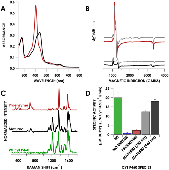
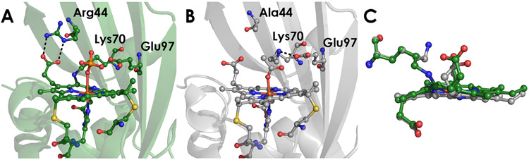
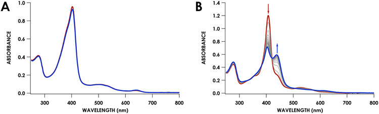
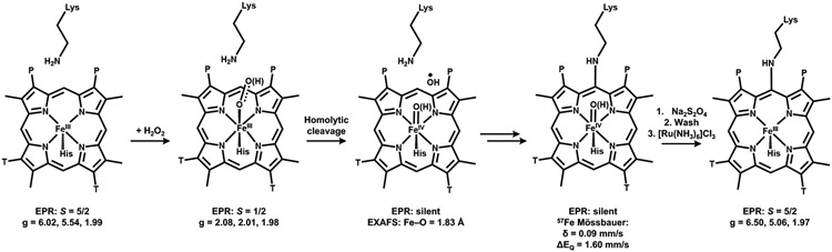
Cytochrome P460s are heme enzymes that oxidize hydroxylamine to nitrous oxide. They bear specialized "heme P460" cofactors that are cross-linked to their host polypeptides by a post-translationally modified lysine residue. Wild-type N. europaea cytochrome P460 may be isolated as a cross-link-deficient proenzyme following anaerobic overexpression in E. coli. When treated with peroxide, this proenzyme undergoes maturation to active enzyme with spectroscopic and catalytic properties that match wild-type cyt P460. This maturation reactivity requires no chaperones─it is intrinsic to the protein. This behavior extends to the broader cytochrome c'β superfamily. Accumulated data reveal key contributions from the secondary coordination sphere that enable selective, complete maturation. Spectroscopic data support the intermediacy of a ferryl species along the maturation pathway.


Figures

Figure 1.
Figure 1.
Known heme-protein cross-links. The purple arrows represent ester bonds to the heme methyl groups, the blue arrows represent cross-links to the vinyl group of the heme, the red arrows represent the tyrosine-heme cross-link in HAO, and the green arrow represents the lysine-heme cross-link in cyt P460. Adapted with permission from Reference . Copyright 2015, Elsevier.
Figure 2.
Figure 2.
The heme P460 active site in N. europaea HAO (PDB 4N4N) and cyt P460 (PDB 2JE3). Both are c-type hemes that feature additional covalent attachments. In HAO, a tyrosine residue forms cross-links from the phenolate O to the pyrrole α-carbon and from Cε to the α-meso-carbon. The heme in cyt P460 contains a cross-link from a lysine N to the γ-meso-carbon.
Figure 3.
Figure 3.
Hypothetical mechanism of peroxide-dependent heme-Lys cross-link formation in cyt P460. A) Possible routes for hydroxylation of the meso-carbon followed by B) condensation by lysine.
Figure 4.
Figure 4.
Characterization of the anaerobically purified proenzyme and matured proenzyme, which was generated by reaction of the proenzyme with 3 equivalents of Li2O2 followed by quenching with sodium dithionite, re-oxidization with [Ru(NH3)6]Cl3, and washing. A) UV/vis absorption spectra of the proenzyme (red) and matured enzyme (black). B) CW X-band EPR spectra collected at 12 K of the proenzyme (red) and matured enzyme (black). Grey traces are the corresponding simulations. C) Resonance Raman spectra obtained via excitation at 405 nm for the proenzyme (red), matured (black), and WT cyt P460 (green). D) Specific activities for WT cyt P460, the proenzyme, and matured proenzyme quantified by the 280 nm extinction coefficient (grey) or the 440 nm extinction coefficient (black). Error bars represent standard deviations of three trials.
Figure 5.
Figure 5.
Reaction of 7 μM proenzyme (red) with 3 equivalents (21 μM) of Li2O2 at 25 °C. Scans were recorded every 0.3 minutes. The scan immediately following Li2O2 addition is shown in black. The final scan is shown in blue, after which the reaction was quenched with Na2S2O4. Time courses of the absorbances corresponding to the proenzyme (404 nm) and product (460 nm) are shown in the inset.
Figure 6.
Figure 6.
Continuous-wave X-band EPR spectrum collected at 10 K of the rapid freeze-quench reaction of 200 μM proenzyme with 400 μM Li2O2 frozen at 1 second (black) with the simulated spectrum (red). The fit gave g-values of 2.08, 2.01, and 1.98. Oxygen background was removed from the raw experimental spectrum and the cavity was subtracted.
Figure 7.
Figure 7.
A) UV/vis spectral time-course for the reaction of the proenzyme with 8 equivalents of Li2O2 in the presence of excess (2 mM) guaiacol. The red trace is before Li2O2 was added, the black trace was immediately after addition, and the blue trace was 30 minutes after addition. Grey traces represent scans every 0.2 minutes. B) UV/vis spectrum from the reaction after addition of Na2S2O2.
Figure 8.
Figure 8.
Fe K-edge XAS XANES region for WT cyt P460 (green) and the reaction of ca. 500 μM proenzyme with 1 equivalent of Li2O2 frozen at 1 second (black).
Figure 9.
Figure 9.
A) Structure of WT N. europaea cyt P460 (PDB 2JE3) B) Structure of the N. europaea cyt P460 Arg44Ala mutant (PDB 8GAR). C) Overlay of the heme groups from the WT (green) and Arg44Ala (grey) cyt P460 structures, highlighting the change in position of the 6-β-pyrrolic propionate and γ-meso-carbon.
Figure 10.
Figure 10.
A) UV/vis spectral time-course for the stopped-flow reaction of 10 μM Arg44Ala with 8 equivalents (80 μM) Li2O2. The red trace is the first spectrum after mixing and the blue trace is the final spectrum at 30 seconds. The inset shows the absorbance at 403 nm over time fit to a double exponential. B) UV/vis spectrum comparing as-isolated Arg44Ala (red) and the intermediate formed during the stopped-flow reaction (black).
Figure 11.
Figure 11.
UV/vis spectral time-course for the reaction of 8 μM Methylococcus capsulatus WT cyt c′β (A) or F61K cyt c′β (B) with 8 equivalents (64 μM) Li2O2. The red trace is before the addition of Li2O2 and the blue trace is the final spectrum after Li2O2 additions. Grey traces represent scans every 0.2 minutes.
Figure 12.
Figure 12.
Summary of species observed during peroxide-driven cyt P460 maturation.

Similar articles

Cited by

References

    1. Macek B; Forchhammer K; Hardouin J; Weber-Ban E; Grangeasse C; Mijakovic I Protein Post-Translational Modifications in Bacteria. Nat. Rev. Microbiol 2019, 17 (11), 651–664. - PubMed
    1. Walsh CT; Garneau-Tsodikova S; Gatto GJ Jr. Protein Posttranslational Modifications: The Chemistry of Proteome Diversifications. Angew. Chem. Int. Ed 2005, 44 (45), 7342–7372. - PubMed
    1. Lin Y-W The Broad Diversity of Heme-Protein Cross-Links: An Overview. Biochim. Biophys. Acta 2015, 1854 (8), 844–859. - PubMed
    1. Bowman SEJ; Bren KL The Chemistry and Biochemistry of Hemec: Functional Bases for Covalent Attachment. Nat. Prod. Rep 2008, 25 (6), 1118–1130. - PMC - PubMed
    1. Coleman RE; Lancaster KM Heme P460: A (Cross) Link to Nitric Oxide. Acc. Chem. Res 2020, 53 (12), 2925–2935. 10.1021/acs.accounts.0c00573. - DOI - PMC - PubMed

Publication types