. 2023 Aug 8;8(15):e168663.
doi: 10.1172/jci.insight.168663.

Diminished responses to mRNA-based SARS-CoV-2 vaccines in individuals with rheumatoid arthritis on immune-modifying therapies

Affiliations

Diminished responses to mRNA-based SARS-CoV-2 vaccines in individuals with rheumatoid arthritis on immune-modifying therapies

Samuel D Klebanoff et al. JCI Insight. .

Abstract

Rheumatoid arthritis (RA) is a chronic inflammatory autoimmune disorder that causes debilitating swelling and destruction of the joints. People with RA are treated with drugs that actively suppress one or more parts of their immune system, and these may alter the response to vaccination against SARS-CoV-2. In this study, we analyzed blood samples from a cohort of patients with RA after receiving a 2-dose mRNA COVID-19 vaccine regimen. Our data show that individuals on the cytotoxic T lymphocyte antigen 4-Ig therapy abatacept had reduced levels of SARS-CoV-2-neutralizing antibodies after vaccination. At the cellular level, these patients showed reduced activation and class switching of SARS-CoV-2-specific B cells, as well as reduced numbers and impaired helper cytokine production by SARS-CoV-2-specific CD4+ T cells. Individuals on methotrexate showed similar but less severe defects in vaccine response, whereas individuals on the B cell-depleting therapy rituximab had a near-total loss of antibody production after vaccination. These data define a specific cellular phenotype associated with impaired response to SARS-CoV-2 vaccination in patients with RA on different immune-modifying therapies and help inform efforts to improve vaccination strategies in this vulnerable population.

Keywords: Adaptive immunity; Autoimmunity; Costimulation; Vaccines.


Conflict of interest statement

Conflict of interest: JHB is a scientific cofounder and Scientific Advisory Board member of GentiBio, is a consultant for Bristol Myers Squibb and Hotspot Therapeutics, and has past and current research projects sponsored by Amgen, Bristol Myers Squibb, Janssen, Novo Nordisk, and Pfizer. CS has been on an advisory board for Vertex Pharmaceuticals. MP is on the Scientific Advisory Board of Vaxart. DJC is on the Scientific Advisory Board of Sonoma Therapeutics.

Figures

Figure 1
Figure 1. Abatacept and rituximab reduce SARS-CoV-2–specific antibody levels after vaccination.
(A) Study schematic. (B) Normalized anti-S antibody levels as measured by ELISA. (C) Pseudovirus neutralization of patients’ sera, as AUC across serum dilutions, with historical/naive (HN) control. (D) Patient age graphed against anti-S antibody levels. (E) Time between each patient’s second vaccine dose and blood draw for the study graphed against anti-S antibody levels. Error bars represent mean ± SD. Linear regression shown with r2 values and P values testing probability of a nonzero slope. Statistics determined by Kruskal-Wallis test with post hoc Dunn’s multiple-comparison test. **P < 0.01, ***P < 0.001.
Figure 2
Figure 2. Abatacept treatment reduces activation and class switching in RBD-specific MBCs after vaccination.
(A) Representative gating on live CD3CD14CD16CD19+CD20+ B cells (left) and number (right) of SARS-CoV-2 RBD-specific B cells (RBD tetramer+decoy tetramer) from PBMCs from control (white), methotrexate-treated (MTX, blue), and abatacept-treated (red) individuals. (B) Representative gating on RBD-specific CD38lo nonplasmablast B cells for naive B cells (CD21+CD27), classical MBCs (CD21+CD27+), activated MBCs (CD21CD27+), and double-negative activated MBCs (CD21CD27). (C) Proportion of RBD-specific B cells that are each phenotype from individuals in the indicated treatment group. (D) Representative gating on RBD-specific CD38lo nonplasmablast B cells for isotypes IgD, IgM, IgG, and IgA. (E) Proportion of RBD-specific B cells expressing the isotypes indicated in the groups indicated. Data combined from 4 individual experiments. Error bars represent mean ± SD. Statistics determined by Kruskal-Wallis test with post hoc Dunn’s multiple-comparison test. All statistically significant comparisons (P < 0.05) are shown. *P < 0.05, **P < 0.01, ***P < 0.001. MBCs, memory B cells.
Figure 3
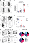
Figure 3. MTX and abatacept reduce S-specific memory T cell responses after vaccination.
(A) Representative gating of CD3+CD45RACD4+ T cells for AIM+ (CD69+CD137+) within indicated stimulation conditions. (B) Quantification of AIM expression by patient groups as percentage of CD3+CD45RACD4+ cells. (C) Representative gating of central memory (CD45RACD27+), effector memory (CD45RACD27), and Temra (CD45RA+CD27+) within non-naive and AIM+ T cells. (D) Quantification of CD4+ memory subsets within S protein–stimulated AIM+ cells. (E) Representative gating of CXCR5+ (containing the Tfh population), Th1 (CXCR3+CCR6), Th17 (CXCR3CCR6+), Th1/17 (CXCR3+CCR6+), and Th2 (CXCR3CCR6CCR4+) cells. (F) Pie charts showing percentage of spike-stimulated AIM+CD4+ T cells falling into each Th subset. Error bars represent mean ± SD. Statistics determined by Kruskal-Wallis test with post hoc Dunn’s multiple-comparison test. All statistically significant comparisons (P < 0.05) between treatment groups are shown. **P < 0.01.
Figure 4
Figure 4. Abatacept-treated patients have reduced Tfh-associated cytokine production by S-specific memory T cells after vaccination.
(A) Representative gating of AIM+ (CD69+CD154+) T cells for intracellular cytokine staining assay coculture. (B) Representative gating of IL-2, IL-21, IL-10, IFN-γ, IL-4, IL-13, and IL-17A expression within AIM+CD4+ T cells. (C) Quantification of the expression of each cytokine by percentage of AIM+CD4+ T cells. (D) Coexpression of IL-2, IL-21, and IFN-γ in each indicated combination. (E) Anti-S antibody level graphed percentage of S-activated AIM+CD4+ T cells expressing IL-21. (F) Anti-S antibody level graphed percentage of S-activated AIM+CD4+ T cells expressing IFN-γ. Error bars represent mean ± SD. Linear regression shown with r2 values and P values testing probability of a nonzero slope. Statistics determined by Kruskal-Wallis test with post hoc Dunn’s multiple-comparison test. All statistically significant comparisons (P < 0.05) are shown. *P < 0.05, **P < 0.01, ***P < 0.001.

References

    1. Baden LR, et al. Efficacy and safety of the mRNA-1273 SARS-CoV-2 vaccine. N Engl J Med. 2021;384(5):403–416. doi: 10.1056/NEJMoa2035389. - DOI - PMC - PubMed
    1. Skowronski DM, De Serres G. Safety and efficacy of the BNT162b2 mRNA Covid-19 vaccine. N Engl J Med. 2021;384(16):1576–1577. doi: 10.1056/NEJMc2036242. - DOI - PubMed
    1. Garcillan B, et al. Response to vaccines in patients with immune-mediated inflammatory diseases: a narrative review. Vaccines (Basel) 2022;10(2):297. doi: 10.3390/vaccines10020297. - DOI - PMC - PubMed
    1. Sonani B, et al. COVID-19 vaccination in immunocompromised patients. Clin Rheumatol. 2021;40(2):797–798. doi: 10.1007/s10067-020-05547-w. - DOI - PMC - PubMed
    1. Rodda LB, et al. Imprinted SARS-CoV-2-specific memory lymphocytes define hybrid immunity. Cell. 2022;185(9):1588–1601. doi: 10.1016/j.cell.2022.03.018. - DOI - PMC - PubMed

Publication types