

In vivo measurement of regional brain and tumor pH using [14C]dimethyloxazolidinedione and quantitative autoradiography. II: Characterization of the extracellular fluid compartment using pH-sensitive microelectrodes and [14C]sucrose
- PMID: 3733903
- PMCID: PMC3047405
- DOI: 10.1038/jcbfm.1986.76
In vivo measurement of regional brain and tumor pH using [14C]dimethyloxazolidinedione and quantitative autoradiography. II: Characterization of the extracellular fluid compartment using pH-sensitive microelectrodes and [14C]sucrose
Abstract
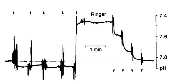
We measured the extracellular (interstitial) pH (pHe) of RG-2 rat gliomas using H+-sensitive microelectrodes and estimated the volume of tumor extracellular space based on the tissue-plasma ratio of [14C]sucrose. The average RG-2 pHe was 7.63 +/- 0.15 (mean +/- SD, n = 6), whereas the average pHe of contralateral brain tissue was 7.34 +/- 0.10 (n = 3) and arterial pH was 7.36 +/- 0.02. RG-2 extracellular space water volume was estimated to be 0.3 ml water/g tissue. In separate experiments in normal, nontumored rats, intracellular pH (pHi) was calculated for nine gray and white matter regions based on measurements of tissue and plasma [14C]dimethyloxazolidinedione concentration. pHi values ranged from 6.80 to 6.94, and no consistent gray-white differences were observed. Our data suggest that tumor pHi is not more acidic than that of normal brain tissue and that the observed alkalinity of primary brain tumors is due to the presence of a large alkaline extracellular space.
Figures

References
-
- Ames A, III, Sakanoue M, Endo S. Na, K, Ca, Mg, and Cl concentrations in choroid plexus fluid and cisternal fluid compared with plasma ultrafiltrate. J Neurophysiol. 1964;27:672–681. - PubMed
-
- Ammann D, Lanter F, Steiner RA, Schulthero P, Shijo Y, Simon W. Neutral carrier based hydrogen ion selective microelectrode for extra- and intracellular studies. Anal Chem. 1981;53:2267–2269. - PubMed
-
- Arnold JB, Junck L, Rottenberg DA. In vivo measurement of regional brain and tumor pH using [14C]dimethyloxazolidinedione and quantitative autoradiography. J Cereb Blood Flow Metab. 1985;5:369–375. - PubMed
-
- Beaney RP, Brooks DJ, Leenders KL, Thomas DGT, Jones T, Halnan KE. Blood flow and oxygen utilisation in the contralateral cerebral cortex of patients with untreated intracranial tumours as studied by positron emission tomography with observations on the effect of decompressive surgery. J Neurol Neurosurg Psychiatry. 1985;48:310–319. - PMC - PubMed
Publication types
MeSH terms
Substances
Grants and funding
LinkOut - more resources
Full Text Sources
Other Literature Sources
Medical
