. 2023 Jun 27;120(26):e2303292120.
doi: 10.1073/pnas.2303292120. Epub 2023 Jun 20.

A broad-spectrum macrocyclic peptide inhibitor of the SARS-CoV-2 spike protein

Affiliations

A broad-spectrum macrocyclic peptide inhibitor of the SARS-CoV-2 spike protein

Vito Thijssen et al. Proc Natl Acad Sci U S A. .

Abstract

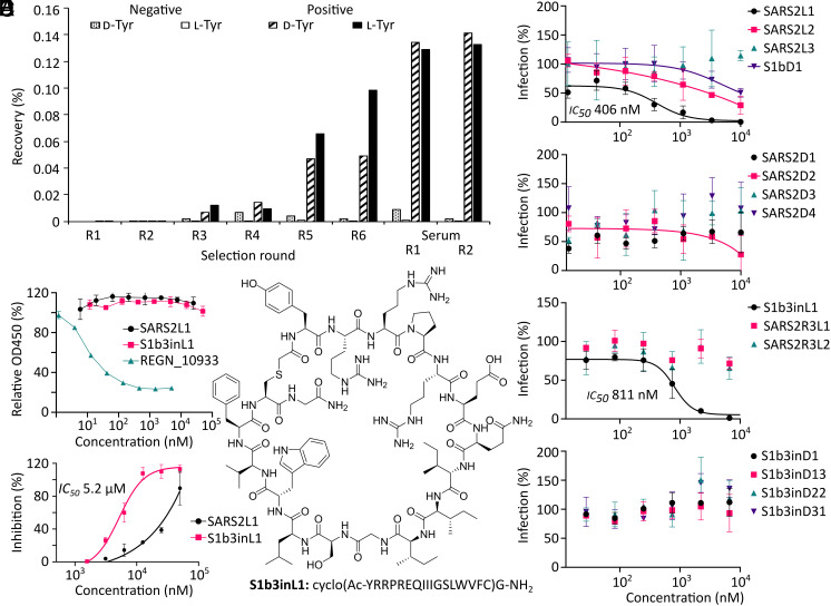
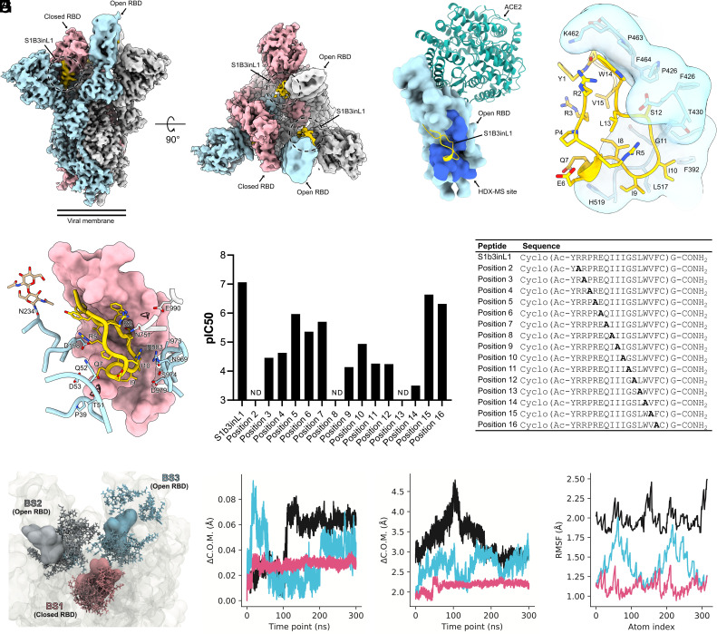
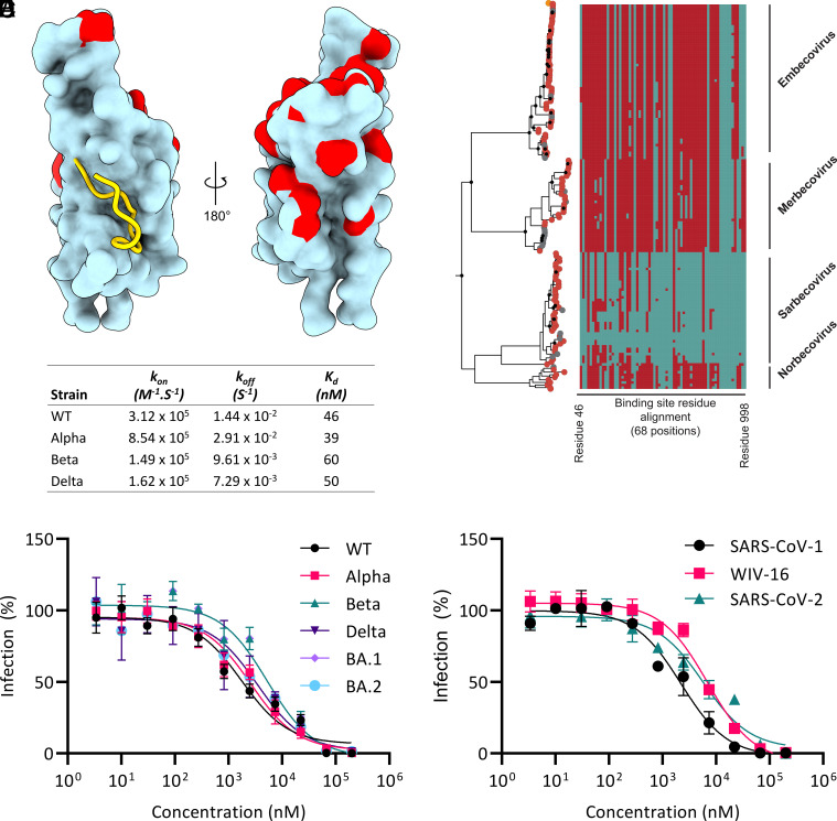
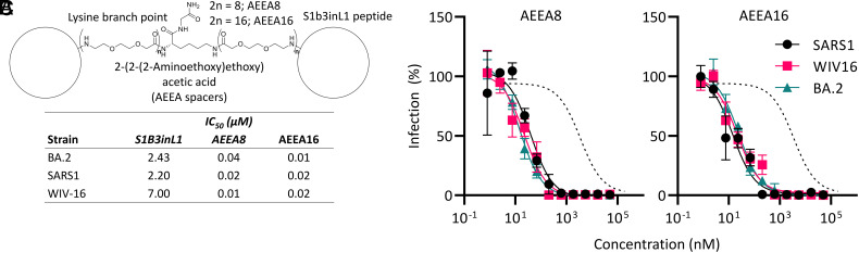
The ongoing COVID-19 pandemic has had great societal and health consequences. Despite the availability of vaccines, infection rates remain high due to immune evasive Omicron sublineages. Broad-spectrum antivirals are needed to safeguard against emerging variants and future pandemics. We used messenger RNA (mRNA) display under a reprogrammed genetic code to find a spike-targeting macrocyclic peptide that inhibits SARS-CoV-2 (severe acute respiratory syndrome coronavirus 2) Wuhan strain infection and pseudoviruses containing spike proteins of SARS-CoV-2 variants or related sarbecoviruses. Structural and bioinformatic analyses reveal a conserved binding pocket between the receptor-binding domain, N-terminal domain, and S2 region, distal to the angiotensin-converting enzyme 2 receptor-interaction site. Our data reveal a hitherto unexplored site of vulnerability in sarbecoviruses that peptides and potentially other drug-like molecules can target.

Keywords: Cryo-EM; SARS-CoV-2; antivirals; mRNA display; macrocyclic peptides.


Conflict of interest statement

I.D. is an employee of Thermo Fisher Scientific. V.T., D.L.H., F.J.M.v.K., and S.A.K.J. are named inventors on a patent application that has been filed on 15 July 2022 entitled: Antiviral cyclic compounds (EP22185235; patent applicants: Universiteit Utrecht Holdings B.V. on behalf of Utrecht University). The other authors declare no competing interests.

Figures

Fig. 1.
Fig. 1.
Macrocyclic peptide selection strategy and selection results. (A) Enrichment of library recovery across rounds for two random 15-mer peptide libraries initiated with chloroacetyl-d-tyrosine and -l-tyrosine. Rounds with fetal calf serum added during incubation are marked as “serum”. (B) Dose–response curves from screening of peptides identified by high throughput sequencing of enriched libraries for inhibition of pseudovirus infection in VeroE6 cells. (C) ELISA showing lack of competition for ACE2 binding to spike protein by the most promising peptide hits, compared to REGN_10933 antibody positive control. (D) Dose–response curve for SARS-CoV-2 inhibition assays in HEK293 ACE2+ TMPRSS2+ cells with the most promising hits from the pseudovirus screen. (E) Structure of most promising hit, peptide S1b3inL1, and its sequence. Error bars are SD of technical triplicates (duplicate for ELISA), with a representative shown of duplicate experiments.
Fig. 2.
Fig. 2.
Characterization of the S1B3inL1 binding site. (A) Composite cryo-EM density map for the SARS-CoV-2 spike ectodomain in complex with S1B3inL1, shown as two orthogonal views. The spike protomers are colored blue, gray, and pink, and the macrocyclic peptide is colored gold. (B) Surface representation of the S1B3inL1-bound S1B overlaid with the S1B-bound ACE2 (PDB ID: 6M0J). Residues 388 to 399, 421 to 431 and 511 to 520, identified by HDX-MS, are colored dark blue. (C) Close-up view showing the interactions between S1B3inL1 and the SARS-CoV-2 S1B. (D) Close-up view of the S1B3inL1-bound closed S1B. Selected S1A and S2 residues are shown as sticks, and putative solvent densities are shown as a mesh. (E) Relative SARS-CoV-2 pseudovirus inhibition activity of peptides with residues at positions 2 to 16 substituted for alanine, with sequences of peptides used in the assay displayed in the table. Activities are shown as pIC50 values, with a higher pIC50 value indicating more potent activity. Alanine mutations for which no inhibition was detected are denoted as “ND”. (F) Initial poses for S1b3inL1 in binding sites (BS) 1-3 (colored by spike protomer as in panel A) are shown in surface representation. Coordinates for final poses after 300 ns are shown in licorice representation. Initial coordinates of the full-length spike are shown in transparent surface representation. (G) Center of mass (COM) evolution for S1B3inL1 BS1-3 from a single representative trajectory. BS2 and 3 peptides (cyan and black) exhibit large COM movements over the 300 ns trajectory, relative to BS1 peptide. (H) Distance between the COM of S1B3inL1 in BS1-3 and the COM of the closest S1B in the initial coordinates. S1B3inL1 COM movements in BS2 and 3 are decoupled from COM movements in the S1B. BS1 remains stable with the closed S1B. (I). Root-mean-square fluctuation (RMSF) over all S1B3inL1 atoms averaged over each sampled trajectory. S1B3inL1 in BS1 remains more conformationally stable than BS2 and 3.
Fig. 3.
Fig. 3.
Sequence conservation of the S1B3inL1 binding site. (A) Close-up view of the S1b3inL1 binding site (gold cartoon) relative to mutation sites (red) in the Alpha, Beta, Gamma, Delta, Lambda, Mu and Omicron (BA.1, BA.2, BA.2.12.1, BA.2.75, and BA.4/5) variants within the S1b domain (cyan surface representation). (B) Maximum likelihood phylogenetic reconstruction of β-coronavirus lineages. The topology is rooted at the midpoint and is reconstructed from 725 nonredundant amino acid sequences. Peptide-binding site residues (designated as SARS-COV2 residue, cyan, or non-SARS-COV2 residue, maroon) are aligned respective tips. Major β-coronavirus lineages are labeled. (C) Characterization of S1b3inL1 binding to S1B by Biacore for wild type and variants. (D) Dose–response curve for inhibition of pseudovirus infection in VeroE6 cells by S1b3inL1 across VOCs. (E) Dose–response curve for inhibition of pseudovirus infection in VeroE6 cells by S1b3inL1 across sarbecoviruses. Error bars are SD of technical triplicates, with a representative shown of duplicate experiments.
Fig. 4.
Fig. 4.
Dimerization for improved potency. (A) Representative structure of S1b3inL1 AEEA dimers. (B) Dose-response curve for inhibition of pseudovirus infection in VeroE6 cells by S1b3inL1 dimers of varying length (total number of AEEA spacers as indicated). Dotted lines indicate fit to monomeric S1b3inL1 peptide with BA.2 (Fig. 3D) (C) IC50 values for S1b3inL1 dimers in the pseudovirus infection assay in panel (B). Error bars are SD of technical triplicates, with a representative shown of duplicate experiments.

References

    1. Eastman R. T., et al. , Remdesivir: A review of its discovery and development leading to emergency use authorization for treatment of COVID-19. ACS Cent. Sci. 6, 672–683 (2020). - PMC - PubMed
    1. Painter G. R., Natchus M. G., Cohen O., Holman W., Painter W. P., Developing a direct acting, orally available antiviral agent in a pandemic: The evolution of molnupiravir as a potential treatment for COVID-19. Curr. Opin. Virol. 50, 17–22 (2021). - PMC - PubMed
    1. Joyce R. P., Hu V. W., Wang J., The history, mechanism, and perspectives of nirmatrelvir (PF-07321332): An orally bioavailable main protease inhibitor used in combination with ritonavir to reduce COVID-19-related hospitalizations. Med. Chem. Res. 31, 1637–1646 (2022). - PMC - PubMed
    1. Pantaleo G., Correia B., Fenwick C., Joo V. S., Perez L., Antibodies to combat viral infections: Development strategies and progress. Nat. Rev. Drug Discov. 21, 676–696 (2022). - PMC - PubMed
    1. Bosch B. J., van der Zee R., de Haan C. A. M., Rottier P. J. M., The Coronavirus spike protein is a class I virus fusion protein: Structural and functional characterization of the fusion core complex. J. Virol. 77, 8801–8811 (2003). - PMC - PubMed

Publication types

Supplementary concepts