. 2023 Jun 27;120(26):e2301606120.
doi: 10.1073/pnas.2301606120. Epub 2023 Jun 20.

Biodegradable lipophilic polymeric mRNA nanoparticles for ligand-free targeting of splenic dendritic cells for cancer vaccination

Affiliations

Biodegradable lipophilic polymeric mRNA nanoparticles for ligand-free targeting of splenic dendritic cells for cancer vaccination

Elana Ben-Akiva et al. Proc Natl Acad Sci U S A. .

Abstract

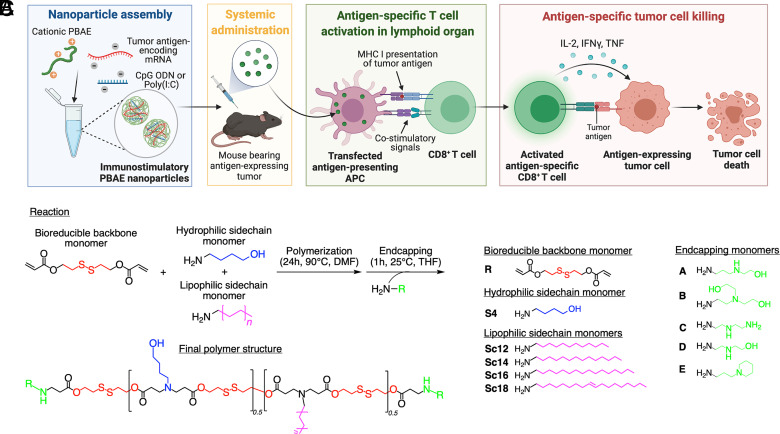
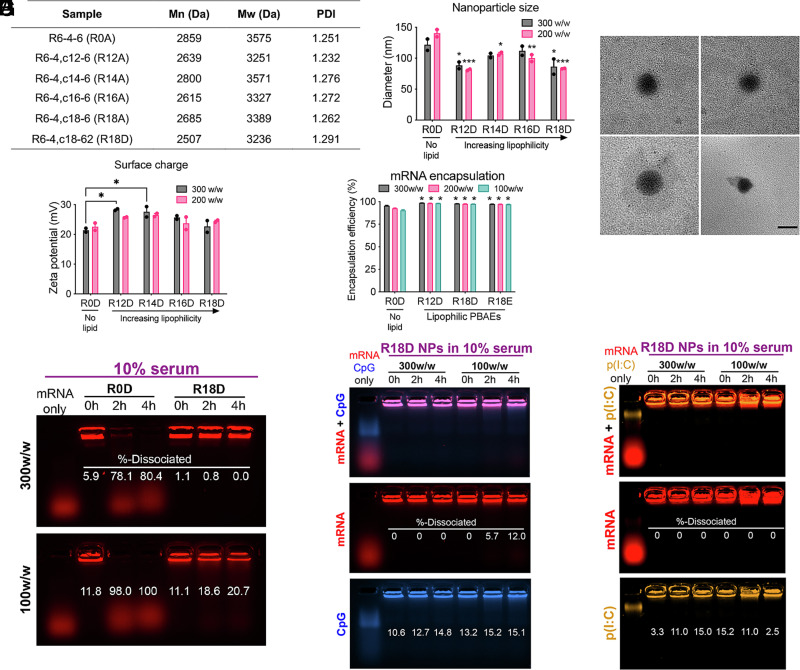
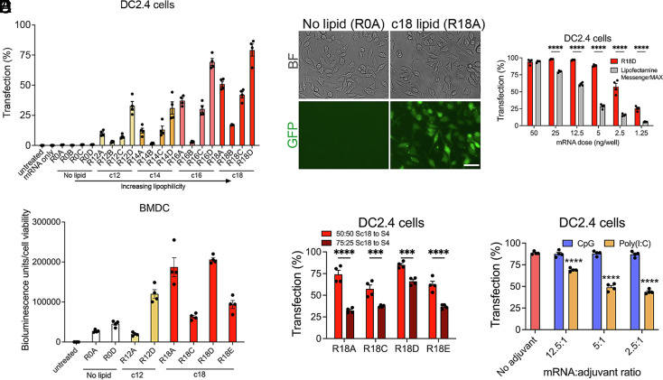
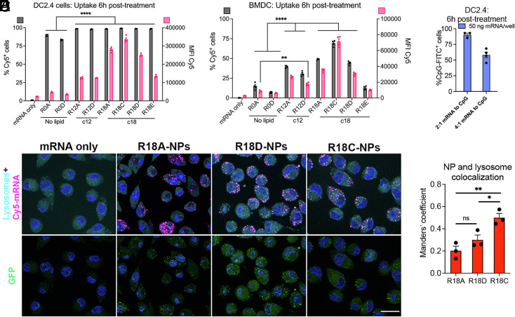
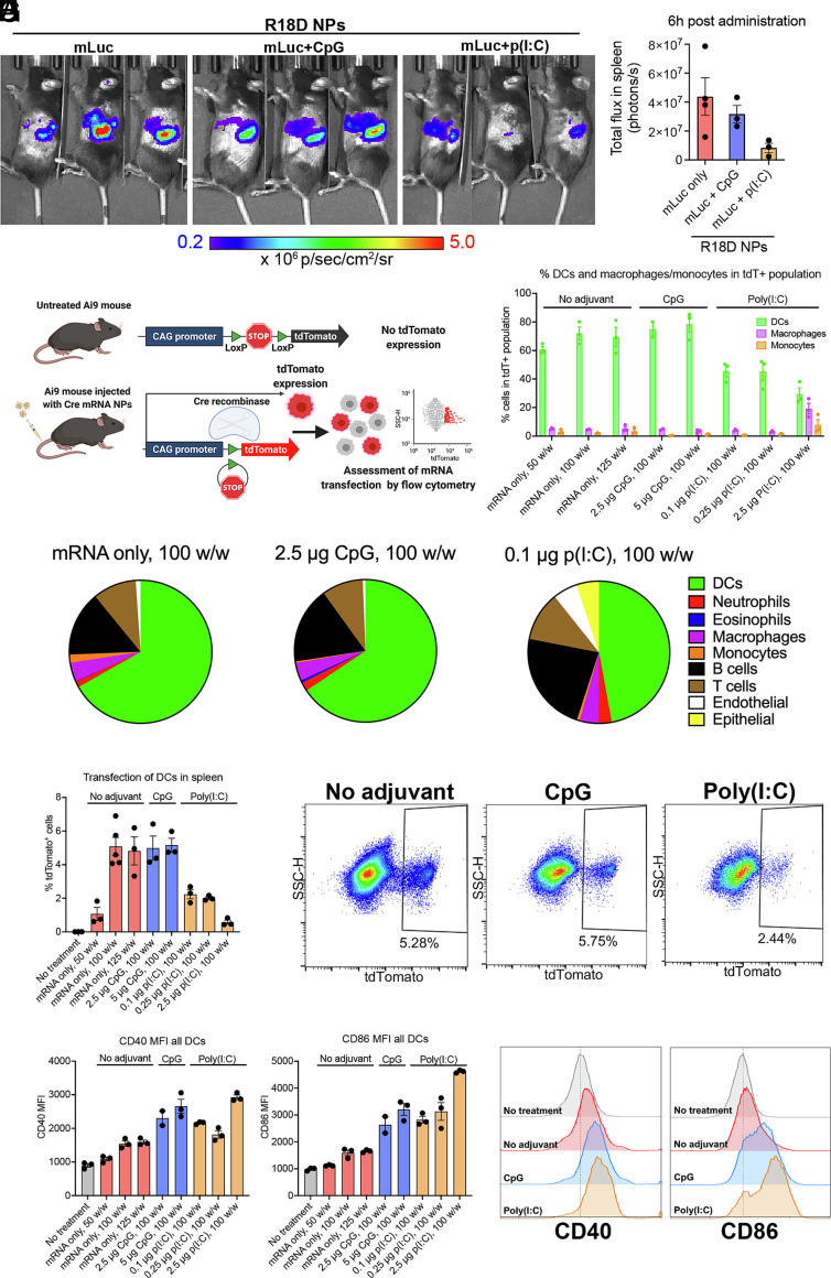
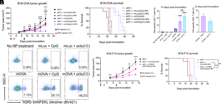
Nanoparticle (NP)-based mRNA cancer vaccines hold great promise to realize personalized cancer treatments. To advance this technology requires delivery formulations for efficient intracellular delivery to antigen-presenting cells. We developed a class of bioreducible lipophilic poly(beta-amino ester) nanocarriers with quadpolymer architecture. The platform is agnostic to the mRNA sequence, with one-step self-assembly allowing for delivery of multiple antigen-encoding mRNAs as well as codelivery of nucleic acid-based adjuvants. We examined structure-function relationships for NP-mediated mRNA delivery to dendritic cells (DCs) and identified that a lipid subunit of the polymer structure was critical. Following intravenous administration, the engineered NP design facilitated targeted delivery to the spleen and preferential transfection of DCs without the need for surface functionalization with targeting ligands. Treatment with engineered NPs codelivering antigen-encoding mRNA and toll-like receptor agonist adjuvants led to robust antigen-specific CD8+ T cell responses, resulting in efficient antitumor therapy in in vivo models of murine melanoma and colon adenocarcinoma.

Keywords: cancer; delivery; mRNA; nanoparticle; vaccine.


Conflict of interest statement

D.M.P. is a consultant for Compugen, Shattuck Labs, Tempest, Immunai, Bristol-Myers Squibb, Amgen, Janssen, Astellas, Rockspring Capital, Immunomic, and Dracen. J.J.G. is a cofounder, manager, and CTO of Dome Therapeutics; cofounder, board member, and CSO of Cove Therapeutics; cofounder and manager of OncoSwitch Therapeutics; cofounder of WyveRNA Therapeutics; scientific advisory board member of Mana Bio; and board member of VasoRx. S.Y.T. is a cofounder and manager of OncoSwitch Therapeutics. D.M.P. owns founder’s equity in manaT Holdings, LLC, Trex, Jounce, Anara, Tizona, Tieza, and RAPT. J.J.G. owns equity in Dome, Cove, OncoSwitch, WyveRNA, Mana Bio, and VasoRx. S.Y.T. owns equity in OncoSwitch. E.B.-A., J.K., S.Y.T., and J.J.G. are a coinventors on patents filed by Johns Hopkins University related to technologies discussed in the manuscript. D.M.P. receives research funding from Compugen, Bristol-Myers Squibb, and Anara and royalties on patents licensed by Compugen, BMS, and Immunomic.

Figures

Fig. 1.
Fig. 1.
Bioreducible lipophilic PBAE–mRNA nanoparticles (NPs) as cancer vaccine. (A) Schematic of the mRNA-based cancer vaccine technology using polymeric (PBAE) NPs. (B) Reaction scheme for bioreducible lipophilic PBAEs. The bioreducible diacrylate backbone monomer (R) is polymerized with a 1:1 mixture of a hydrophilic amine sidechain monomer (S4) and a lipophilic amine side chain monomer (Sc12-18) via Michael addition. The obtained diacrylate-terminated random copolymer is endcapped with an amine-containing monomer (AE) to form the final polymer structure. (C) Monomers used in the combinatorial library synthesis to form bioreducible lipophilic PBAEs.
Fig. 2.
Fig. 2.
Characterization of bioreducible lipophilic poly(beta-amino ester) (PBAE) polymers and mRNA nanoparticles (NPs). (A) Molecular weights of PBAEs of varying lipophilicity assessed by GPC. (B) Hydrodynamic diameter of PBAE NPs formed at a 300 or 200 w/w ratio of polymer to mRNA assessed via DLS (n = 2). Significance indicates comparison to nonlipophilic PBAE nanoparticles (R0D) at the respective w/w ratio. (C) Representative TEM images of R18D mRNA NPs (Scale bar, 200 nm). (D) Surface charge of mRNA PBAE NPs in PBS (n = 2). (E) Encapsulation efficiency of mRNA assessed by the RiboGreen assay (n = 3). Significance indicates comparison to nonlipophilic PBAE nanoparticles (R0D) at the respective w/w ratio. (F) Encapsulation and dissociation of fluorescently labeled mRNA for nonlipophilic (R0D) and lipophilic (R18D) NPs formed at 300 and 100 w/w ratios after incubation in 10% serum over 4 h assessed by a gel electrophoresis assay. (G) mRNA and CpG ODN, (H) mRNA and poly(I:C) dissociation in 10% serum from R18D-based NPs formed at 300 and 100 w/w ratios over 4 h, respectively. Error bars represent SEM. *P < 0.05, **P < 0.01, and ***P < 0.001.
Fig. 3.
Fig. 3.
Transfection of dendritic cells (DCs) in vitro by bioreducible liphophilic PBAE mRNA nanoparticles (NPs). (A) Polymer library was evaluated for transfection of the murine dendritic cell line DC2.4 using mRNA encoding GFP. Cells were treated with NPs formed at 200 w/w and a dose of 50 ng mRNA/well, and transfection efficiency was assessed via flow cytometry after 24 h. (B) Representative bright-field (BF) and fluorescent microscopy images of DC2.4 cells transfected with nonlipophilic R0A or lipophilic R18A GFP mRNA NPs (Scale bar, 50 nm). (C) Transfection of DC2.4 cells by top-performing R18D NPs was assessed at various mRNA doses and compared to leading commercial mRNA transfection reagent Lipofectamine MessengerMAX. (D) A subset of the polymer library was evaluated on murine BMDCs using luciferase-encoding mRNA. Cells were treated with NPs at a dose of 25 ng mRNA/well, and bioluminescence activity was assessed after 24 h to determine transfection levels normalized to cell viability. (E) Polymers with the Sc18 monomer were synthesized with a 50:50 or 75:25 ratio of lipophilic side chain monomer Sc18 to hydrophilic side chain monomer S4. DC2.4 cells were treated with GFP mRNA NPs with varied lipophilicity at a dose of 25 ng mRNA/well, and transfection was assessed after 24 h. (F) The transfection efficiency in DC2.4s was examined following treatment with R18D NPs coencapsulating GFP mRNA and CpG or poly(I:C) adjuvants with varied mRNA to adjuvant ratios where the mRNA dose was kept constant (25 ng/well) after 24 h. Significance indicates comparison to no adjuvant control. Error bars represent SEM (n = 4). ***P < 0.001 and ****P < 0.0001.
Fig. 4.
Fig. 4.
Cellular uptake and endosomal escape following mRNA nanoparticle (NP) design. (A) DC2.4 cells and (B) murine primary BMDCs were treated with NPs carrying Cy5-mRNA at a dose of 50 ng mRNA/well, and NP uptake was assessed at 6 h posttreatment by flow cytometry (n = 4). (C) DC2.4 cells were treated with R18D NPs coencapsulating mRNA at 50 ng mRNA/well and FITC-CpG, and uptake of CpG was assessed 6 h posttreatment by flow cytometry (n = 4). (D) Representative images of DC2.4 cells labeled with lysosome/endosome dye 6 h posttreatment with NPs carrying Cy5-labeled GFP-encoding mRNA to visualize cellular uptake, NP colocalization with endosomes/lysosomes, and GFP transfection (Scale bars, 20 μm). (E) Manders’ coefficient was determined using ImageJ to quantify the degree of colocalization between NPs and endosomes/lysosomes (n = 3). Error bars represent SEM. *P < 0.05, **P < 0.01, and ****P < 0.0001.
Fig. 5.
Fig. 5.
In vivo transfection in spleen following systemic administration of R18D mRNA nanoparticles (NPs). (A) R18D NPs carrying luciferase mRNA (mLuc) (10 μg/mouse) and CpG (2.5 μg/mouse) or poly(I:C) (0.1 μg/mouse) were assembled at a polymer-to-nucleic acid ratio of 100 w/w and administered intravenously to C57BL/6J mice. Whole-animal bioluminescence imaging was performed 6 h after administration. (B) Image analysis was used to assess total flux in the spleen. (C) Schematic of the Ai9 mouse model used to assess transfected cell types in vivo following systemic administration of mRNA NPs carrying Cre mRNA. Cells that are transfected undergo Cre recombinase–mediated recombination, resulting in tdTomato expression that is detected by flow cytometry. (D–H) R18D Cre mRNA NPs were administered intravenously to Ai9 mice at 10 μg mRNA/mouse, and tdTomato expression in key cell populations in the spleen was assessed after 24 h. (D) Percent of all tdTomato+ (tdT+) cells in the spleen that are DCs, macrophages, or monocytes. (E) Pie charts indicating average share of transfected cells in the spleen belonging to each cell population shown for NP treatments carrying no adjuvant, 2.5 μg CpG/mouse, or 0.1 μg poly(I:C)/mouse. (F) Percent of DCs in the spleen that are transfected. (G) Representative flow cytometry plots showing transfected tdTomato+ DCs treated with mRNA-NP formulations coencapsulating no adjuvant, 2.5 μg CpG, or 0.1 μg poly(I:C). (H) Geometric mean fluorescent intensity (MFI) of CD40 and CD86 expression in all splenic DCs. (I) Representative histograms of CD40 and CD86 expression in no-treatment control, and following NP treatment coencapsulating no adjuvant, 2.5 μg CpG/mouse, or 0.1 μg poly(I:C)/mouse. Error bars represent SEM.
Fig. 6.
Fig. 6.
In vivo therapeutic efficacy of PBAE mRNA nanoparticle (NP) vaccination in B16-OVA and B16-F10 mouse melanoma models. (A–E) 3 × 10⁵ B16-OVA cells were inoculated subcutaneously on day 0, and R18D NPs encapsulating luciferase-encoding mRNA or OVA-encoding mRNA were administered intravenously (I.V.) on days 4 and 9 at 10 μg mRNA/mouse and 2.5 μg CpG/mouse or 0.1 μg poly(I:C)/mouse (n = 7 to 8 mice/group). Then, 200 μg of aPD-1 was injected intraperitoneally (I.P.) on day 5. (A) Tumor growth measurements showing in vivo therapeutic effects between treatments. *P < 0.05, **P < 0.01, and ****P < 0.0001 for comparison between aPD-1 + mOVA/CpG NP treatment and respective controls (indicated by color). #P < 0.05, ##P < 0.01, and ####P < 0.0001 for comparison between aPD-1 + mOVA/p(I:C) NP treatment and respective controls (indicated by color). (B) Mice were euthanized once tumors reached 200 mm2, and survival curves are shown. (C) Mice were bled on day 14 postinoculation, and the percent of OVA-specific CD8+ T cells out of total CD8+ T cells was assessed using H2Kb-SIINFEKL tetramer staining. Significance indicates comparison of mOVA/CpG and mOVA/p(I:C) NP treatment compared to all respective controls. (D) On day 32 postinoculation, surviving mice in mOVA/CpG and mOVA/p(I:C) NP treatment groups were bled, and the presence of OVA-specific CD8+ T cells was assessed via tetramer staining. (E) Representative flow cytometry plots showing H2Kb-SIINFEKL tetramer staining in CD3+ CD8+ cells on day 14. (F and G) 3 × 10⁵ B16-F10 cells were inoculated subcutaneously on day 0, and R18D NPs encapsulating luciferase mRNA or a 1:1 mixture of TRP2 and GP100-encoding mRNA (10 μg total mRNA/mouse) and CpG were administered following the previously described treatment scheme (n = 7 to 8 mice/group). (F) Tumor growth measurements showing in vivo therapeutic effects between treatments. **P < 0.01, ***P < 0.001, and ****P < 0.0001 for comparison between aPD-1 + mTRP2/mGP100/CpG NP treatment and aPD-1 control (black) or aPD-1 + mLuc/CpG NP group (pink). #P < 0.05 for comparison between the aPD-1 + mLuc/CpG NP group and aPD-1 group. (G) Mice were euthanized once tumors reached 200 mm2, and survival curves are shown. Error bars represent SEM.
Fig. 7.
Fig. 7.
In vivo therapeutic efficacy of PBAE mRNA nanoparticle (NP) vaccination in the MC38-OVA mouse colon carcinoma model. (A) 1 × 10⁶ MC38-OVA cells were inoculated subcutaneously in the right flank of C57BL/6J mice on day 0, and R18D NPs encapsulating luciferase-encoding mRNA or OVA-encoding mRNA (10 μg mRNA/mouse) and CpG (2.5 μg/mouse) were administered intravenously on days 9 and 14 (n = 7 to 8 mice/group). Then, 200 μg of aPD-1 was injected intraperitoneally on day 10. Tumor growth measurements showing the in vivo therapeutic effects between the treatment groups. Significance indicates comparison of the aPD-1 + mOVA/CpG NP treatment group to the aPD-1 group (black) or aPD-1 + mLuc/CpG NP group (pink). (B) Mice were euthanized once tumors reached 200 mm2, and survival curves are shown. (C) Four mice were randomly selected from each group to be bled on day 21 postinoculation, and the percent of OVA-specific CD8+ T cells out of total CD8+ T cells in the blood was assessed using H2Kb SIINFEKL tetramer staining. (D) The percent of CD8+ T cells out of total CD3+ T cells in blood is shown. (E) Representative flow cytometry plots showing BV421 H2Kb SIINFEKL tetramer staining in CD3+ CD8+ cells in all groups. Error bars represent SEM.

Similar articles

Cited by

References

    1. Blass E., Ott P. A., Advances in the development of personalized neoantigen-based therapeutic cancer vaccines. Nat. Rev. Clin. Oncol. 18, 215–229 (2021). - PMC - PubMed
    1. Whiteside T. L., Demaria S., Rodriguez-Ruiz M. E., Zarour H. M., Melero I., Emerging opportunities and challenges in cancer immunotherapy. Clin. Cancer Res. 22, 1845–1855 (2016). - PMC - PubMed
    1. Vormehr M., Türeci Ö., Sahin U., Harnessing tumor mutations for truly individualized cancer vaccines. Annu. Rev. Med. 70, 395–407 (2019). - PubMed
    1. Wang R. F., Wang H. Y., Immune targets and neoantigens for cancer immunotherapy and precision medicine. Cell Res. 27, 11–37 (2017). - PMC - PubMed
    1. Hu Z., Ott P. A., Wu C. J., Towards personalized, tumour-specific, therapeutic vaccines for cancer. Nat. Rev. Immunol. 18, 168–182 (2018). - PMC - PubMed

Publication types

LinkOut - more resources