. 2023 Jun;13(6):230058.
doi: 10.1098/rsob.230058. Epub 2023 Jun 21.

The structural basis of the talin-KANK1 interaction that coordinates the actin and microtubule cytoskeletons at focal adhesions

Affiliations

The structural basis of the talin-KANK1 interaction that coordinates the actin and microtubule cytoskeletons at focal adhesions

Xingchen Li et al. Open Biol. 2023 Jun.

Abstract

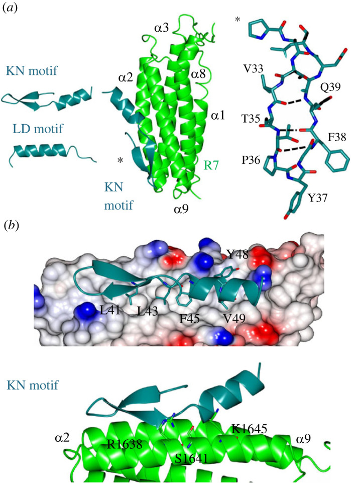
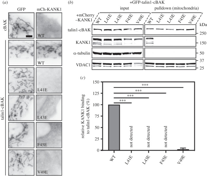
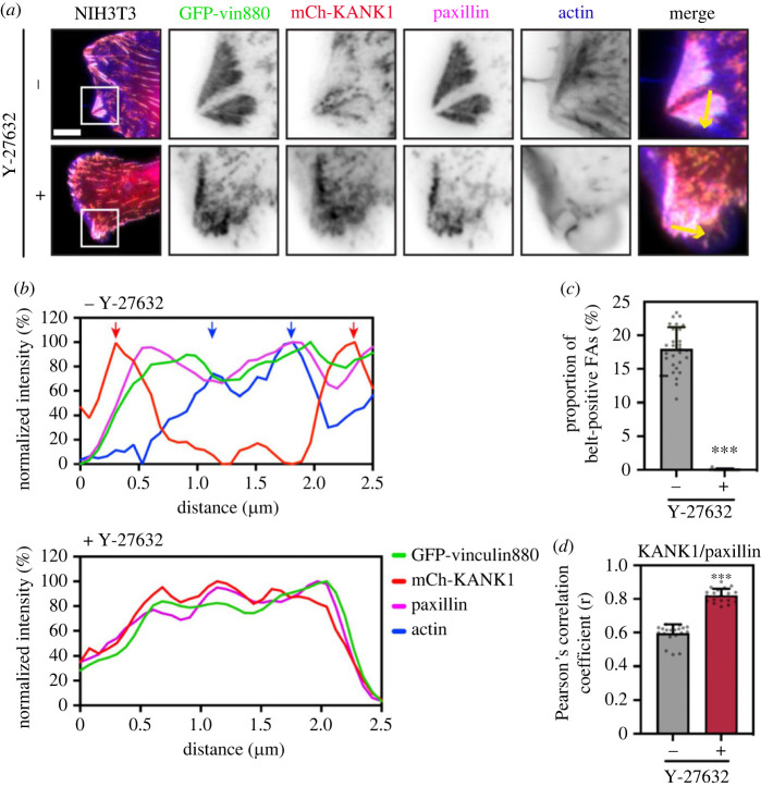
Adhesion between cells and the extracellular matrix is mediated by heterodimeric (αβ) integrin receptors that are intracellularly linked to the contractile actomyosin machinery. One of the proteins that control this link is talin, which organizes cytosolic signalling proteins into discrete complexes on β-integrin tails referred to as focal adhesions (FAs). The adapter protein KANK1 binds to talin in the region of FAs known as the adhesion belt. Here, we adapted a non-covalent crystallographic chaperone to resolve the talin-KANK1 complex. This structure revealed that the talin binding KN region of KANK1 contains a novel motif where a β-hairpin stabilizes the α-helical region, explaining both its specific interaction with talin R7 and high affinity. Single point mutants in KANK1 identified from the structure abolished the interaction and enabled us to examine KANK1 enrichment in the adhesion belt. Strikingly, in cells expressing a constitutively active form of vinculin that keeps the FA structure intact even in the presence of myosin inhibitors, KANK1 localizes throughout the entire FA structure even when actomyosin tension is released. We propose a model whereby actomyosin forces on talin eliminate KANK1 from talin binding in the centre of FAs while retaining it at the adhesion periphery.

Keywords: KANK1; cell adhesion; talin.


Conflict of interest statement

We declare we have no competing interests.

Figures

Figure 1.
Figure 1.
The structure of talin R7 in complex with the KANK1 KN motif. (a) (Top) Talin contains an N-terminal FERM domain connected to a rod region (R1–R13) composed of thirteen 4- and 5-helix bundles. The KANK1 binding site is on the R7 helical bundle. KANK1 domain structure (middle) and (bottom) the three protein units used in the crystallization strategy were (i) a KN-BBD (BCL6 binding domain) synthetic peptide, (ii) the R7R8 module, and (iii) the BCL6–BTB homodimer. (b) The KN motif (dark green) binds to talin R7 (green) between helices α2 and α9 with no change in any of the helical positions. (c) The BTB chaperone works by capturing the talin–KANK1 complex in a readily crystallizable lattice. The N-terminal KN motif binds to R7 and the BBD engages the BCL6 lateral groove.
Figure 2.
Figure 2.
The structure of the talin–KANK1 complex reveals a novel interface. (a) (Left) The KN motif is novel and different from other LD motifs. The LD motif of DLC1 is shown for comparison. (Middle) The crystal structure of the talin–KANK1 complex reveals a novel arrangement where intramolecular hydrogen bonds maintain the compact shape of the KN motif. (Right) The KN motif β-hairpin hydrogen bonds. (b) The anti-parallel β-strand maintains a rigid hydrophobic interface mediated by L41, L43, F45 and V49 side chains, and with carbonyl side chain bonding donated from talin R1638, S1641 and K1645.
Figure 3.
Figure 3.
HSQC mapping of the talin–KANK1 interface. 1H,15N HSQC spectra of 400 µM R7R8 (blue) titrated with a 2:1 molar excess of synthetic KANK1 WT peptide (red) (top). The locations of L41E, L43E, F45E and V49E mutations are shown in the cartoon of the KN motif (right). Bottom: spectra of R7R8 on its own (blue) and in the presence of 2:1 KANK1 peptides (red).
Figure 4.
Figure 4.
MTS assay and biochemical quantification of talin-KANK1 interactions in cells. (a) Co-expression of GFP- and GFP–talin–cBAK with mCherry–KANK1 WT, L41E, L43E, F45E and V49E in NIH3T3 fibroblasts, respectively. The mCherry–KANK1 is recruited to mitochondria as shown by the colocalization with WT GFP–talin–cBAK. Note that all mutations abolish the mitochondrial recruitment of mCherry–KANK1. Scale bar, 5 µm. (b) Mitochondria pulldown of KANK1 WT or point mutations in HEK293T cells. Whole-cell lysates (input) and isolated mitochondria were analysed by Western blot with antibodies against GFP, mCherry, α-tubulin and VDAC. (c) Quantification of KANK1 mitochondrial pulldown from triplicate experiments. Data are normalized to WT. Error bar is s.d. *** indicates p < 0.001 (ordinary one-way ANOVA with Dunnett's multiple comparison test).
Figure 5.
Figure 5.
Point mutations in KANK1 abolish adhesion localization. (a) Talin null cells were transfected with GFP–talin1 (green), mCherry–KANK1 (red) and immunostained against paxillin (magenta) and actin (phalloidin, blue). Scale bar, 5 µm. (b) Line profile (shown by the yellow arrow in a) indicates normalized fluorescence intensity levels of proteins from a FA in (a). (c) NIH3T3 fibroblasts expressing GFP–paxillin and mCherry–KANK1 wild-type and point mutations L41E, L43E, F45E and V49E. The wild-type mCherry–KANK1 is recruited to the FA belt but the mutations abolish this localization. Scale bar, 10 µm.
Figure 6.
Figure 6.
Actomyosin controls the localization of talin–KANK1. (a) NIH3T3 fibroblasts expressing GFP–vin880, mCherry–KANK1, and immunostained against paxillin and actin. Cells were treated for 60 min with Y27632 (+) or water (−) with associated line profiles (shown by yellow arrows) shown in (b). Distinct peaks of KANK1 and actin are indicated by red or blue arrows. Scale bar, 10 µm. (c) Percentage of belt-positive FAs in Y27632 (+) or water (−) treated cells. FAs with a size over 0.3 µm2 were counted. −: 17.90 ± 3.27% (n = 28); +: 0.02 ± 0.10% (n = 17). (d) Pearson's mean correlation coefficient of KANK1/paxillin overlap in Y27632 (+) or water (−) treated cells. 20 individual images (40 × 40 µm2) of FA area from each group were measured. −: r = 0.59 ± 0.05; +: r = 0.82 ± 0.04. Error bars are s.d. *** indicates p < 0.001 (Welch's t-test).
Figure 7.
Figure 7.
The talin–KANK1 connection controls CMSC assembly. NIH3T3 fibroblasts were co-transfected with GFP–paxillin and either mCherry–KANK1 WT or F45E, and immunostained against either α- or β-liprin (magenta) or F-actin (phalloidin, blue). Scale bar, 5 µm.

References

    1. Calderwood DA, Campbell ID, Critchley DR. 2013. Talins and kindlins: partners in integrin-mediated adhesion. Nat. Rev. Mol. Cell Biol. 14, 503-517. (10.1038/nrm3624) - DOI - PMC - PubMed
    1. Kanchanawong P, Calderwood DA. 2023. Organization, dynamics and mechanoregulation of integrin-mediated cell-ECM adhesions. Nat. Rev. Mol. Cell Biol. 24, 142-161. (10.1038/s41580-022-00531-5) - DOI - PMC - PubMed
    1. Critchley DR. 2000. Focal adhesions: the cytoskeletal connection. Curr. Opin. Cell Biol. 12, 133-139. (10.1016/S0955-0674(99)00067-8) - DOI - PubMed
    1. Goult BT, et al. 2013. RIAM and vinculin binding to talin are mutually exclusive and regulate adhesion assembly and turnover. J. Biol. Chem. 288, 8238-8249. (10.1074/jbc.M112.438119) - DOI - PMC - PubMed
    1. Sun Z, et al. 2016. Kank2 activates talin, reduces force transduction across integrins and induces central adhesion formation. Nat. Cell Biol. 18, 941-953. (10.1038/ncb3402) - DOI - PMC - PubMed

Publication types