

Transcriptomic and in vivo approaches introduced human iPSC-derived microvesicles for skin rejuvenation
- PMID: 37339980
- PMCID: PMC10282097
- DOI: 10.1038/s41598-023-36162-9
Transcriptomic and in vivo approaches introduced human iPSC-derived microvesicles for skin rejuvenation
Abstract
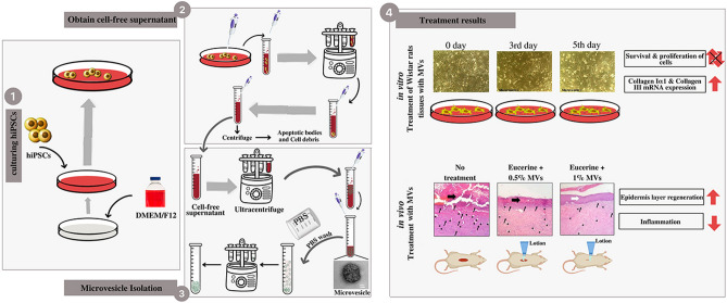
The skin undergoes the formation of fine lines and wrinkles through the aging process; also, burns, trauma, and other similar circumstances give rise to various forms of skin ulcers. Induced pluripotent stem cells (iPSCs) have become promising candidates for skin healing and rejuvenation due to not stimulating inflammatory responses, low probability of immune rejection, high metabolic activity, good large-scale production capacity and potentials for personalized medicine. iPSCs can secrete microvesicles (MVs) containing RNA and proteins responsible for the normal repairing process of the skin. This study aimed to evaluate the possibility, safety and effectiveness of applying iPSCs-derived MVs for skin tissue engineering and rejuvenation applications. The possibility was assessed using the evaluation of the mRNA content of iPSC-derived MVs and the behavior of fibroblasts after MV treatment. Investigating the effect of microvesicle on stemness potential of mesenchymal stem cells was performed for safety concerns. In vivo evaluation of MVs was done in order to investigate related immune response, re-epithelialization and blood vessel formation to measure effectiveness. Shedding MVs were round in shape distributed in the range from 100 to 1000 nm in diameter and positive for AQP3, COL2A, FGF2, ITGB, and SEPTIN4 mRNAs. After treating dermal fibroblasts with iPSC-derived MVs, the expressions of collagens Iα1 and III transcripts (as the main fibrous extracellular matrix (ECM) proteins) were upregulated. Meanwhile, the survival and proliferation of MV treated fibroblasts did not change significantly. Evaluation of stemness markers in MV treated MSCs showed negligible alteration. In line with in vitro results, histomorphometry and histopathology findings also confirmed the helpful effect of MVs in skin regeneration in the rat burn wound models. Conducting more investigations on hiPSCs-derived MVs may lead to produce more efficient and safer biopharmaceutics for skin regeneration in the pharmaceutical market.
© 2023. The Author(s).
Conflict of interest statement
The authors declare no competing interests.
Figures


Similar articles
-
Induced pluripotent stem cells-derived microvesicles accelerate deep second-degree burn wound healing in mice through miR-16-5p-mediated promotion of keratinocytes migration.Theranostics. 2020 Aug 8;10(22):9970-9983. doi: 10.7150/thno.46639. eCollection 2020. Theranostics. 2020. PMID: 32929328 Free PMC article.
-
Human Induced Pluripotent Stem Cell-Derived Microvesicles Transmit RNAs and Proteins to Recipient Mature Heart Cells Modulating Cell Fate and Behavior.Stem Cells. 2015 Sep;33(9):2748-61. doi: 10.1002/stem.2078. Epub 2015 Jun 24. Stem Cells. 2015. PMID: 26031404
-
Characterization of Induced Pluripotent Stem Cell Microvesicle Genesis, Morphology and Pluripotent Content.Sci Rep. 2016 Jan 22;6:19743. doi: 10.1038/srep19743. Sci Rep. 2016. PMID: 26797168 Free PMC article.
-
Applications of Mesenchymal Stem Cells in Skin Regeneration and Rejuvenation.Int J Mol Sci. 2021 Feb 27;22(5):2410. doi: 10.3390/ijms22052410. Int J Mol Sci. 2021. PMID: 33673711 Free PMC article. Review.
-
Aging in the mouse and perspectives of rejuvenation through induced pluripotent stem cells (iPSCs).Results Probl Cell Differ. 2012;55:413-27. doi: 10.1007/978-3-642-30406-4_21. Results Probl Cell Differ. 2012. PMID: 22918818 Review.
Cited by
-
Unveiling advanced strategies for therapeutic stem cell interventions in severe burn injuries: a comprehensive review.Int J Surg. 2024 Oct 1;110(10):6382-6401. doi: 10.1097/JS9.0000000000001812. Int J Surg. 2024. PMID: 38869979 Free PMC article. Review.
-
The application potential of iMSCs and iMSC-EVs in diseases.Front Bioeng Biotechnol. 2024 Jul 29;12:1434465. doi: 10.3389/fbioe.2024.1434465. eCollection 2024. Front Bioeng Biotechnol. 2024. PMID: 39135947 Free PMC article. Review.
-
MSC-Derived Extracellular Vesicles: Roles and Molecular Mechanisms for Tissue Repair.Int J Nanomedicine. 2025 Jun 21;20:7953-7974. doi: 10.2147/IJN.S525394. eCollection 2025. Int J Nanomedicine. 2025. PMID: 40568355 Free PMC article. Review.
-
Incorporation of iPSCs together with TERT-immortalized keratinocytes and fibroblasts into reconstructed human gingiva enhances phenotype of gingival epithelium.PLoS One. 2025 Jul 21;20(7):e0327728. doi: 10.1371/journal.pone.0327728. eCollection 2025. PLoS One. 2025. PMID: 40690428 Free PMC article.
-
Human-Induced Pluripotent Stem Cells in Plastic and Reconstructive Surgery.Int J Mol Sci. 2024 Feb 3;25(3):1863. doi: 10.3390/ijms25031863. Int J Mol Sci. 2024. PMID: 38339142 Free PMC article. Review.
References
Publication types
MeSH terms
LinkOut - more resources
Full Text Sources
