. 2023 Jun 20;13(1):9964.
doi: 10.1038/s41598-023-36844-4.

CHI3L1 induces autophagy through the JNK pathway in lung cancer cells

Affiliations

CHI3L1 induces autophagy through the JNK pathway in lung cancer cells

Da Eun Hong et al. Sci Rep. .

Abstract

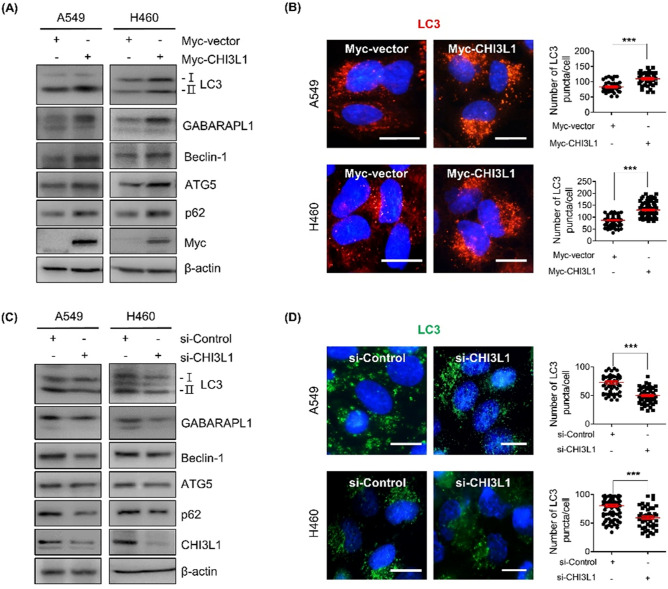
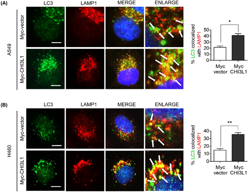
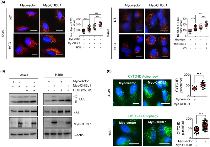
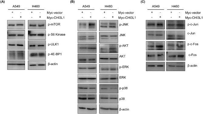
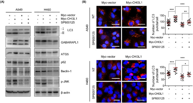
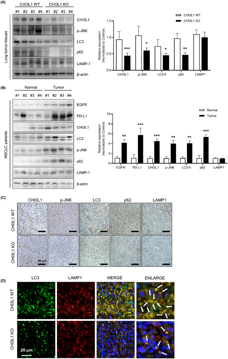
CHI3L1 is closely related to the molecular mechanisms of cancer cell migration, growth, and death. According to recent research, autophagy regulates tumor growth during various stages of cancer development. This study examined the association between CHI3L1 and autophagy in human lung cancer cells. In CHI3L1-overexpressing lung cancer cells, the expression of LC3, an autophagosome marker, and the accumulation of LC3 puncta increased. In contrast, CHI3L1 depletion in lung cancer cells decreased the formation of autophagosomes. Additionally, CHI3L1 overexpression promoted the formation of autophagosomes in various cancer cell lines: it also increased the co-localization of LC3 and the lysosome marker protein LAMP-1, indicating an increase in the production of autolysosomes. In mechanism study, CHI3L1 promotes autophagy via activation of JNK signaling. JNK may be crucial for CHI3L1-induced autophagy since pretreatment with the JNK inhibitor reduced the autophagic effect. Consistent with the in vitro model, the expression of autophagy-related proteins was downregulated in the tumor tissues of CHI3L1-knockout mice. Furthermore, the expression of autophagy-related proteins and CHI3L1 increased in lung cancer tissues compared with normal lung tissues. These findings show that CHI3L1-induced autophagy is triggered by JNK signals and that CHI3L1-induced autophagy could be a novel therapeutic approach to lung cancer.


Conflict of interest statement

The authors declare no competing interests.

Figures

Figure 1
Figure 1
CHI3L1 enhances formation of autophagosomes in human lung cancer cells. (A) A549 and H460 cells were transfected with either Myc-vector or Myc-CHI3L1 for 24 h. The expression of autophagosome-related proteins such as LC3, GABARAPL1, Beclin-1, ATG5 and p62 was evaluated by Western blotting. (B) Transfected cells were stained with LC3 antibody. Using fluorescent microscopy, LC3 puncta formation was detected. The number of LC3 puncta per cell was calculated. The data was the average of three independent experiment and error bars were mean ± SD. ***, p < 0.001. Scale bar, 10 μm. (C) A549 and H460 cells were transfected with either siRNA Control or CHI3L1 siRNA for 48 h. The expression of autophagosome-related proteins such as LC3, GABARAPL1, Beclin-1, ATG5 and p62 was evaluated by Western blotting. (D) Transfected cells were stained with LC3 antibody. Using fluorescent microscopy, LC3 puncta formation was detected. The number of LC3 puncta per cell was calculated. The data was the average of three independent experiment and error bars were mean ± SD. ***, p < 0.001. Scale bar, 10 μm.
Figure 2
Figure 2
CHI3L1 enhances formation of autolysosomes in human lung cancer cells. (A) A549 and (B) H460 cells were transfected with either Myc-vector or Myc-CHI3L1 for 24 h. The cells were fixed and permeabilized. Cell were immunostained with LC3 (Green) and LAMP-1 (Red) during fusion with autophagosome and lysosome. Cell nucleus was stained with Hoechst 33,342 (blue). Autolysosome localization observed by fluorescence microscopy. The data was the average of three independent experiment and error bars were mean ± SD. *, p < 0.05, **, p < 0.01. Scale bar, 10 μm.
Figure 3
Figure 3
CHI3L1 induces autophagic flux in human lung cancer cells. (A) A549 and H460 cells were transfected with either Myc-vector or Myc-CHI3L1 for 24 h with or without HCQ (25 μM) for 6 h. The cells were fixed, permeabilized and then stained with LC3 antibody. Using fluorescent microscopy, LC3 puncta formation was detected. The number of LC3 puncta per cell was calculated. The data was the average of three independent experiment and error bars were mean ± SD. ***, p < 0.001. Scale bar, 10 μm. (B) The expression of LC3 and p62 levels were evaluated by Western blotting. (C) Transfected cells were stained with CYTO-ID for 1 h at 37 °C in the dark. Cell nucleus was stained with Hoechst 33,342 (blue). The data was the average of three independent experiment and error bars were mean ± SD. ***, p < 0.001. Scale bar, 10 μm.
Figure 4
Figure 4
Activation of the JNK pathway involved in CHI3L1-induced autophagy. A549 and H460 cells were transfected with either Myc-vector or Myc-CHI3L1 for 24 h. (A) The expression of mTOR pathway related proteins such as p-mTOR, p-S6 kinase, p-ULK1 and p-4E-BP1 was evaluated by Western blotting. (B) The expression of MAPK pathway related proteins such as p-JNK, JNK, p-AKT, AKT, p-ERK, ERK, p-p38, and p38 was evaluated by Western blotting. (C) The expression of JNK downstream pathway related proteins such as p-c-Jun, c-Jun, p-c-Fos, and c-Fos was evaluated by Western blotting. Western blotting was performed two times with duplicate samples.
Figure 5
Figure 5
Blocking of JNK signaling inhibited CHI3L1-induced autophagy. (A) A549 and H460 cells were transfected with either Myc-vector or Myc-CHI3L1 for 24 h with or without JNK inhibitor, SP600125, pre-treatment (20 μM) for 2 h. The indicated protein expression levels were evaluated by Western blotting. (B) Transfected cells were fixed, permeabilized and then stained with LC3 antibody. Using fluorescent microscopy, LC3 puncta formation was detected. The number of LC3 puncta per cell was calculated. The data was the average of three independent experiment and error bars were mean ± SD. ***, p < 0.001; **, p < 0.01; *, p < 0.05. Scale bar, 20 μm.
Figure 6
Figure 6
CHI3L1-induced autophagy in vivo. (A) A549 human lung cancer cells were subcutaneously injected in CHI3L1-WT and -KO mice, and the formation of xenografted tumors was confirmed later. After 4 weeks, they were sacrificed to obtain tumor tissues. The indicated protein expression levels were evaluated by Western blotting. The relative protein expression in the CHI3L1-KO mice tumor tissue group compared with that in the control group is shown in the graphs. ***, p < 0.001; **, p < 0.01; *, p < 0.05 (vs. Control). (B) The indicated protein expression levels from human patients tissue lysates was evaluated by Western blotting. The relative protein expression in the NSCLC human patients group compared with that in the control group is shown in the graphs. ***, p < 0.001; **, p < 0.01 (vs. Control). (C) Immunohistochemical analysis for CHI3L1, p-JNK, LC3, p62, and LAMP-1 in CHI3L1-WT and -KO mice tumor tissue sections. Scale bar, 50 μm. (D) Immunofluorescence analysis for anti-LC3 and anti-LAMP-1 in CHI3L1-WT and -KO mice tumor tissue sections. The sections were immunostained with LC3 (Green) and LAMP-1 (Red) during fusion with autophagosome and lysosome. Cell nucleus was stained with Hoechst 33,342 (blue). The data was the average of three independent experiment. Scale bar, 20 μm.

References

    1. Zhao T, Su Z, Li Y, Zhang X, You Q. Chitinase-3 like-protein-1 function and its role in diseases. Signal Transduct. Target. Ther. 2020;5:1–20. - PMC - PubMed
    1. Libreros S, Garcia-Areas R, Iragavarapu-Charyulu V. CHI3L1 plays a role in cancer through enhanced production of pro-inflammatory/pro-tumorigenic and angiogenic factors. Immunol. Res. 2013;57:99–105. doi: 10.1007/s12026-013-8459-y. - DOI - PMC - PubMed
    1. Chiang Y-C, et al. Overexpression of CHI3L1 is associated with chemoresistance and poor outcome of epithelial ovarian carcinoma. Oncotarget. 2015;6:39740. doi: 10.18632/oncotarget.5469. - DOI - PMC - PubMed
    1. Johansen J, Cintin C, Jørgensen M, Kamby C, Price P. Serum YKL-40: A new potential marker of prognosis and location of metastases of patients with recurrent breast cancer. Eur. J. Cancer. 1995;31:1437–1442. doi: 10.1016/0959-8049(95)00196-P. - DOI - PubMed
    1. Furuhashi K, et al. Increased expression of YKL-40, a chitinase-like protein, in serum and lung of patients with idiopathic pulmonary fibrosis. Respir. Med. 2010;104:1204–1210. doi: 10.1016/j.rmed.2010.02.026. - DOI - PubMed

Publication types

Substances