

Application of additional three-dimensional materials for education in pediatric anatomy
- PMID: 37340064
- PMCID: PMC10282057
- DOI: 10.1038/s41598-023-36912-9
Application of additional three-dimensional materials for education in pediatric anatomy
Abstract
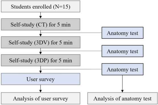
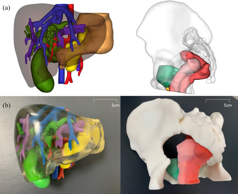
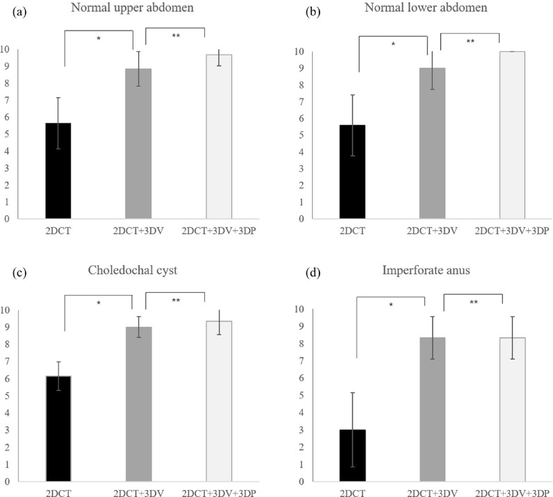
We conducted this study to investigate the effects of additional education using 3D visualization (3DV) and 3D printing (3DP) after applying 2D images for anatomical education in normal pediatric structures and congenital anomalies. For the production of 3DV and 3DP of the anatomical structures, computed tomography (CT) images of the four topics (the normal upper/lower abdomen, choledochal cyst, and imperforate anus) were used. Anatomical self-education and tests were administered to a total of 15 third-year medical students with these modules. Following the tests, surveys were conducted in order to evaluate satisfaction from students. In all four topics, there were significant increases in the test results with additional education with 3DV after initial self-study with CT (P < 0.05). The difference in scores was highest for the imperforate anus when 3DV supplemented the self-education. In the survey on the teaching modules, the overall satisfaction scores for 3DV and 3DP were 4.3 and 4.0 out of 5, respectively. When 3DV was added to pediatric abdominal anatomical education, we found an enhancement in understanding of normal structures and congenital anomalies. We can expect the application of 3D materials to become more widely used in anatomical education in various fields.
© 2023. The Author(s).
Conflict of interest statement
The authors declare no competing interests.
Figures
Similar articles
-
Do Three-dimensional Visualization and Three-dimensional Printing Improve Hepatic Segment Anatomy Teaching? A Randomized Controlled Study.J Surg Educ. 2016 Mar-Apr;73(2):264-9. doi: 10.1016/j.jsurg.2015.10.002. J Surg Educ. 2016. PMID: 26868314 Clinical Trial.
-
Evaluation by medical students of the educational value of multi-material and multi-colored three-dimensional printed models of the upper limb for anatomical education.Anat Sci Educ. 2018 Jan;11(1):54-64. doi: 10.1002/ase.1703. Epub 2017 May 19. Anat Sci Educ. 2018. PMID: 28544582
-
Application of three-dimensional reconstruction and printing as an elective course for undergraduate medical students: an exploratory trial.Surg Radiol Anat. 2019 Oct;41(10):1193-1204. doi: 10.1007/s00276-019-02248-1. Epub 2019 Apr 27. Surg Radiol Anat. 2019. PMID: 31030233
-
Three-dimensional printing in anatomy teaching: current evidence.Surg Radiol Anat. 2020 Jul;42(7):835-841. doi: 10.1007/s00276-020-02470-2. Epub 2020 Apr 28. Surg Radiol Anat. 2020. PMID: 32346753 Review.
-
Role of 3D printing technology in paediatric teaching and training: a systematic review.BMJ Paediatr Open. 2021 Dec;5(1):e001050. doi: 10.1136/bmjpo-2021-001050. BMJ Paediatr Open. 2021. PMID: 35290958 Free PMC article.
Cited by
-
Advanced Strategies for the Fabrication of Multi-Material Anatomical Models of Complex Pediatric Oncologic Cases.Bioengineering (Basel). 2023 Dec 27;11(1):31. doi: 10.3390/bioengineering11010031. Bioengineering (Basel). 2023. PMID: 38247908 Free PMC article.
-
The novel technique for surgical simulation training of patient-specific silicone models of pediatric congenital choledochal cysts.3D Print Med. 2025 Jul 16;11(1):37. doi: 10.1186/s41205-025-00252-3. 3D Print Med. 2025. PMID: 40668454 Free PMC article.
References
Publication types
MeSH terms
LinkOut - more resources
Full Text Sources
