

Contemplating the necessity of surgical treatment for posterior lenticonus: a case report
- PMID: 37340338
- PMCID: PMC10283238
- DOI: 10.1186/s12886-023-03042-9
Contemplating the necessity of surgical treatment for posterior lenticonus: a case report
Abstract
Background: Posterior lenticonus is an uncommon congenital abnormality that causes a progressive, localized spherical or conical bulging of the posterior capsular membrane, resulting in an abnormal shape of the lens.
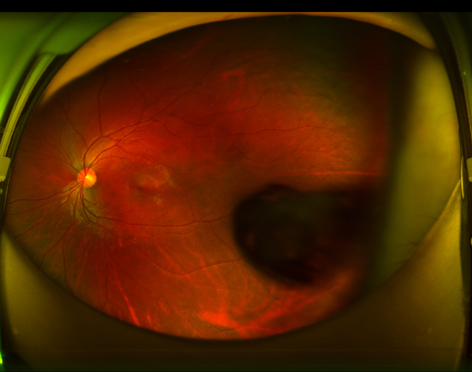
Case presentation: A 13-year-old girl presented with ametropia in both eyes. After mydriasis, examination revealed an oval bubble-shaped alteration with a distinct boundary above the temporal region on the center of the posterior capsule of her left lens. The subcortical region surrounding the alteration appeared feathery and turbid. The patient had no history of trauma or family history of visual impairment. Systemic investigations were normal. A thorough eye examination was performed, which included optometry, ultrasound biomicroscopy, ocular B-Scan, and anterior segment optical coherence, to assess the disease. The patient was diagnosed with posterior lenticonus in the left eye, as well as ametropia and anisometropia in both eyes. Conservative treatment was initiated since the patient's current best corrected visual acuity was good, and regular monitoring of the condition's progression was scheduled.
Conclusions: This case report presents a rare instance of posterior lenticonus. The findings of this report raise new considerations regarding the necessity of surgical intervention for this condition.
Keywords: Case report; Lens and anterior segment optical coherence; Lenticonus.
© 2023. The Author(s).
Conflict of interest statement
The authors declare no conflict of interest.
Figures


References
Publication types
MeSH terms
LinkOut - more resources
Full Text Sources
Medical
