. 2023 Jun 20;21(1):402.
doi: 10.1186/s12967-023-04235-y.

CircNFATC3 promotes the proliferation of gastric cancer through binding to IGF2BP3 and restricting its ubiquitination to enhance CCND1 mRNA stability

Affiliations

CircNFATC3 promotes the proliferation of gastric cancer through binding to IGF2BP3 and restricting its ubiquitination to enhance CCND1 mRNA stability

Feifei Yang et al. J Transl Med. .

Abstract

Background: Insulin like growth factor II mRNA binding protein 3 (IGF2BP3) is an RNA binding protein with multiple roles in regulation of gene expression at the post-transcriptional level and is implicated in tumorigenesis and progression of numerous cancers including gastric cancer (GC). Circular RNAs (circRNAs) are a diverse endogenous noncoding RNA population that have important regulatory roles in cancer. However, circRNAs that regulate the expression of IGF2BP3 in GC is largely unknown.

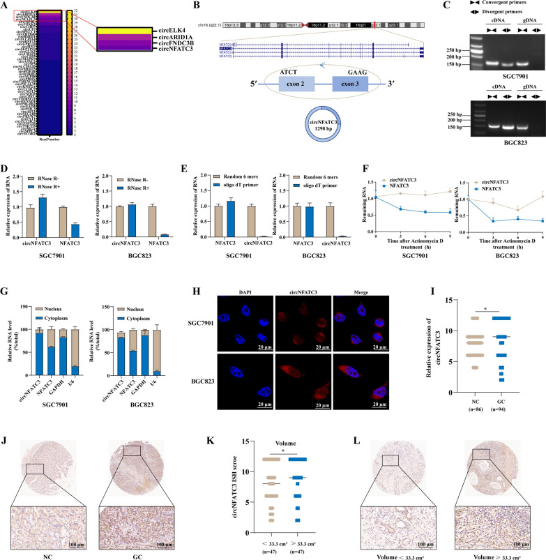
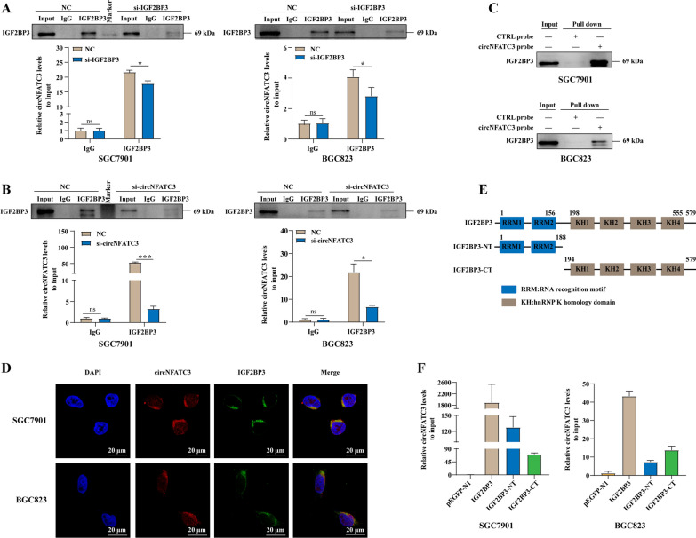
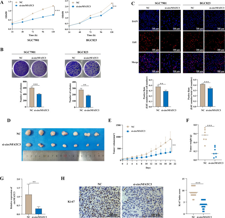
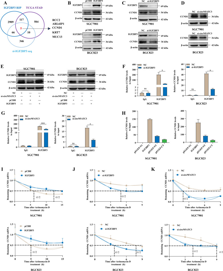
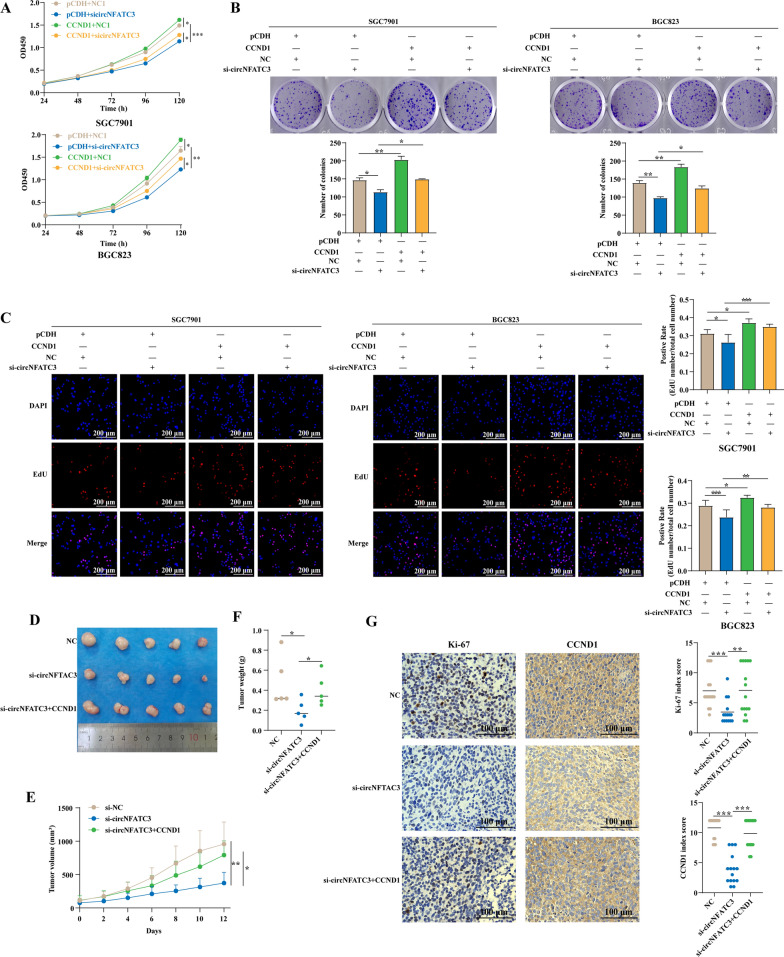
Methods: CircRNAs that bound to IGF2BP3 were screened in GC cells using RNA immunoprecipitation and sequencing (RIP-seq). The identification and localization of circular nuclear factor of activated T cells 3 (circNFATC3) were identified using Sanger sequencing, RNase R assays, qRT-PCR, nuclear-cytoplasmic fractionation and RNA-FISH assays. CircNFATC3 expression in human GC tissues and adjacent normal tissues were measured by qRT-PCR and ISH. The biological role of circNFATC3 in GC was confirmed by in vivo and in vitro experiments. Furthermore, RIP, RNA-FISH/IF, IP and rescue experiments were performed to uncover interactions between circNFATC3, IGF2BP3 and cyclin D1 (CCND1).

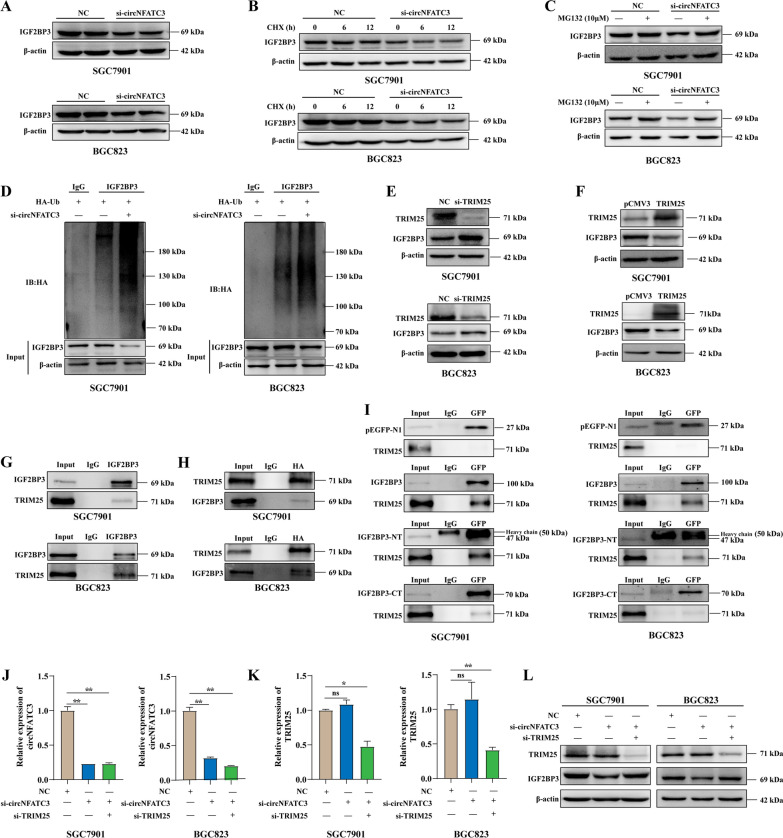
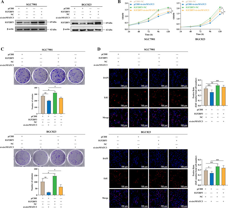
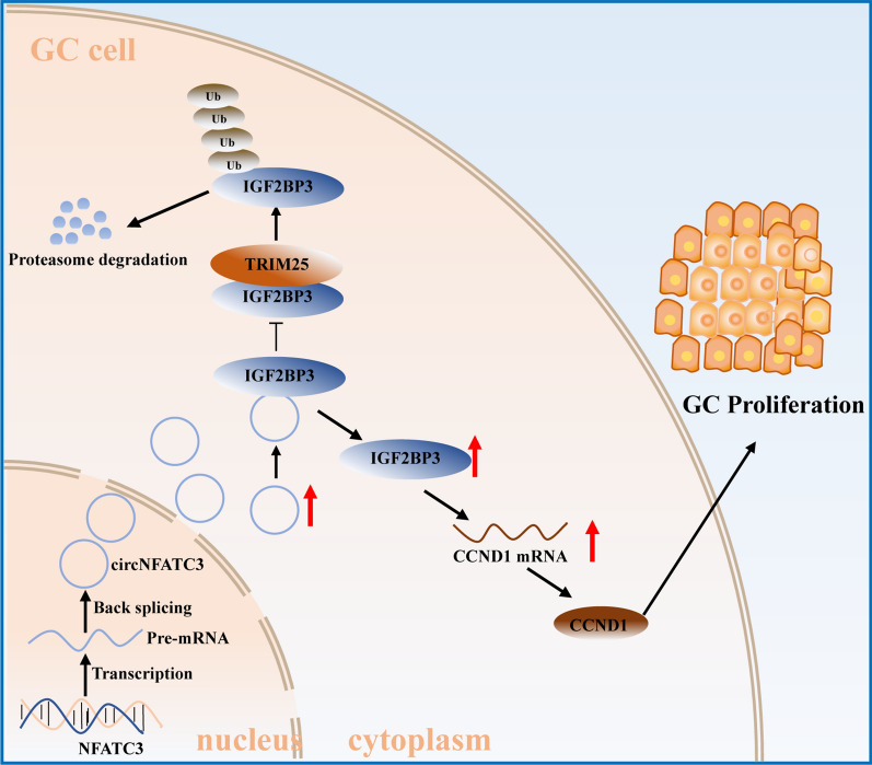
Results: We identified a GC-associated circRNA, circNFATC3, that interacted with IGF2BP3. CircNFATC3 was significantly overexpressed in GC tissues and was positively associated with tumor volume. Functionally, the proliferation of GC cells decreased significantly after circNFATC3 knockdown in vivo and in vitro. Mechanistically, circNFATC3 bound to IGF2BP3 in the cytoplasm, which enhanced the stability of IGF2BP3 by preventing ubiquitin E3 ligase TRIM25-mediated ubiquitination, thereby enhancing the regulatory axis of IGF2BP3-CCND1 and promoting CCND1 mRNA stability.

Conclusions: Our findings demonstrate that circNFATC3 promotes GC proliferation by stabilizing IGF2BP3 protein to enhance CCND1 mRNA stability. Therefore, circNFATC3 is a potential novel target for the treatment of GC.

Keywords: CCND1; Gastric cancer; IGF2BP3; TRIM25; circNFATC3.


Conflict of interest statement

All the authors declare no potential competing interests.

Figures

Fig. 1
Fig. 1
CircNFATC3, an IGF2BP3-related circRNA is upregulated in GC. A CircRNAs that bind to IGF2BP3 in SGC7901 cells. B Schematic illustration of circNFATC3 formation from its parental gene NFATC3. C Existence of circNFATC3 in cDNA or genomic DNA (gDNA) of SGC7901 and BGC823 cells. D Expression of circNFATC3 and NFATC3 amplified from total RNA of SGC7901 and BGC823 cells treated with RNase R. E Expression of circNFATC3 and NFATC3 amplified from total RNA of SGC7901 and BGC823 cells reverse transcribed by random 6 mers or oligo dT 18 primers. F Stabilities of circNFATC3 and NFATC3 in SGC7901 and BGC823 cells treated with actinomycin D. G qRT-PCR for the distribution of circNFATC3, NFATC3, GAPDH, and U6 in the cytoplasmic and nuclear fractions of SGC7901 and BGC823. H RNA-FISH for circNFATC3 in SGC7901 and BGC823 cells. CircNFATC3 was shown in red and nuclei were stained with DAPI. I ISH analysis of circNFATC3 expression in TMA of GC patients (adjacent tissues (NC) = 86 cases; GC tissues = 94 cases). J Representative images of circNFATC3 staining in GC and adjacent tissues (NC). K Expression of circNFATC3 in GC patients with tumor volumes < 33.3 cm3 or ≥ 33.3 cm3. L Representative images of circNFATC3 staining in tumor tissues of GC patients with tumor volumes < 33.3 cm3 or ≥ 33.3 cm3. The Mann–Whitney U test was used to calculate p-values. *P < 0.05
Fig. 2
Fig. 2
CircNFATC3 directly binds to IGF2BP3 protein in GC. A-B RIP analyses of circNFATC3 enrichment pull-downs by IGF2BP3 antibodies in SGC7901and BGC823 cells following IGF2BP3 (A) or circNFATC3 (B) knockdown. C RNA pull-down validation of the interaction between circNFATC3 and IGF2BP3 protein in SGC7901and BGC823 cells. D RNA-FISH-immunofluorescence showing the co-localization of circNFATC3 (red) with IGF2BP3 (green) in SGC7901 and BGC823 cells. E Structural diagram of IGF2BP3 protein and IGF2BP3 truncations. F RIP analysis of circNFATC3 enrichment pulled down by GFP antibodies in SGC7901 and BGC823 cells transfected with full-length or IGF2BP3 truncations. The Student’s t test was used to calculate p-values. ns, not significant, *P < 0.05, ***P < 0.001
Fig. 3
Fig. 3
Knockdown of circNFATC3 inhibits the proliferation of GC cells in vitro and in vivo. A-C The proliferation of GC cells transfected with circNFATC3 siRNAs and evaluated by CCK-8 (A), plate colony formation (B), and EdU assays (C). D-F Tumor images (D), growth curves (E), and tumor weights (F) were obtained from xenograft tumor derived from HGC-27 cells which treated with cholesterol modified siRNAs targeting circNFATC3. G Relative expression of circNFATC3 in xenografted tumor tissues. H Representative images of Ki-67 expression evaluated by IHC in xenografted tumor tissues. Three different visual fields were randomly selected for each section for staining scoring. The Student’s t test (A-C, E–G) and Mann–Whitney U test (H) were used to calculate p-values. ** P < 0.01, and *** P < 0.001
Fig. 4
Fig. 4
CircNFATC3 enhances the stability of IGF2BP3 by preventing TRIM25-mediated ubiquitination. A Western blot analysis of IGF2BP3 in SGC7901 and BGC823 cells transfected with siRNAs targeting circNFATC3. B Stability of IGF2BP3 protein in SGC7901 and BGC823 cells after circNFATC3 knockdown evaluated by cycloheximide (CHX) chase assays. C Protein expression of IGF2BP3 in SGC7901 and BGC823 cells treated with proteasome inhibitor MG132 after transfection with si-circNFATC3. D Immunoprecipitation analysis for ubiquitination modification of IGF2BP3 in SGC7901 and BGC823 cells with circNFATC3 knockdown. E–F Protein expression of IGF2BP3 in SGC7901 and BGC823 cells after TRIM25 knockdown (E) or overexpression(F). G-H Immunoprecipitation analyses of TRIM25 pulled down by IGF2BP3 (G) and IGF2BP3 pulled down by TRIM25 (HA-tagged) (H) in SGC7901 and BGC823 cells. I Immunoprecipitation analysis of TRIM25 pulled down by GFP in SGC7901 and BGC823 cells transfected with full-length of IGF2BP3 or truncations. J-K The expression of circNFATC3 (J) or TRIM25 (K) in SGC7901 and BGC823 cells transfected with si-TRIM25 and/or si-circNFATC3. L Protein levels for IGF2BP3 in SGC7901 and BGC823 cells transfected with si-TRIM25 or si-circNFATC3 alone and in combination. The Student’s t test was used to calculate p-values. ns, not significant, *P < 0.05, **P < 0.01
Fig. 5
Fig. 5
Overexpression of IGF2BP3 can attenuate the proliferation inhibition caused by circNFATC3 knockdown. A Protein expression of IGF2BP3 in SGC7901 and BGC823 cells transfected with blank vector or IGF2BP3 overexpression plasmids and co-transfected with NC or si-circNFATC3. B-D CCK-8 (B), plate colony formation (C), and EdU assays (D) of SGC7901 and BGC823 cells transfected with blank vector or IGF2BP3 overexpression plasmids and co-transfected with NC or si-circNFATC3 as indicated. One-way ANOVA was used to calculate p-values. *P < 0.05, **P < 0.01, ***P < 0.001
Fig. 6
Fig. 6
CircNFATC3 binds to IGF2BP3 to regulate the expression of CCND1 in GC. A Potential target screening strategy of the circNFATC3-IGF2BP3 axis. B-D The protein levels of CCND1 in SGC7901 and BGC823 cells following IGF2BP3 overexpression (B) or knockdown (C) or circNFATC3 knockdown (D). E Protein expression of CCND1 in SGC7901 and BGC823 cells transfected with NC, si-circNFATC3 or si-circNFATC3 combined with IGF2BP3 overexpression. F-G RIP analyses of CCND1 enrichment pull-downs by IGF2BP3 in SGC7901 and BGC823 cells following IGF2BP3 (F) and circNFATC3 (G) knockdowns. H RIP analysis of CCND1 enrichment pulled down by GFP in SGC7901 and BGC823 cells transfected with full-length or IGF2BP3 truncations. I-K Stability of CCND1 mRNA at indicated times following IGF2BP3 overexpression (I) or knockdown (J) or circNFATC3 knockdown (K) in SGC7901and BGC823 cells. The Student’s t test was used to calculate p-values. ns, not significant, *P < 0.05, ***P < 0.001
Fig. 7
Fig. 7
CircNFATC3 promotes the proliferation of GC via regulating CCND1. A−C CCK-8 (A), plate colony formation (B), and EdU assays (C) of SGC7901 and BGC823 cells transfected with blank vector or CCND1 overexpression plasmids and co-transfected with NC or si-circNFATC3 as indicated. D−F CCND1 lentivirus or si-circNFATC3 modified by cholesterol were injected into the xenograft tumor derived from HGC-27 cells. Tumor images (D), growth curves (E) and weights (F) were obtained from xenograft tumors derived from HGC-27 cells treated with cholesterol modified siRNAs targeting circNFATC3 or combined with CCND1 lentivirus. G Representative images of Ki-67 and CCND1 expression evaluated by IHC in xenografted tumor tissues. One-way ANOVA (A−C), the Student’s t test (E, F) and Mann–Whitney U test (G) were used to calculate p-values. *P < 0.05, **P < 0.01, ***P < 0.001
Fig. 8
Fig. 8
A schematic illustration of the molecular mechanism of circNFATC3 in regulating the proliferation of GC. CircNFATC3 expression is up-regulated in GC. CircNFATC3 enhances the stability of IGF2BP3 by preventing TRIM25-mediated ubiquitination, subsequently elevating the stability of CCND1 and promoting GC proliferation

Similar articles

Cited by

References

    1. Sung H, Ferlay J, Siegel RL, Laversanne M, Soerjomataram I, Jemal A, et al. Global Cancer Statistics 2020: GLOBOCAN estimates of incidence and mortality worldwide for 36 cancers in 185 countries. CA Cancer J Clin. 2021;71(3):209–249. doi: 10.3322/caac.21660. - DOI - PubMed
    1. Li GZ, Doherty GM, Wang J. Surgical management of gastric cancer: a review. JAMA Surg. 2022;157(5):446–454. doi: 10.1001/jamasurg.2022.0182. - DOI - PubMed
    1. Smyth EC, Nilsson M, Grabsch HI, van Grieken NC, Lordick F. Gastric cancer. Lancet. 2020;396(10251):635–648. doi: 10.1016/S0140-6736(20)31288-5. - DOI - PubMed
    1. Qin H, Ni H, Liu Y, Yuan Y, Xi T, Li X, et al. RNA-binding proteins in tumor progression. J Hematol Oncol. 2020;13(1):90. doi: 10.1186/s13045-020-00927-w. - DOI - PMC - PubMed
    1. Zhang N, Shen Y, Li H, Chen Y, Zhang P, Lou S, et al. The m6A reader IGF2BP3 promotes acute myeloid leukemia progression by enhancing RCC2 stability. Exp Mol Med. 2022;54(2):194–205. doi: 10.1038/s12276-022-00735-x. - DOI - PMC - PubMed

Publication types