

Correlation exploration among CT imaging, pathology and genotype of pulmonary ground-glass opacity
- PMID: 37340599
- PMCID: PMC10339074
- DOI: 10.1111/jcmm.17797
Correlation exploration among CT imaging, pathology and genotype of pulmonary ground-glass opacity
Abstract
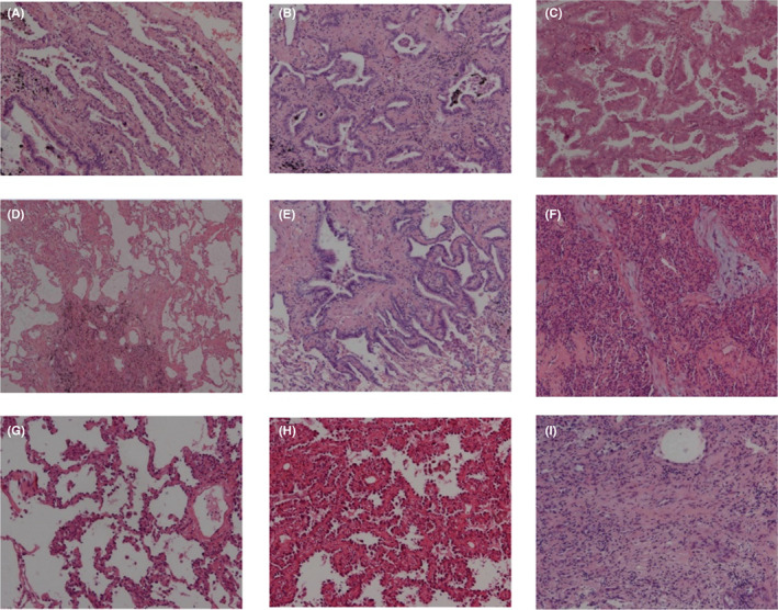
To analyse the clinical features, imaging manifestation, pathological typing and genetic testing results of patients undergoing surgery for ground-glass opacity (GGO) nodules, and explore the reasonable diagnosis and treatment program for GGO patients as to provide the basis for the establishment of GGO treatment process. This study is an exploratory study. 465 cases with GGO confirmed by HRCT, undergoing surgery and approved by pathologic diagnosis in Shanghai pulmonary hospital were enrolled in this study. All the patients with GGO were cases with single lesion. The relationship between the clinical, imaging, pathological and molecular biological data of single GGO were statistically studied. (1) Among 465 cases, the median age was 58 years and females were 315 (67.7%); there were 397 (85.4%) non-smoking, and 354 cases (76.1%) had no clinical symptoms. There were 33 cases of benign and 432 cases of malignant GGO. Significant differences were observed on the size, vacuole sign, pleural indentation and blood vessel sign of GGO between two groups (p < 0.05). Of 230 mGGO, there were no AAH, 13 cases of AIS, 25 cases of MIA and 173 cases of invasive adenocarcinoma. The probability of solid nodules in invasive adenocarcinoma was higher than that in micro invasive carcinoma, and the difference was statistically significant (p < 0.05). 360 cases were followed up with the average follow-up time of 6.05 months, and GGO of 34 cases (9.4%) increased. (2) In 428 adenocarcinoma samples approved by pathologic diagnosis, there were 262 (61.2%) lesions of EGFR mutation, 14 (3.3%) lesions of KRAS mutation, 1 (0.2%) lesion of Braf mutation, 9 (2.1%) lesions of EML4-ALK gene fusion and 2 (0.5%) lesions of ROS1 fusion. The detection rate of gene mutation in mGGO was higher than that of pGGO. During the follow-up period, genetic testing results of 32 GGO showed that EGFR mutation rate was 53.1%, ALK positive rate of 6.3%, KRAS mutation rate of 3.1% and no ros1 and BRAF gene mutation. No statistically significant difference was observed in comparison with unchanged GGO. (3) EGFR mutation rate of invasive adenocarcinoma was the highest (168/228, 73.7%), mainly in the 19Del and L858R point mutations. No KRAS mutation was found in atypical adenoma hyperplasia. No significant difference was observed on the mutation rate of KRAS between different types of GGO (p = 0.811). EML4-ALK fusion gene was mainly detected in invasive adenocarcinoma (7/9). GGO tends to occur in young, non-smoking women. The size of GGO is related to the degree of malignancy. Pleural depression sign, vacuole sign and vascular cluster sign are all characteristic images of malignant GGO. pGGO and mGGO reflect the pathological development of GGO. During the follow-up, it is found that GGO increases and solid components appear, which is the indication of surgical resection. The detection rate of EGFR mutations in mGGO and invasive adenocarcinoma is high. pGGO has heterogeneity in imaging, pathology and molecular biology. Heterogeneity research helps to formulate correct individualized diagnosis and treatment plans.
Keywords: X-ray computed; ground-glass opacity (GGO); lung/pathology; oncogenes; tomography.
© 2023 The Authors. Journal of Cellular and Molecular Medicine published by Foundation for Cellular and Molecular Medicine and John Wiley & Sons Ltd.
Conflict of interest statement
The authors declared that they have no competing interests.
Figures
Similar articles
-
EGFR L858R mutation is associated with lung adenocarcinoma patients with dominant ground-glass opacity.Lung Cancer. 2015 Mar;87(3):272-7. doi: 10.1016/j.lungcan.2014.12.016. Epub 2015 Jan 5. Lung Cancer. 2015. PMID: 25582278
-
Genetic features of pulmonary adenocarcinoma presenting with ground-glass nodules: the differences between nodules with and without growth.Ann Oncol. 2015 Jan;26(1):156-161. doi: 10.1093/annonc/mdu505. Epub 2014 Oct 30. Ann Oncol. 2015. PMID: 25361983
-
Impact of the International Association for the Study of Lung Cancer/American Thoracic Society/European Respiratory Society classification of stage IA adenocarcinoma of the lung: Correlation between computed tomography images and EGFR and KRAS gene mutations.Exp Ther Med. 2015 Jun;9(6):2095-2103. doi: 10.3892/etm.2015.2422. Epub 2015 Apr 14. Exp Ther Med. 2015. PMID: 26136941 Free PMC article.
-
Ground-glass opacity nodules: histopathology, imaging evaluation, and clinical implications.J Thorac Imaging. 2011 May;26(2):106-18. doi: 10.1097/RTI.0b013e3181fbaa64. J Thorac Imaging. 2011. PMID: 21508733 Review.
-
Management of Ground-Glass Opacities in the Lung Cancer Spectrum.Ann Thorac Surg. 2020 Dec;110(6):1796-1804. doi: 10.1016/j.athoracsur.2020.04.094. Epub 2020 Jun 7. Ann Thorac Surg. 2020. PMID: 32525031 Review.
Cited by
-
Enhanced management strategy of synchronous percutaneous biopsy and microwave ablation in patients with lung ground-glass opacities undergoing antithrombotic treatment: a clinical perspective on our experience.Front Oncol. 2025 Jul 23;15:1554365. doi: 10.3389/fonc.2025.1554365. eCollection 2025. Front Oncol. 2025. PMID: 40772032 Free PMC article.
-
An artificial intelligence algorithm for the detection of pulmonary ground-glass nodules on spectral detector CT: performance on virtual monochromatic images.BMC Med Imaging. 2024 Oct 29;24(1):293. doi: 10.1186/s12880-024-01467-2. BMC Med Imaging. 2024. PMID: 39472819 Free PMC article.
-
CT morphological features and histogram parameters to predict micropapillary or solid components in stage IA lung adenocarcinoma.Front Oncol. 2024 Jul 24;14:1448333. doi: 10.3389/fonc.2024.1448333. eCollection 2024. Front Oncol. 2024. PMID: 39114305 Free PMC article.
-
Artificial intelligence-measured nodule mass for determining the invasiveness of neoplastic ground glass nodules.Quant Imaging Med Surg. 2024 Sep 1;14(9):6698-6710. doi: 10.21037/qims-24-665. Epub 2024 Aug 28. Quant Imaging Med Surg. 2024. PMID: 39281163 Free PMC article.
-
The clinical, radiological, postoperative pathological, and genetic features of nodular lung adenocarcinoma: a real-world single-center data.J Thorac Dis. 2024 May 31;16(5):3228-3250. doi: 10.21037/jtd-24-510. Epub 2024 May 21. J Thorac Dis. 2024. PMID: 38883620 Free PMC article.
References
-
- Lee HY, Lee KS. Ground‐glass opacity nodules: histopathology, imaging evaluation, and clinical implications. J Thorac Imaging. 2011;26(2):106‐118. - PubMed
-
- Kim HY, Shim YM, Lee MS, et al. Persistent pulmonary nodular ground‐glass opacity at thin–section CT: histopathologic comparisons. Radiology. 2007;245:267‐275. - PubMed
-
- Haro A, Yano T, Kohno M, Yoshida T, Okamoto T, Maehara Y. Ground‐glass opacity lesions on computed tomography during postoperative surveillance for primary non‐small cell lung cancer. Lung Cancer. 2012;76(1):56‐60. - PubMed
-
- Uehara H, Tsutani Y, Okumura S. Prognostic role of positron emission tomography and high‐resolution computed toography in clinical stage IA lung adenocarcinoma. Ann Thorac Surg. 2013;96(6):1958‐1965. - PubMed
Publication types
MeSH terms
Substances
LinkOut - more resources
Full Text Sources
Medical
Research Materials
Miscellaneous
