. 2024 Apr 2;12(2):2222632.
doi: 10.1080/21688370.2023.2222632. Epub 2023 Jun 21.

Culture media and format alter cellular composition and barrier integrity of porcine colonoid-derived monolayers

Affiliations

Culture media and format alter cellular composition and barrier integrity of porcine colonoid-derived monolayers

Alicia M Barnett et al. Tissue Barriers. .

Abstract

Intestinal organoid technology has revolutionized our approach to in vitro cell culture due in part to their three-dimensional structures being more like the native tissue from which they were derived with respect to cellular composition and architecture. For this reason, organoids are becoming the new gold standard for undertaking intestinal epithelial cell research. Unfortunately, their otherwise advantageous three-dimensional geometry prevents easy access to the apical epithelium, which is a major limitation when studying interactions between dietary or microbial components and host tissues. To overcome this problem, we developed porcine colonoid-derived monolayers cultured on both permeable Transwell inserts and tissue culture treated polystyrene plates. We found that seeding density and culture format altered the expression of genes encoding markers of specific cell types (stem cells, colonocytes, goblets, and enteroendocrine cells), and barrier maturation (tight junctions). Additionally, we found that changes to the formulation of the culture medium altered the cellular composition of colonoids and of monolayers derived from them, resulting in cultures with an increasingly differentiated phenotype that was similar to that of their tissue of origin.

Keywords: Barrier integrity; Porcine colonoids; colonoid-derived monolayers; differentiation; medium formulation; stem cells.

Plain language summary

In vitro models of the intestine are used to study the complex in vivo intestinal processes in a simplified context. As such, these models need to be representative of their tissue of origin. Here, we demonstrate that porcine colonoids and colonoid-derived monolayers that have comparable stem cells and differentiated cell types to those of the native tissue can be developed but are influenced by cell seeding density, culture format, and medium formulation.


Conflict of interest statement

No potential conflict of interest was reported by the author(s).

Figures

Figure 1.
Figure 1.
Microscopic imaging and immunohistochemistry of colonoid-derived monolayers. Cells were seeded at low (a, d), medium (b, e, and g-i), and high (c, f) (7.6 ×10⁴, 1.5 × 105 and 2.3 × 105 cells/cm2, respectively) densities and cultured for two (a-c) or six (d-i) days with 50+/+ medium. Cultures were imaged using an inverted microscope at 10× magnification (a-f) or confocal microscope (g-i). Immunoreactivity for the epithelial-specific structural protein cytokeratin − 18 (G-green) and tight-junction protein zonula occludens 1 (Zo1; H-red), and merged image of both cytokeratin − 18 and Zo1 (I). Scale bars = 50 µm.
Figure 2.
Figure 2.
Influence of seeding density of colonoid-derived monolayer resistance. Cells were seeded at low, medium, and high (7.6 ×10⁴, 1.5 × 105 and 2.3 × 105/cm2, respectively) densities and cultured in 50+/+ medium. Resistance, presented as ohms (Ω).cm2. Data are presented as the mean ± SD of data pooled from three independent experiments (n = 3). Data at the same time point were analyzed using the Kruskal-Wallis test with Tukey post-hoc test for multiple comparisons. Statistical significance is indicated as *p < 0.05.
Figure 3.
Figure 3.
Differential gene expression in colonoid-derived monolayers seeded at different cell density. The relative expression of markers of stem cells (Lgr5 and Sox9), differentiated colonocytes (Sglt1and Ca2), enteroendocrine and goblet cells (Cga and Muc2, respectively), and barrier maturation (Ocln, Zo1, and Cldn) in colonoid-derived monolayers seeded at medium (1.5 ×10⁵ cells per cm2) and high (2.3×10⁵ cells per cm2) densities relative to those seeded at low density (7.6 ×10⁴ cells per cm2) into inserts (a) or tissue culture (TC) plates (c) or at high density relative to those seeded at medium density into inserts (b) or TC plates (d) (n = 3 independent experiments). Data are presented as log2-fold change normalized to respective monolayers seeded at low cell seeding density (a and c) or medium density (b and d) where the mean is shown, and whiskers represent SD. Data was analyzed using Mann–Whitney U test. Values below − 0.585 and above 0.585 (corresponding to 1.5-fold change) are indicated by the dashed lines and have statistical significance (p < 0.05) denoted by*. Ca2, carbonic anhydrase 2; Cga, chromogranin A; Cldn, claudin; Lgr5, leucine-rich repeat-containing G protein-coupled receptor; Muc2, mucin 2; Ocln, occludin; Sox9, sex-determining region Y-box 9; Sglt1, sodium-glucose transporter 1; Zo1, zonula occludens 1.
Figure 4.
Figure 4.
Differential gene expression in colonoid-derived epithelial monolayers seeded into TC treated plates at low, medium, and high cell density relative to inserts. The relative expression of mRNA transcripts of markers of stem cells (Leucine-rich repeat-containing G protein-coupled receptor 5 (Lgr5) and Sex-determining region Y-box 9 (Sox9)), colonocytes (Sodium-glucose transporter 1 (Sglt1) and Carbonic anhydrase (Ca2)) enteroendocrine cells (Chromogranin a (Cga)) goblet cells (Mucin 2 (Muc2)), and barrier maturation (Occludin (Ocln), Zonula occludens 1 (Zo1), and Claudin (Cldn)) in monolayers seeded at low (a), medium (b), and high (c) (7.6 ×10⁴, 1.5 × 105 and 2.3 × 105 cells per cm2, respectively) densities in TC plates relative to those seeded at the comparable densities into inserts (n = 3 independent experiments). Data represent log2-fold change in TC monolayers normalized to insert monolayers at comparable cell seeding densities where the mean is shown, and whiskers represent SD. Data was analyzed using Mann–Whitney U test. Values below − 0.585 and above 0.585 (corresponding to 1.5-fold change) are indicated by the dashed lines and have statistical significance (p < 0.05) denoted by *.
Figure 5.
Figure 5.
Differential gene expression in sub-cultured, colonoid-derived epithelial monolayers. The relative expression of mRNA transcripts of markers of stem cell (Lgr5 and Sox9), specific, differentiated colonocytes (Sglt1 and Ca2), enteroendocrine and goblet cells (Cga and Muc2, respectively), and barrier maturation (Ocln, Zo1, and Cldn) in sub-cultured colonoid-derived monolayers at passage 1, 2 and 3, relative to passage 0 monolayers as determined from qPCR. Data are presented as log2-fold change normalized to P0 monolayers and shown as scatter dot plots. The mean is shown, and whiskers represent SD from three independent experiments (n = 3). Data was analyzed using one-way ANOVA with Dunnett’s multiple comparison test. Values below − 0.585 and above 0.585 (corresponding to 1.5-fold change) are indicated by the dashed lines and have statistical significance (p < 0.05) denoted by*. Ca2, carbonic anhydrase 2; Cga, chromogranin A; Cldn, claudin; Lgr5, leucine-rich repeat-containing G protein-coupled receptor; Muc2, mucin 2; Ocln, occludin; Sox9, sex-determining region Y-box 9; Sglt1, sodium-glucose transporter 1; Zo1, zonula occludens 1.
Figure 6.
Figure 6.
Differential gene expression in porcine colonoids and colonoid-derived monolayers cultured with different medium formulations. The relative expression of the stem cell markers (Lgr5 and Sox9), colonocyte markers (Sglt1 and Ca2), and secretory cell lineage markers (Muc2 and Cga) in colonoids (a), and colonoid-derived monolayers cultured on either Transwell inserts (b), or tissue culture (TC) treated plates (c) with alternative medium formulations relative to the respective cultures grown with 50+/+ medium as determined from qPCR (n = 3 independent experiments). Initially all cultures were grown with 50+/+ medium for 72 h post-seeding, and then cultured with alternative media for an additional 72 h. Data are presented as log2-fold change normalized to respective cultures grown with 50+/+ medium and are shown as scatter dot plots. The mean is shown, and whiskers represent SD from three independent experiments (n = 3). Data was analyzed using one-way ANOVA with Dunnett’s multiple comparisons test. Values below − 0.585 and above 0.585 (corresponding to 1.5-fold change) are indicated by the dashed lines and have statistical significance (p < 0.05) denoted by*. Mediums used: 50+/+ medium (50% conditioned media (CM) + 50% basal media (BM) supplemented with epidermal growth factor (EGF) and inhibitors (p38 Mapk and TGF-β)); 50+/- (50% CM + 50% BM, +EGF – inhibitors; 50-/- (50% CM + 50% BM, -EGF – inhibitors); 5+/+ (5% CM + 95% BM, +EGF +inhibitors); 5+/- (5% CM + 95% BM, +EGF -inhibitors); and 5-/- (5% CM + 95% BM, -EGF -inhibitors). Ca2, carbonic anhydrase 2; Cga, chromogranin A; Lgr5, leucine-rich repeat-containing G protein-coupled receptor 5; Muc2, mucin 2; Sox9, sex-determining region Y-box 9; Sglt1, sodium-glucose transporter 1.
Figure 7.
Figure 7.
Immunofluorescent staining of porcine colonoids identifies stem and secretory cell lineages. the existence of stem cells and differentiated secretory cell lineages were confirmed in colonoids when cultured with alternative media formulations. Colonoids were cultured with 50+/+ medium for 72 hpost-seeding and then cultured with the same (a) 50+/+ medium or alternative media (b) 50+/-, (c) 5+/-, and (d) 5-/- for an additional 72 h. Sox9 (green) was used to identify stem cells while mucin 2 (Muc2 - red) and chromogranin-A (Cga – blue) was used to identify secretory goblet and enteroendocrine cells, respectively. Scale bar = 10 µm. Mediums used: 50+/+ medium (50% conditioned media (CM) + 50% basal media (BM) supplemented with epidermal growth factor (EGF) and inhibitors (p38 Mapk and TGF-β)); 50+/- (50% CM + 50% BM, +EGF – inhibitors; 5+/- (5% CM + 95% BM, +EGF -inhibitors); and 5-/- (5% CM + 95% BM, -EGF -inhibitors).
Figure 8.
Figure 8.
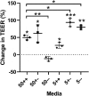
Influence of media composition on resistance of colonoid-derived monolayers. A single cell suspension from porcine colonoids was seeded at medium density (1.5 ×10⁵ per cm2) onto Transwell inserts and cultured with 50+/+ medium. After 72 h, resistance for each insert was recorded (initial TEER value) and respective inserts replenished with alternative media formulations as indicated in the graph. Inserts were cultured for an additional 72 h, and resistance for each insert recorded again. Data are presented as the percentage change in TEER compared to initial TEER for each insert. Data were analyzed using two-tailed Welch’s t test. The mean is shown, and whiskers represent SD from three independent experiments (n = 3). Inserts cultured with alternative media were also compared to those cultured with 50+/+ media and data analyzed using one-way ANOVA with Holm-Sidak’s multiple comparison test. Statistical significance is indicated as *p < 0.05, **p < 0.01 and ***p < 0.001. Media formulations: 50+/+ medium (50% conditioned media (CM) + 50% basal media (BM) supplemented with epidermal growth factor (EGF) and inhibitors (p38 Mapk and TGF-β)); 50+/- (50% CM + 50% BM, +EGF – inhibitors; 50-/- (50% CM + 50% BM, -EGF – inhibitors); 5+/+ (5% CM + 95% BM, +EGF +inhibitors); 5+/- (5% CM + 95% BM, +EGF -inhibitors); and 5-/- (5% CM + 95% BM, -EGF -inhibitors).

Similar articles

Cited by

References

    1. Altay G, Larrañaga E, Tosi S, Barriga FM, Batlle E, Fernández-Majada V, Martínez E.. Self-organized intestinal epithelial monolayers in crypt and villus-like domains show effective barrier function. Sci Rep. 2019;9(1):1. doi:10.1038/s41598-019-46497-x. - DOI - PMC - PubMed
    1. Gillet JP, Varma S, Gottesman MM, Gillet J-P. The clinical relevance of cancer cell lines. J Natl Cancer Inst. 2013;105(7):452–27. doi:10.1093/jnci/djt007. - DOI - PMC - PubMed
    1. Liu Y, Mi Y, Mueller T, Kreibich S, Williams EG, Van Drogen A, Borel C, Frank M, Germain P-L, Bludau I, et al. Multi-omic measurements of heterogeneity in HeLa cells across laboratories. Nat Biotechnol. 2019;37(3):314–322. doi:10.1038/s41587-019-0037-y. - DOI - PubMed
    1. Aldhous MC, Shmakov AN, Bode J, Ghosh S, Aldhous MC. Characterization of conditions for the primary culture of human small intestinal epithelial cells. Clin Exp Immunol. 2001;125(1):32–40. doi:10.1046/j.1365-2249.2001.01522.x. - DOI - PMC - PubMed
    1. Grossmann J, Walther K, Artinger M, Kiessling S, Steinkamp M, Schmautz WK, Stadler F, Bataille F, Schultz M, Schölmerich J, et al. Progress on isolation and short-term ex-vivo culture of highly purified non-apoptotic human intestinal epithelial cells (IEC). Eur J Cell Biol. 2003;82(5):262–270. doi:10.1078/0171-9335-00312. - DOI - PubMed

Publication types