. 2023 Jul 26;43(7):BSR20230846.
doi: 10.1042/BSR20230846.

Structural polymorphism of the PH domain in TFIIH

Affiliations

Structural polymorphism of the PH domain in TFIIH

Masahiko Okuda et al. Biosci Rep. .

Abstract

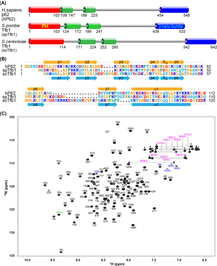
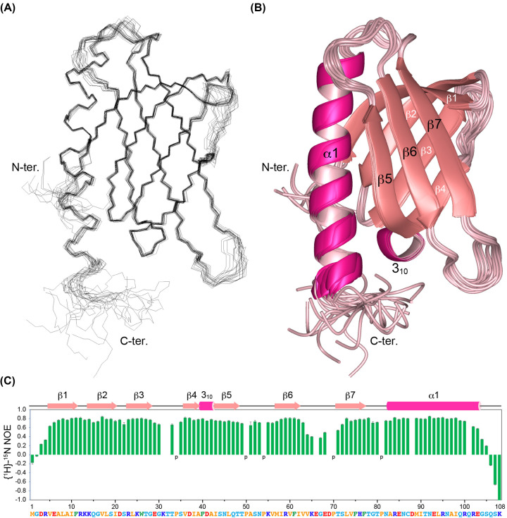
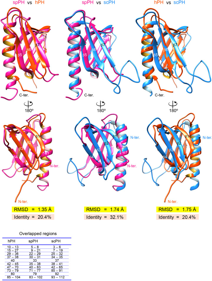
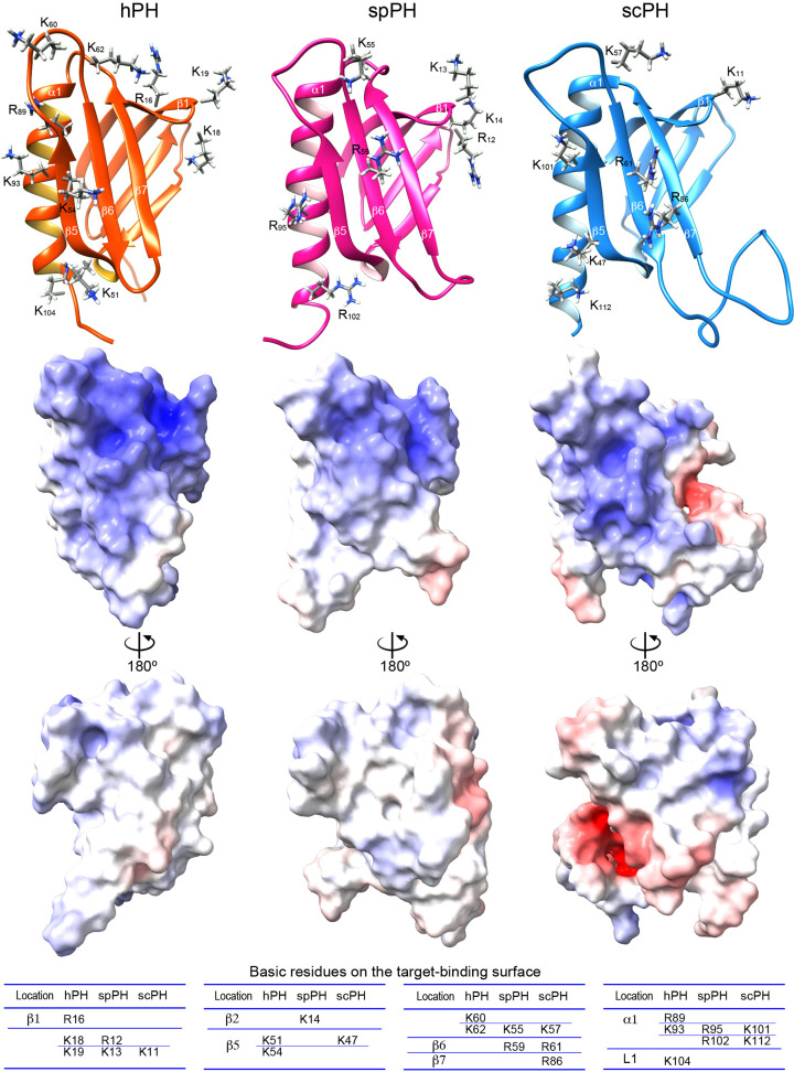
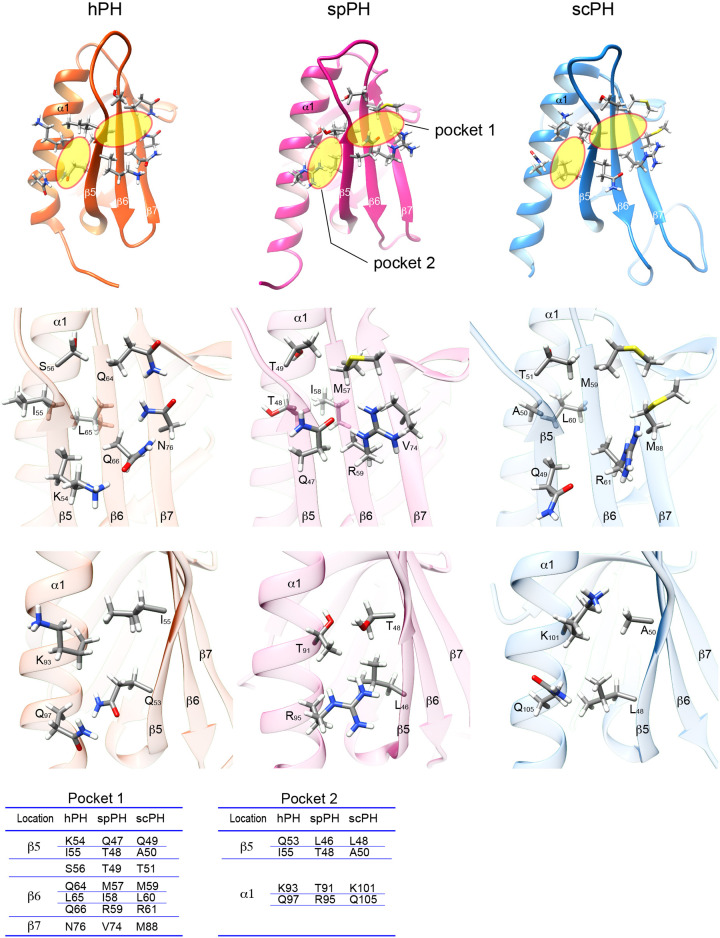
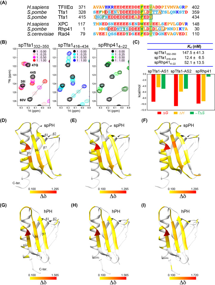
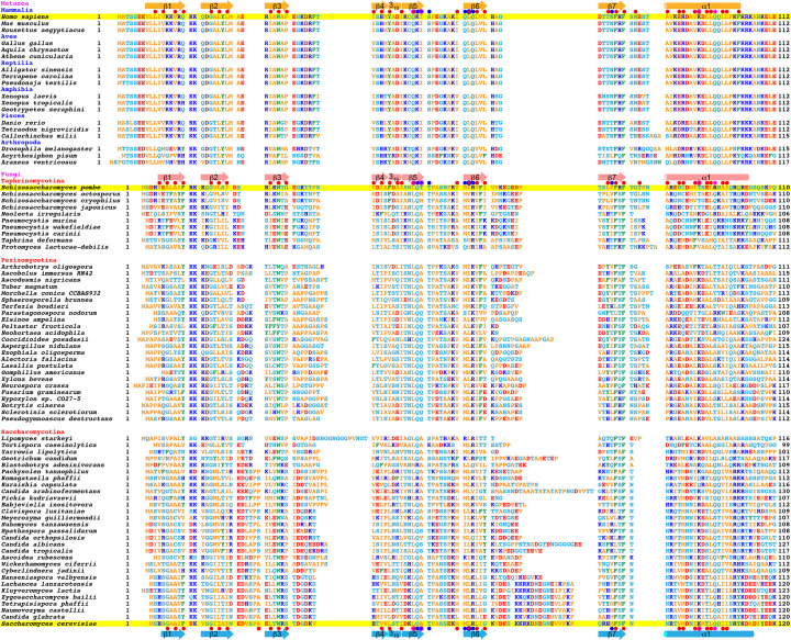
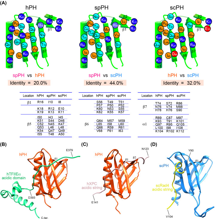
The general transcription factor TFIIH is a multi-subunit complex involved in transcription, DNA repair, and cell cycle in eukaryotes. In the human p62 subunit and the budding yeast Saccharomyces cerevisiae Tfb1 subunit of TFIIH, the pleckstrin homology (PH) domain (hPH/scPH) recruits TFIIH to transcription-start and DNA-damage sites by interacting with an acidic intrinsically disordered region in transcription and repair factors. Whereas metazoan PH domains are highly conserved and adopt a similar structure, fungal PH domains are divergent and only the scPH structure is available. Here, we have determined the structure of the PH domain from Tfb1 of fission yeast Schizosaccharomyces pombe (spPH) by NMR. spPH holds an architecture, including the core and external backbone structures, that is closer to hPH than to scPH despite having higher amino acid sequence identity to scPH. In addition, the predicted target-binding site of spPH shares more amino acid similarity with scPH, but spPH contains several key residues identified in hPH as required for specific binding. Using chemical shift perturbation, we have identified binding modes of spPH to spTfa1, a homologue of hTFIIEα, and to spRhp41, a homologue of the repair factors hXPC and scRad4. Both spTfa1 and spRhp41 bind to a similar but distinct surface of spPH by modes that differ from those of target proteins binding to hPH and scPH, revealing that the PH domain of TFIIH interacts with its target proteins in a polymorphic manner in Metazoa, and budding and fission yeasts.

Keywords: NMR spectroscopy; PH domain; TFIIH; budding yeast; general transcription factor.


Conflict of interest statement

The authors declare that there are no competing interests associated with the manuscript.

Figures

Figure 1
Figure 1. The PH domain of the TFIIH subunit spTfb1
(A) Domain organization of the homologous hP62, spTfb1, and scTfb1 subunits. (B) Amino acid sequence alignment of the PH domains. Secondary structure elements of the hPH and scPH domains are shown respectively above and below their sequences. Arrow, β-strand; cylinder, helix. (C) 1H-15N-HSQC spectrum of the expressed spPH domain.
Figure 2
Figure 2. Solution structure of the spPH domain
(A,B) Overlay of the 20 best structures, shown as line (A) and ribbon (B) diagrams, respectively. (C) 15N-{1H} NOE of the spPH domain. Proline residues are indicated by ‘p’. For some residues, no NOE value was determined due to an overlapping or unreliable weak amide peak.
Figure 3
Figure 3. Comparison of the backbone structures of the hPH, spPH, and scPH domains
hPH domain is colored orange-red, spPH domain deep pink, and scPH domain blue. Pairwise RMSD and amino acid sequence identity are indicated. The overlapped regions are listed below.
Figure 4
Figure 4. Comparison of the electrostatic potential surfaces of the hPH, spPH, and scPH domains
Upper panel, backbone structure with side-chains of basic amino acids involved in target binding. Middle and lower panels, electrostatic potential surface. Positive potential is shown in blue, and negative potential in red. Residues that form the basic surface are listed below.
Figure 5
Figure 5. Comparison of the target-binding pockets of the hPH, spPH, and scPH domains
Upper panel, target-binding pockets 1 and 2 (yellow circles). Middle panel, close up of pocket 1. Lower panel, close up of pocket 2. Residues that form the respective pockets are listed below.
Figure 6
Figure 6. Target-binding activity and the deduced binding surface of the spPH domain
(A) Amino acid sequence alignment of p62/Tfb1 target proteins. Pocket 1- and 2-inserted residues of hTFIIEα, hXPC, and scRad4, and the corresponding residues in other target proteins, are highlighted in yellow and light green, respectively. The sequences of peptides used in chemical shift perturbation (CSP) are indicated by boxes. (B) NMR CSP experiment. Overlay of four 1H-15N-HSQC spectra of the spPH domain alone (black) and titrated with target peptide at molar ratios of 1:0.25, 1:0.50, and 1:1.00. Selected regions are shown here (see Supplementary Figure S4 for the full regions). (C) Estimated Kd values and thermodynamic parameters. (D-I) CSP mapping. Residues showing a chemical shift change (Δδ) greater than 0.100 upon addition of the target peptide are mapped on the structure of the PH domain: spTfa1332-350 (D); spTfa1416-434 (E); spRhp414-22 (F); hTFIIEα378-439 (acidic domain) (G); hTFIIEα378-396 (H); and hXPC109-156 (I). Residues are colored according to the magnitude of Δδ.
Figure 7
Figure 7. Amino acid sequence alignment of metazoan p62 and fungal Tfb1 PH domains
Secondary structure elements of the hPH, spPH, and scPH domains are shown above and below their sequences, which are highlighted in yellow. Arrow, β-strand; cylinder, helix. Red dots, residues that form a hydrophobic core; blue dots, residues that form pocket 1; magenta dots, residues that form pocket 2.
Figure 8
Figure 8. Comparison of the target-binding surfaces of the hPH, spPH, and scPH domains
(A) Schematic target-binding surfaces of the PH domains. Pairwise amino acid identity is indicated. Residues that form the target binding surface and the corresponding residues are listed below. (B-D) Structures of the complex of the PH domain and its target. (B) Complex of the hPH domain (orange-red) with the hTFIIEα acidic domain (green) [PDB ID 2RNR]. (C) Complex of the hPH domain (orange-red) with the hXPC acidic string (pale pink) [PDB ID 2RVB]. (D) Complex of the scPH domain (blue) with the scRad4 acidic string (yellow) [PDB ID 2M14].

Similar articles

References

    1. Conaway R.C. and Conaway J.W. (1989) An RNA polymerase II transcription factor has an associated DNA-dependent ATPase (dATPase) activity strongly stimulated by the TATA region of promoters. Proc. Natl. Acad. Sci. U. S. A. 86, 7356–7360 10.1073/pnas.86.19.7356 - DOI - PMC - PubMed
    1. Schaeffer L., Roy R., Humbert S., Moncollin V., Vermeulen W., Hoeijmakers J.H.et al. . (1993) DNA repair helicase: a component of BTF2 (TFIIH) basic transcription factor. Science 260, 58–63 10.1126/science.8465201 - DOI - PubMed
    1. Roy R., Adamczewski J.P., Seroz T., Vermeulen W., Tassan J.P., Schaeffer L.et al. . (1994) The MO15 cell cycle kinase is associated with the TFIIH transcription-DNA repair factor. Cell 79, 1093–1101 10.1016/0092-8674(94)90039-6 - DOI - PubMed
    1. Compe E. and Egly J.M. (2016) Nucleotide excision repair and transcriptional regulation: TFIIH and beyond. Annu. Rev. Biochem. 85, 265–290 10.1146/annurev-biochem-060815-014857 - DOI - PubMed
    1. Rimel J.K. and Taatjes D.J. (2018) The essential and multifunctional TFIIH complex. Protein Sci. 27, 1018–1037 10.1002/pro.3424 - DOI - PMC - PubMed

Publication types

MeSH terms

Substances