. 2023 Jul 18;62(14):2115-2127.
doi: 10.1021/acs.biochem.3c00109. Epub 2023 Jun 21.

Irreversible Inactivation of SARS-CoV-2 by Lectin Engagement with Two Glycan Clusters on the Spike Protein

Affiliations

Irreversible Inactivation of SARS-CoV-2 by Lectin Engagement with Two Glycan Clusters on the Spike Protein

Aakansha Nangarlia et al. Biochemistry. .

Abstract

Host cell infection by SARS-CoV-2, similar to that by HIV-1, is driven by a conformationally metastable and highly glycosylated surface entry protein complex, and infection by these viruses has been shown to be inhibited by the mannose-specific lectins cyanovirin-N (CV-N) and griffithsin (GRFT). We discovered in this study that CV-N not only inhibits SARS-CoV-2 infection but also leads to irreversibly inactivated pseudovirus particles. The irreversibility effect was revealed by the observation that pseudoviruses first treated with CV-N and then washed to remove all soluble lectin did not recover infectivity. The infection inhibition of SARS-CoV-2 pseudovirus mutants with single-site glycan mutations in spike suggested that two glycan clusters in S1 are important for both CV-N and GRFT inhibition: one cluster associated with the RBD (receptor binding domain) and the second with the S1/S2 cleavage site. We observed lectin antiviral effects with several SARS-CoV-2 pseudovirus variants, including the recently emerged omicron, as well as a fully infectious coronavirus, therein reflecting the breadth of lectin antiviral function and the potential for pan-coronavirus inactivation. Mechanistically, observations made in this work indicate that multivalent lectin interaction with S1 glycans is likely a driver of the lectin infection inhibition and irreversible inactivation effect and suggest the possibility that lectin inactivation is caused by an irreversible conformational effect on spike. Overall, lectins' irreversible inactivation of SARS-CoV-2, taken with their breadth of function, reflects the therapeutic potential of multivalent lectins targeting the vulnerable metastable spike before host cell encounter.


Conflict of interest statement

The authors declare no competing financial interest.

Figures
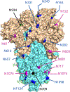
Figure 1.
Figure 1.
Structure of SARS-CoV-2 spike complex, composed of three S1–S2 subunit dimers, showing the locations of 22 glycans. The PDB structure file shown is 6VXX, with subunit S1 in brown and subunit S2 in cyan. Glycan labels are color-coded according to % oligomannose content: black – 80–100% (least processed glycans), magenta – 30–79% (hybrid), blue – 0–29% (most processed or complex). Glycan residues that are not shown include N17, N74, N149, N1158, N1173, and N1194.
Figure 2.
Figure 2.
Infection inhibition of SARS-CoV-2 by lectins cyanovirin-N (CV-N), griffithsin (GRFT), and microvirin (MVN). CV-N and GRFT both showed dose-dependent inhibition of infection by the wild-type SARS-CoV-2 Wuhan parental strain. Treatment with MVN did not show any inhibitory effect against wild-type SARS-CoV-2. The data represent the mean of three independent experiments, and the error bars depict the standard deviation around the mean.
Figure 3.
Figure 3.
Irreversible inactivation of SARS-CoV-2 pseudoviruses by CV-N, as shown by loss of infectivity after lectin washout. (A) SARS-CoV-2 pseudovirus samples were tested for infectivity after CV-N-treatment at various doses, followed by washout with 10 buffer suspension-centrifugation cycles. The data represent the mean of three independent experiments, and the error bars depict the standard deviation around the mean. (B) Western blot (WB) analysis of the washes confirms the removal of unbound CV-N from the residual pseudovirus samples by the 10th wash as seen for the 10 μM flowthrough lanes.
Figure 4.
Figure 4.
CV-N does not cause S1 shedding but S complex entrapment based on shedding analysis. (A) Dose-dependent western blot detection of the S1 protein in the first wash (of 10) flowthrough after ACE2 treatment of SARS-CoV-2 WT pseudoviruses. (B) No detection of the S1 protein in the first wash (of 10) flowthrough after CV-N treatment of SARS-CoV-2 WT pseudoviruses. (C) Detection of S1 protein in virus samples, after CV-N treatment and 10 washes, of SARS-CoV-2 WT pseudoviruses, showing no dose dependence.
Figure 5.
Figure 5.
Identification and location of CV-N-critical glycans in the SARS-CoV-2 spike. IC50 values for CV-N infection inhibition of SARS-CoV-2 pseudo-viruses mutated at single glycan sites. Reduction in CV-N inhibition was observed for N61D, N122D, N331D, N343D, N603D, and N657D mutants, as shown in Table 1. (A) Structure showing the clustering of CV-N functional glycans in RBD and furin cleavage site associated locations (PDB: 6VXX). (B) Washout experiments (after 2-hour lectin preincubation) showed that “CV-N treated” single-site pseudovirus mutants (N61D, N122D, N331D, N343D, and N657D, denoted by asterisks (*)) retained infectivity. The data represent the mean of three independent experiments, and the error bars depict the standard deviation around the mean.
Figure 6.
Figure 6.
Infection Inhibition of WT SARS-CoV-2 by Dimer-disfavoring CV-N [P51G] Mutant. CV-N [P51G] exhibits dose-dependent inhibition of WT SARS-CoV-2 infection, although at an order of magnitude worse IC50 when compared to inhibition with WT CV-N. The data represent the mean of three independent experiments, and the error bars depict the standard deviation around the mean.
Figure 7.
Figure 7.
SARS-CoV-2 breadth and fully infectious MHV-A59 assays. (A) Preliminary screen of CV-N inhibition of SARS-CoV-2 pseudovirus variant infection of CHO-ACE2 cells. (B) Plaque assay evaluation of CV-N inhibition of fully infectious MHV-A59 virus infection. CV-N was capable of inhibiting infection of the virus at various multiplicities of infection (MOIs). The data represent the mean of three independent experiments, and the error bars depict the standard deviation around the mean.

References

    1. Watanabe Y; Allen JD; Wrapp D; McLellan JS; Crispin M Site-specific glycan analysis of the SARS-CoV-2 spike. Science 2020, 369, 330–333. - PMC - PubMed
    1. Zhao X; Chen H; Wang H Glycans of SARS-CoV-2 Spike Protein in Virus Infection and Antibody Production. Front. Mol. Biosci. 2021, 8, No. 629873. - PMC - PubMed
    1. Ahan RE; Hanifehnezhad A; Kehribar EŞ; Oguzoglu TC; Földes K; Özçelik CE; Filazi N; Öztop S; Palaz F; Onder S; Bozkurt EU; Ergünay K; Özkul A; Şeker UÖŞ A Highly Potent SARS-CoV-2 Blocking Lectin Protein. ACS Infect. Dis. 2022, 8, 1253–1264. - PMC - PubMed
    1. Cai Y; Xu W; Gu C; Cai X; Qu D; Lu L; Xie Y; Jiang S Griffithsin with A Broad-Spectrum Antiviral Activity by Binding Glycans in Viral Glycoprotein Exhibits Strong Synergistic Effect in Combination with A Pan-Coronavirus Fusion Inhibitor Targeting SARS-CoV-2 Spike S2 Subunit. Virol. Sin. 2020, 35, 857–860. - PMC - PubMed
    1. Walls AC; Park Y-J; Tortorici MA; Wall A; McGuire AT; Veesler D Structure, Function, and Antigenicity of the SARS-CoV-2 Spike Glycoprotein. Cell 2020, 181, 281.e6–292.e6. - PMC - PubMed

Publication types

Supplementary concepts