. 2023 Jul 5;145(26):14307-14315.
doi: 10.1021/jacs.3c02742. Epub 2023 Jun 21.

Design of Heme Enzymes with a Tunable Substrate Binding Pocket Adjacent to an Open Metal Coordination Site

Affiliations

Design of Heme Enzymes with a Tunable Substrate Binding Pocket Adjacent to an Open Metal Coordination Site

Indrek Kalvet et al. J Am Chem Soc. .

Abstract

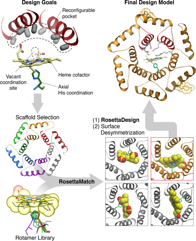
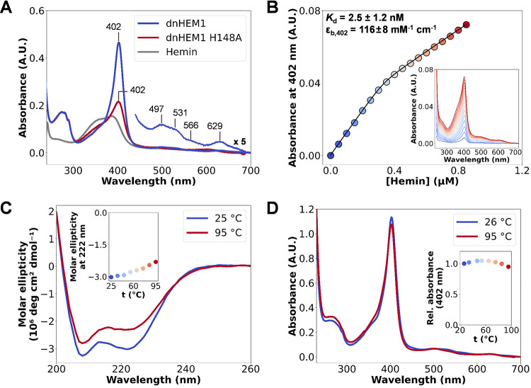
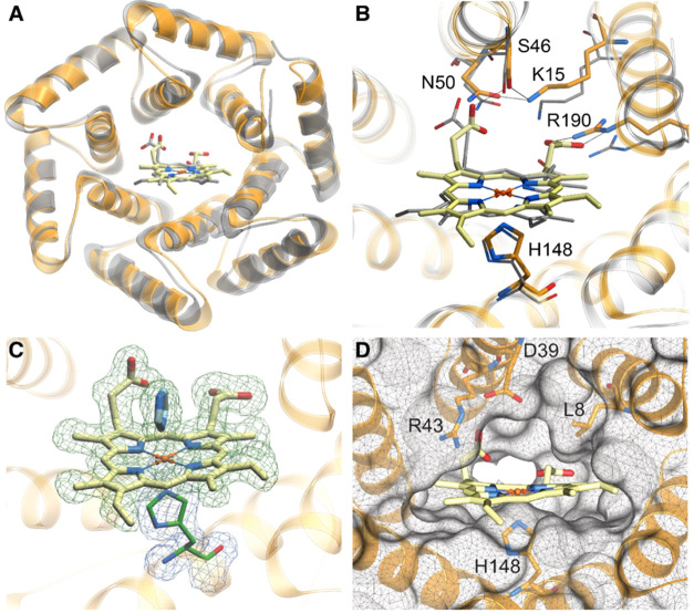
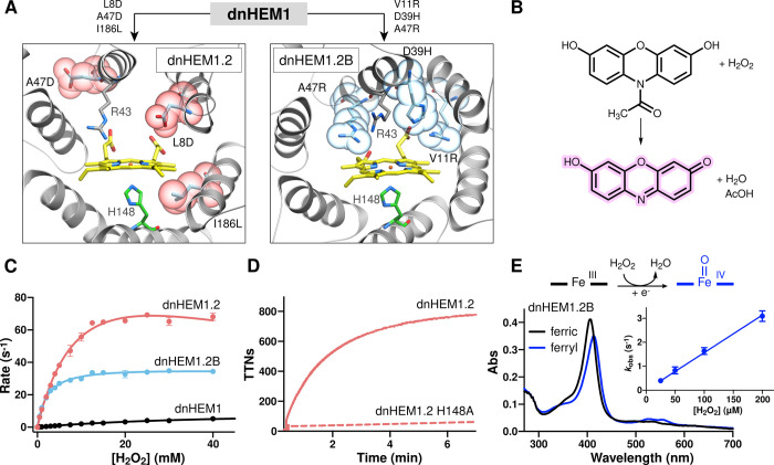
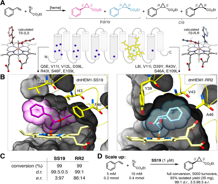
The catalytic versatility of pentacoordinated iron is highlighted by the broad range of natural and engineered activities of heme enzymes such as cytochrome P450s, which position a porphyrin cofactor coordinating a central iron atom below an open substrate binding pocket. This catalytic prowess has inspired efforts to design de novo helical bundle scaffolds that bind porphyrin cofactors. However, such designs lack the large open substrate binding pocket of P450s, and hence, the range of chemical transformations accessible is limited. Here, with the goal of combining the advantages of the P450 catalytic site geometry with the almost unlimited customizability of de novo protein design, we design a high-affinity heme-binding protein, dnHEM1, with an axial histidine ligand, a vacant coordination site for generating reactive intermediates, and a tunable distal pocket for substrate binding. A 1.6 Å X-ray crystal structure of dnHEM1 reveals excellent agreement to the design model with key features programmed as intended. The incorporation of distal pocket substitutions converted dnHEM1 into a proficient peroxidase with a stable neutral ferryl intermediate. In parallel, dnHEM1 was redesigned to generate enantiocomplementary carbene transferases for styrene cyclopropanation (up to 93% isolated yield, 5000 turnovers, 97:3 e.r.) by reconfiguring the distal pocket to accommodate calculated transition state models. Our approach now enables the custom design of enzymes containing cofactors adjacent to binding pockets with an almost unlimited variety of shapes and functionalities.


Conflict of interest statement

The authors declare no competing financial interest.

Figures

Figure 1
Figure 1
Schematic of the approach to computational design. To generate heme-binding proteins containing a reconfigurable pocket near a free coordination site on the Fe atom (top left), a Heme-HIS moiety was matched into the pore of helical solenoid scaffolds of appropriate size (bottom left and right). The sequence around the heme cofactor was then optimized using Rosetta (top right).
Figure 2
Figure 2
Characterization of Heme binding. (A) UV/Vis spectra of dnHEM1 (blue) and its H148A mutant (red), after mixing 10 μM protein with 2 μM hemin, indicating the importance of H148 for heme binding. (B) Kd determination by heme titration into dnHEM1 (0.4 μM) and following absorbance changes at 402 nm, which were plotted against heme concentration and fitted to a one-site binding equation (see SI). (C) CD spectra of holo-dnHEM1 at increasing temperatures. (D) UV/Vis spectra of holo-dnHEM1 collected at increasing temperatures indicate that heme-binding ability is retained at temperatures up to 95 °C.
Figure 3
Figure 3
The crystal structure of dnHEM1 (PDB id: 8C3W) closely matches the design model. (A) Crystal structure of dnHEM1 (orange) overlaid with the design model (silver). (B) Superposition of the heme-binding site of the dnHEM1 design model (silver) and the crystal structure (orange). Hydrogen bond interactions with heme are indicated with dashed lines. (C) Crystal structure of dnHEM1 showing the electron density corresponding to heme and the exogenous imidazole in blue (Fo – Fc omit map contoured at 1σ) and His148 shown in green (2Fo – Fc map contoured at 1σ). (D) Open substrate binding pocket at the distal site upon omission of co-crystallized imidazole.
Figure 4
Figure 4
Directed evolution converts dnHEM1 into a proficient peroxidase with a stable high valent ferryl intermediate. (A) AlphaFold2 predicted models of dnHEM1.2 and dnHEM1.2B indicating the mutations accumulated as a result of divergent directed evolution. Sites of mutation for dnHEM1.2 and dnHEM1.2B are shown as atom colored ball and sticks with purple or blue carbons, respectively. The heme cofactor is shown as gray atom colored ball and sticks and CPK spheres. (B) Chemical scheme showing the conversion of Amplex Red dye to resorufin mediated by hydrogen peroxide and dnHEM1 variants. (C) Michaelis–Menten plots under saturating concentrations of Amplex Red for dnHEM1 (black, kcat = 9.5 ± 0.2 s–1, KM[H2O2] = 36.7 ± 1.6 mM), dnHEM1.2 (red, kcat = 129.5 ± 8.7 s–1, KM[H2O2] = 11.5 ± 1.2 mM, KI = 58.9 ± 10.7 mM) and dnHEM1.2B (blue, kcat = 37.0 ± 0.3 s–1, KM[H2O2] = 2.0 ± 0.1 mM). Error bars represent SD n = 3. (D) Time course of resorufin formation by dnHEM1.2 (red solid line, TTNs = 788 ± 18) and dnHEM1.2 H148A (red dotted line). (E) Formation of a ferryl intermediate (blue) in dnHEM1.2B upon oxidation of the ferric enzyme (black line) with H2O2. The observed neutral ferryl heme state is most likely formed via rapid single electron transfer to a transient porphyrin-π cation radical species, with a redox active amino acid side chain being the most likely electron donor. Inset: A linear fit of kobs vs [H2O2] was used to derive a bimolecular rate constant of (1.6 ± 0.05) × 104 M–1 s–1 for ferryl heme formation. Error bars represent SD n = 3.
Figure 5
Figure 5
Computational redesign of dnHEM1 for enantioselective olefin cyclopropanation activity. (A) Enantiocomplementary transition states for R,R-and S,S-cyclopropane formation were computed with DFT and selected positions in the distal pocket were redesigned with Rosetta. (B) Complementarity of the designed pocket with most selective enzymes for S,S (left) and R,R (right) transition state models. (C) Selectivities obtained with best variants for the synthesis of S,S and R,R cyclopropanes. Standard reaction condition: 1 μM catalyst, 1 mM styrene, 10 mM EDA, 100 μM dithionite, under N2 in aqueous potassium phosphate buffer (50 mM, NaCl 200 mM, pH 7.2) and 5% MeCN cosolvent for 2 h at 25 °C. See SI for the results with other designs. (D) Preparative scale synthesis of the S,S stereoisomer, catalyzed by dnHEM1-SS19 over 2 h at r.t.

References

    1. Rittle J.; Green M. T. Cytochrome P450 Compound I: Capture, Characterization, and C-H Bond Activation Kinetics. Science 2010, 330, 933–937. 10.1126/science.1193478. - DOI - PubMed
    1. Poulos T. L. Heme Enzyme Structure and Function. Chem. Rev. 2014, 114, 3919–3962. 10.1021/cr400415k. - DOI - PMC - PubMed
    1. Casadei C. M.; Gumiero A.; Metcalfe C. L.; Murphy E. J.; Basran J.; Concilio M. G.; Teixeira S. C. M.; Schrader T. E.; Fielding A. J.; Ostermann A.; Blakeley M. P.; Raven E. L.; Moody P. C. E. Heme Enzymes. Neutron Cryo-Crystallography Captures the Protonation State of Ferryl Heme in a Peroxidase. Science 2014, 345, 193–197. 10.1126/science.1254398. - DOI - PubMed
    1. Moody P. C. E.; Raven E. L. The Nature and Reactivity of Ferryl Heme in Compounds I and II. Acc. Chem. Res. 2018, 51, 427–435. 10.1021/acs.accounts.7b00463. - DOI - PubMed
    1. Huang X.; Groves J. T. Oxygen Activation and Radical Transformations in Heme Proteins and Metalloporphyrins. Chem. Rev. 2018, 118, 2491–2553. 10.1021/acs.chemrev.7b00373. - DOI - PMC - PubMed

Publication types