. 2023 Sep;34(3):453-463.
doi: 10.1007/s00335-023-09999-8. Epub 2023 Jun 21.

The spontaneous mouse mutant low set ears (Lse) is caused by tandem duplication of Fgf3 and Fgf4

Affiliations

The spontaneous mouse mutant low set ears (Lse) is caused by tandem duplication of Fgf3 and Fgf4

Alana Luzzio et al. Mamm Genome. 2023 Sep.

Abstract

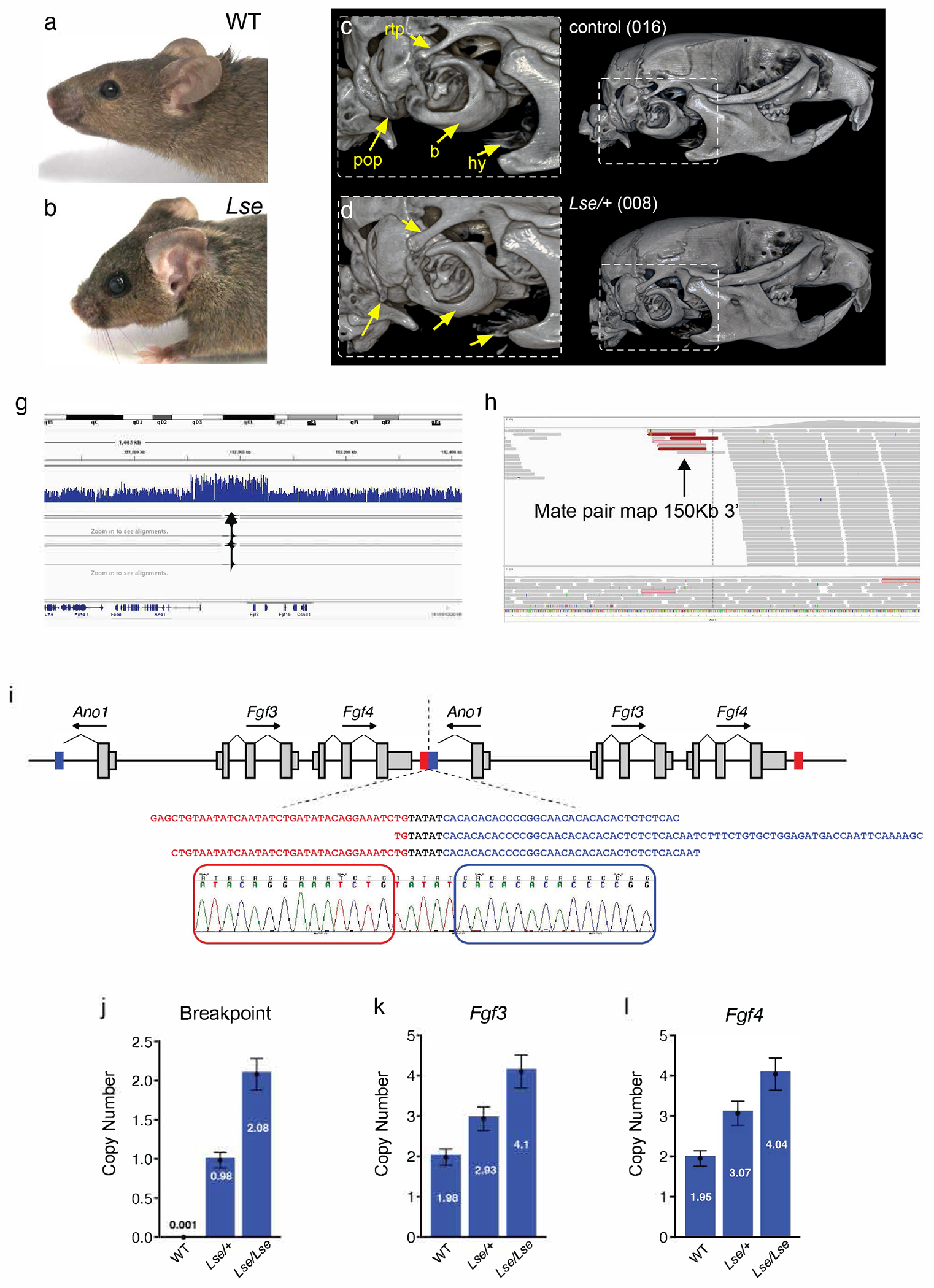
The external ear develops from an organized convergence of ventrally migrating neural crest cells into the first and second branchial arches. Defects in external ear position are often symptomatic of complex syndromes such as Apert, Treacher-Collins, and Crouzon Syndrome. The low set ears (Lse) spontaneous mouse mutant is characterized by the dominant inheritance of a ventrally shifted external ear position and an abnormal external auditory meatus (EAM). We identified the causative mutation as a 148 Kb tandem duplication on Chromosome 7, which includes the entire coding sequences of Fgf3 and Fgf4. Duplications of FGF3 and FGF4 occur in 11q duplication syndrome in humans and are associated with craniofacial anomalies, among other features. Intercrosses of Lse-affected mice revealed perinatal lethality in homozygotes, and Lse/Lse embryos display additional phenotypes including polydactyly, abnormal eye morphology, and cleft secondary palate. The duplication results in increased Fgf3 and Fgf4 expression in the branchial arches and additional discrete domains in the developing embryo. This ectopic overexpression resulted in functional FGF signaling, demonstrated by increased Spry2 and Etv5 expression in overlapping domains of the developing arches. Finally, a genetic interaction between Fgf3/4 overexpression and Twist1, a regulator of skull suture development, resulted in perinatal lethality, cleft palate, and polydactyly in compound heterozygotes. These data indicate a role for Fgf3 and Fgf4 in external ear and palate development and provide a novel mouse model for further interrogation of the biological consequences of human FGF3/4 duplication.


Conflict of interest statement

Competing Interests: The corresponding author, Stephen A. Murray, is an editor-in-chief of the journal. There are no other competing interests.

Figures

Figure 1:
Figure 1:. The low set ears (Lse) mouse mutant is caused by a tandem duplication which includes Fgf3 and Fgf4.
(a,b) Lse mutant mice display abnormal external ear morphology, including both the position and shape of the external auditory meatus (EAM) and the overall appearance of the pinnae. Mutant mice also display variably penetrant bulging eyes. (c,d) MicroCT scans of adult Lse/+ and WT skulls revealed a number of subtle bony anomalies, including altered morphology of the bulla (b), hypoplasia of the paraoccipital process (pop), and abnormal morphology of hyoid bone (hy). The retrotympanic process (rtp) appeared to be thicker in heterozygotes than controls, although ~40% of controls had processes of comparable thickness. (e) IGV visualization of array-captured sequencing of the region around the D7Nds4 marker showing the increase in apparent read number mapping to the region, which suggests a copy number variation. (f) IGV visualization of individual sequencing reads with mate pairs that map to the 3’ end of the 148Kb interval. (g) Reconstruction of the duplicated region, color coded to indicate sequences at the 5’ (blue) and 3’ (red) of the interval. Breakpoint reads which define the tail-head fusion were identified from unmapped mate pairs (gray with red outline in f), and confirmed by Sanger sequencing. (h) A digital droplet PCR (ddPCR) breakpoint assay was used to genotype the Fgf3/4 duplication, and the duplication was confirmed by ddPCR copy number assessment of the Fgf3 (i) and Fgf4 (j) genes in WT, Lse/+, and Lse/Lse embryos.
Figure 2:
Figure 2:. Gross morphology of Lse mutants.
Appearance of WT, Lse/+, and Lse/Lse embryos at E14.5 (a-c) and E17.5 (d-f). The malpositioned external ear is evident in both mutant genotypes at each stage (arrows) and polydactyly is noted in Lse/Lse embryos (arrowheads in c and f). (g,h) MicroCT imaging reveals cleft secondary palate and bulging eyes in Lse/Lse mutants and surface renderings (i, j) illustrate the abnormal shape of the pinnae in mutants.
Figure 3:
Figure 3:. Skeletal abnormalities in Lse embryos.
(a) Alizarin red/alcian blue staining at E18.5 reveal minor abnormalities in the palatal process and tympanic ring in Lse/+ embryos, while Lse/Lse skulls showed cleft secondary palate and a hypomorphic tympanic ring remnant (yellow arrows). (b) Staining of limbs show postaxial polydactyly in Lse/Lse mutants only (arrow).
Figure 4:
Figure 4:. Increased and ectopic expression of Fgf3 and Fgf4 in Lse mutant embryos.
Whole mount in situ hybridization for Fgf3 (a-c) at E10.5 reveals highly increased expression levels in the second branchial arch in Lse/+ embryos (arrow in b and b’), with ectopic expression in the urogenital ridge and adjacent to the forelimb (arrowheads b and b’’). Lse/Lse embryos show a similar pattern but with higher expression levels (c, c’, c’’), with some expansion of the expression domain into the first branchial arch. Otocyst expression is indicated by *. (d-f) Fgf4 expression at E10.5 shows a modest increase in expression in the caudal aspect of the second arch with similar levels in both Lse/+ and Lse/Lse embryos. At E9.5 (g-i), similar levels of Fgf4 expression are seen in mutants and controls in the arches (arrowheads), but an additional domain of ventral expression is observed in both mutants, with higher levels in Lse/Lse embryos (arrows in h’ and i’).
Figure 5:
Figure 5:. The Lse duplication leads to increased FGF signaling.
Whole mount in situ hybridization reveals increased expression of FGF pathway targets Spry2 (a) and Etv5 (b) in Lse/+ mutants versus controls. This is observed in domains which overlap expanded Fgf3 expression, including the branchial arches (inset, white arrow), and the urogenital ridge (yellow arrow).
Figure 6:
Figure 6:. Genetic interaction between Lse and Twist1.
(a) Surface rendering of a microCT scan of a Lse/+; Twist +/− E18.5 embryo showing the expected low set ear phenotype and polydactyly on both limbs (red arrows). (b) Gross appearance of Lse/+;Twist+/− neonates with distention of the abdomen and lack of a milk spot, consistent with ingestion of air while feeding. (c) Sagittal sections of a microCT scan showing apparent posterior, soft-tissue palate cleft, which was confirmed in gross dissections (d).

Similar articles

References

    1. Arnold JS, Braunstein EM, Ohyama T, Groves AK, Adams JC, Brown MC, Morrow BE, 2006. Tissue-specific roles of Tbx1 in the development of the outer, middle and inner ear, defective in 22q11DS patients. Hum Mol Genet 15, 1629–1639. - PMC - PubMed
    1. Bourgeois P, Bolcato-Bellemin AL, Danse JM, Bloch-Zupan A, Yoshiba K, Stoetzel C, Perrin-Schmitt F, 1998. The variable expressivity and incomplete penetrance of the twist-null heterozygous mouse phenotype resemble those of human Saethre-Chotzen syndrome. Hum Mol Genet 7, 945–957. - PubMed
    1. Brewer JR, Mazot P, Soriano P, 2016. Genetic insights into the mechanisms of Fgf signaling. Genes Dev 30, 751–771. - PMC - PubMed
    1. Carlton MB, Colledge WH, Evans MJ, 1998. Crouzon-like craniofacial dysmorphology in the mouse is caused by an insertional mutation at the Fgf3/Fgf4 locus. Dev Dyn 212, 242–249. - PubMed
    1. Carver EA, Oram KF, Gridley T, 2002. Craniosynostosis in Twist heterozygous mice: a model for Saethre-Chotzen syndrome. Anat Rec 268, 90–92. - PubMed

Publication types

Substances

LinkOut - more resources