. 2023 Aug;146(2):191-210.
doi: 10.1007/s00401-023-02600-1. Epub 2023 Jun 21.

Tau seeding and spreading in vivo is supported by both AD-derived fibrillar and oligomeric tau

Affiliations

Tau seeding and spreading in vivo is supported by both AD-derived fibrillar and oligomeric tau

Anastasie Mate De Gerando et al. Acta Neuropathol. 2023 Aug.

Abstract

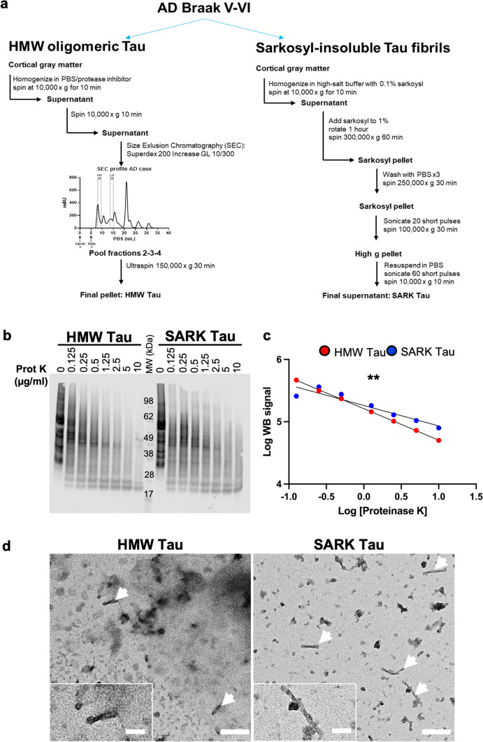
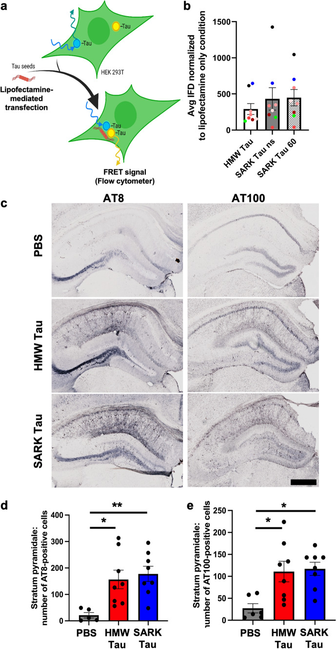
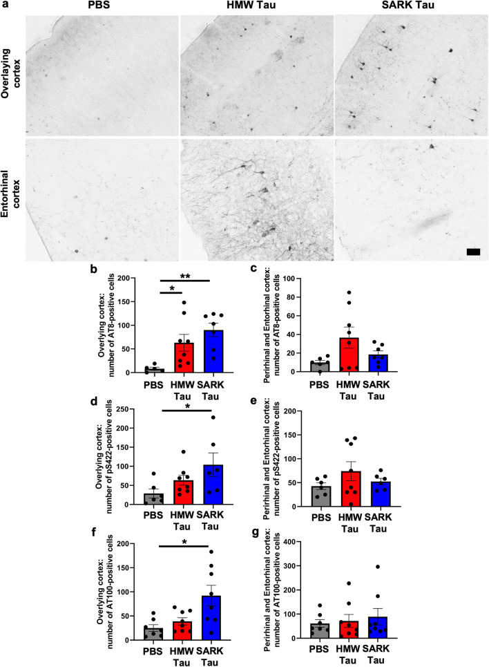
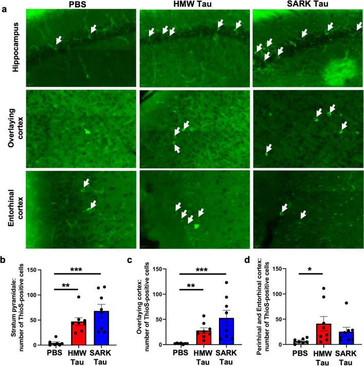
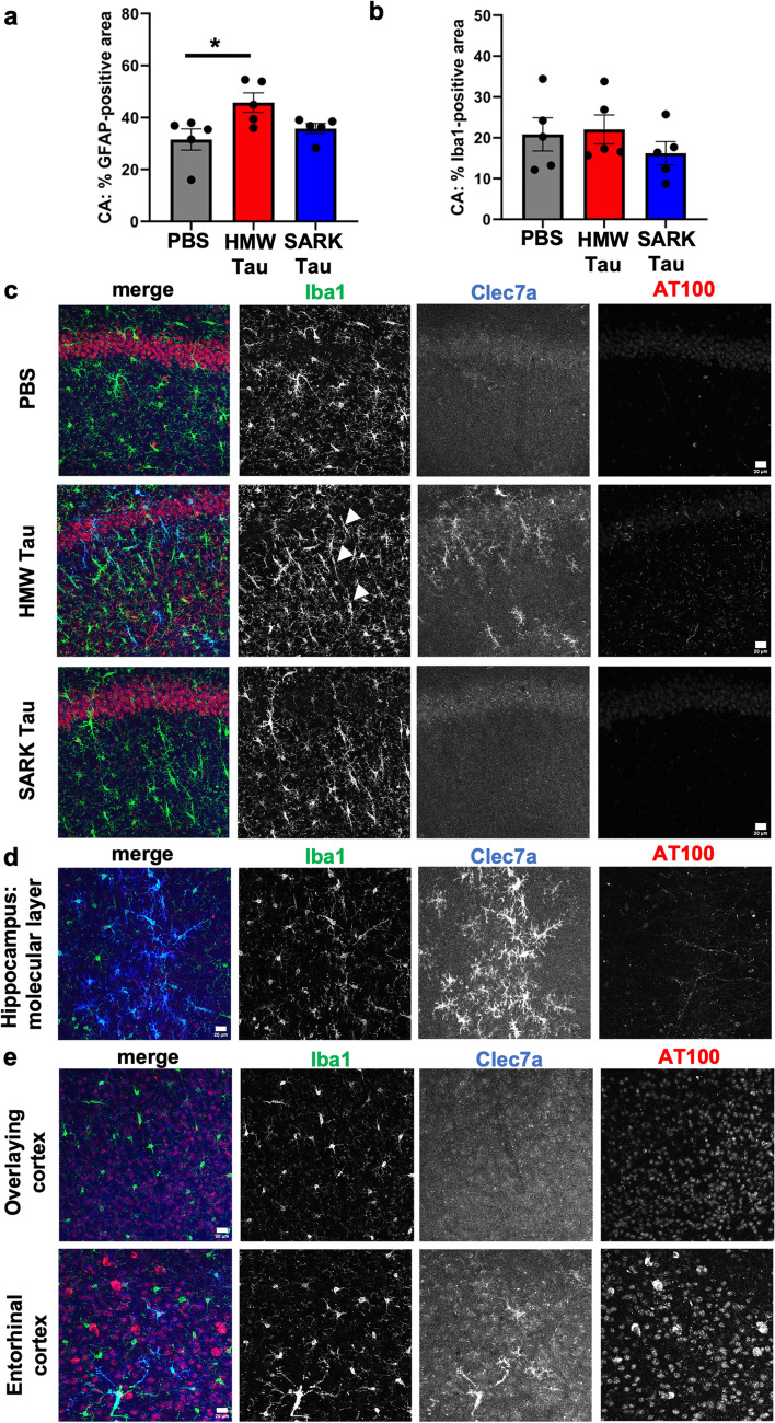
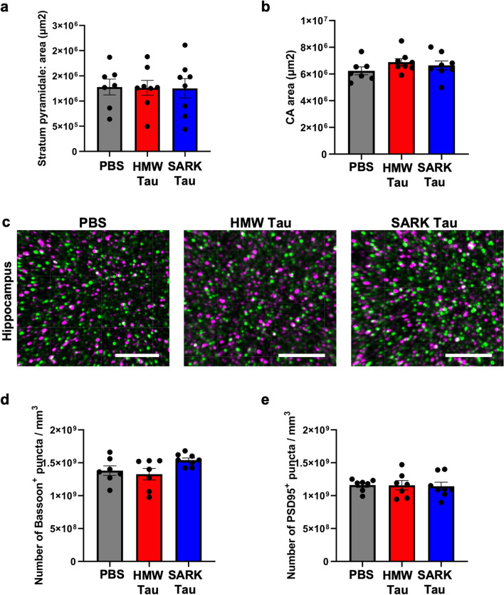
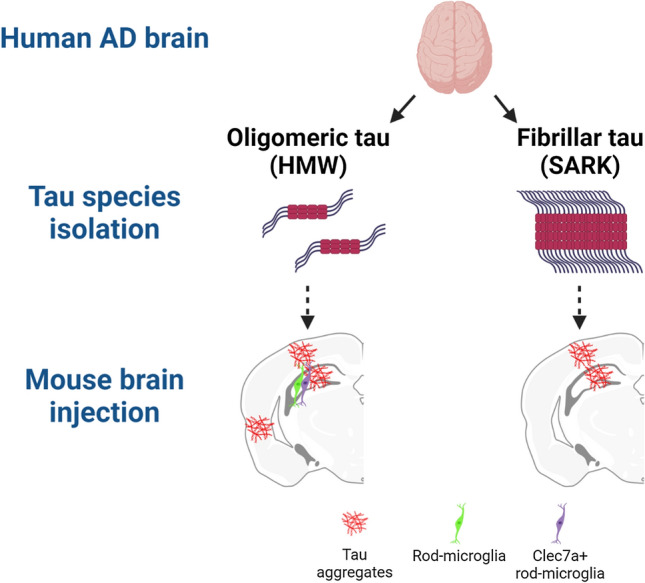
Insoluble fibrillar tau, the primary constituent of neurofibrillary tangles, has traditionally been thought to be the biologically active, toxic form of tau mediating neurodegeneration in Alzheimer's disease. More recent studies have implicated soluble oligomeric tau species, referred to as high molecular weight (HMW), due to their properties on size-exclusion chromatography, in tau propagation across neural systems. These two forms of tau have never been directly compared. We prepared sarkosyl-insoluble and HMW tau from the frontal cortex of Alzheimer patients and compared their properties using a variety of biophysical and bioactivity assays. Sarkosyl-insoluble fibrillar tau comprises abundant paired-helical filaments (PHF) as quantified by electron microscopy (EM) and is more resistant to proteinase K, compared to HMW tau, which is mostly in an oligomeric form. Sarkosyl-insoluble and HMW tau are nearly equivalent in potency in HEK cell bioactivity assay for seeding aggregates, and their injection reveals similar local uptake into hippocampal neurons in PS19 Tau transgenic mice. However, the HMW preparation appears to be far more potent in inducing a glial response including Clec7a-positive rod microglia in the absence of neurodegeneration or synapse loss and promotes more rapid propagation of misfolded tau to distal, anatomically connected regions, such as entorhinal and perirhinal cortices. These data suggest that soluble HMW tau has similar properties to fibrillar sarkosyl-insoluble tau with regard to tau seeding potential, but may be equal or even more bioactive with respect to propagation across neural systems and activation of glial responses, both relevant to tau-related Alzheimer phenotypes.

Keywords: Alzheimer; Microglia; Seeding; Spreading; Tau.


Figures

Fig. 1
Fig. 1
SARK tau is mainly fibrillar, while HMW tau is mainly oligomeric. a Schematic of the experimental workflow to extract tau oligomers [17] (HMW Tau) and tau fibrils [42] (SARK tau) from the cortical gray matter of eight AD cases of Braak V–VI. b Total tau WB (DAKO) of SARK and HMW tau samples after incubation with increasing concentrations of proteinase K. c Quantification of the WB signal intensity after proteinase K treatment. Log-transformed data were plotted and the slopes compared. Linear regression testing for the equality of slopes, **p < 0.01. d Representative negative stain EM images of non-sonicated samples from AD case #2399 used for in vivo injections. HMW tau samples present mainly as amorphous material and a few PHFs (white arrows), while PHFs are more abundant in the SARK tau samples. Scale bar = 200 nm and 100 nm for inserts. MW molecular weight
Fig. 2
Fig. 2
Tau oligomers and tau fibrils have similar bioactivities in vitro and in vivo. a Schematic of the FRET-biosensor seeding assay. HEK293T cells stably express the tau repeat domain fused to either CFP or YFP. When transfected with bioactive tau seeds, these constructs aggregate and become close enough to generate a FRET signal quantifiable by flow cytometry. Here, SARK and HMW tau samples from eight AD cases were normalized to total tau amounts (8 ng monomer equivalent per well) before being added to the cells. Artwork was created using BioRender. b Quantification of the seeding activity by flow cytometry 24 h after transfection. Integrated FRET densities were normalized to the lipofectamine only negative control. Data represented as mean ± SEM from three independent experiments, one color per case, Wilcoxon matched-pairs test, non-significant. Cases further used for in vivo experiments have been highlighted in green for #1892 and blue for #2399. ns non-sonicated; 60 = sonicated 60 pulses. c Representative images of phosphorylated AT8 and AT100 tau pathology in the dorsal hippocampus of injected PS19 mice 3 months after injection. Scale bar = 500 µm. d Quantification of the number of AT8-positive cells in the pyramidal layer (Stratum pyramidale) of the dorsal hippocampus of PS19 mice 3 months after injection. e Quantification of the number of AT100-positive cells in the pyramidal layer (Stratum pyramidale) of the dorsal hippocampus of PS19 mice 3 months after injection. Data represented as mean ± SEM, Kruskal–Wallis, Dunn’s multiple comparison, *p < 0.05
Fig. 3
Fig. 3
Tau oligomers and tau fibrils both spread to interconnected distal brain regions. a Representative images of phosphorylated AT8 tau pathology in the overlying isocortex (upper panel) and entorhinal cortex (lower panel) of PS19 mice 3 months after injection. Scale bar = 50 µm. b, c Quantification of the number of AT8-positive cells in the cortical areas overlying the dorsal hippocampus and in the perirhinal/entorhinal cortices, respectively, in PS19 mice 3 months after injection. The overlying isocortex comprises the retrosplenial, motor and somatosensory cortices. d, e Quantification of the number of pS422-positive cells in the cortical areas overlying the dorsal hippocampus and in the perirhinal/entorhinal cortices, respectively, in PS19 mice 3 months after injection. f, g Quantification of the number of AT100-positive cells in the cortical areas overlying the dorsal hippocampus and in the perirhinal/entorhinal cortices, respectively, in PS19 mice 3 months after injection. Data represented as mean ± SEM, Kruskal–Wallis, Dunn’s multiple comparison, *p < 0.05, **p < 0.01
Fig. 4
Fig. 4
Tau oligomers and tau fibrils accelerate tau pathology maturation. a Representative images of ThioS-positive staining (green) in the dorsal hippocampus, iso- and entorhinal cortices in PS19 mice 3 months after injection. White arrows show ThioS-positive neurons. Scale bars = 20 µm. b–d Quantification of the number of ThioS-positive cells in the pyramidal layer of the dorsal hippocampus, the isocortex and the peri-/entorhinal cortices, respectively, in PS19 mice 3 months after injection. Data represented as mean ± SEM, Kruskal–Wallis, Dunn’s multiple comparison, *p < 0.05, **p < 0.01, ***p < 0.001
Fig. 5
Fig. 5
Only tau oligomers induce rod-like and Clec7a-positive microglia after injection. a Quantification of the percentage of GFAP-positive astrocyte covered area in the dorsal hippocampus of PS19 mice 3 months after injection. b Quantification of the percentage of Iba1-positive microglia covered area in the dorsal hippocampus of PS19 mice 3 months after injection. c Representative images of Clec7a-positive (blue) and Iba1-positive (green) rod-shaped microglia in the dorsal hippocampus of PS19 mice 3 months after injection. Rod-microglia (white arrowheads) is not necessarily along pathological AT100-positive neurons (red), and Clec7a-positive microglia are only present in the HMW tau-injected animals. d Representative images of Clec7a- (blue) and Iba1- (green) double-positive microglia and their localization to AT100-positive neurons (red) in the hippocampus of HMW tau-injected PS19 mice 3 months after injection. Clec7a-positive microglia are particularly abundant in the molecular layer of the hippocampus containing the axons of CA1 pyramidal neurons. e Representative images of Clec7a- (blue) and Iba1- (green) double-positive microglia in relation to AT100-positive cells (red) in the cortex overlying the dorsal hippocampus and in the entorhinal cortex of HMW tau-injected PS19 mice 3 months after injection. Some Clec7a-positive microglia are observed in the entorhinal cortex, direct projection site of the hippocampal neurons suggesting Clec7a-positive microglia as a readout for oligomeric HMW tau propagation. Scale bars = 20 µm. Data represented as mean ± SEM, Kruskal–Wallis, Dunn’s multiple comparison, *p < 0.05
Fig. 6
Fig. 6
Tau oligomers and tau fibrils do not trigger evident neurodegeneration. a Quantification of the area of the DAPI-positive pyramidal layer in the dorsal hippocampus of PS19 mice 3 months after injection. b Quantification of the CA area of the dorsal hippocampus of PS19 mice 3 months after injection. c Representative images of Bassoon-positive pre-synapses (green) and PSD95-positive post-synapses (magenta) in the stratum radiatum of PS19 mice 3 months after injection. Scale bar = 4 µm. d Quantification of Bassoon-positive pre-synaptic density in the stratum radiatum of PS19 mice 3 months after injection. e Quantification of PSD95-positive post-synaptic density in the stratum radiatum of PS19 mice 3 months after injection. Data represented as mean ± SEM, Kruskal–Wallis, non-significant for (a, b). Data represented as mean ± SEM, one-way ANOVA, non-significant for (d, e)
Fig. 7
Fig. 7
Distinct tau species induce distinct biological effects. Schematic representation of the main findings using BioRender. AD brain-derived tau species were isolated using PBS solubility and SEC (HMW) or sarkosyl insolubility (SARK), obtaining mostly oligomeric forms of tau and mostly fibrillar tau, respectively, after biochemical and EM characterization. Tau samples were then injected into the hippocampus of PS19 and Tau22 tau mouse models. Histological analysis of injected mouse brains revealed extensive neuronal tau pathology (AT8, pS422, AT100 and ThioS positive) in the hippocampus and the overlying cortex in both injection groups, but abundant tau pathology in the peri-/entorhinal cortices as well as Clec7a-positive rod microglia at the injection site only in the HMW tau-injected animals, attributing distinct biological effects to distinct tau species

Update of

Comment in

References

    1. Alzheimer A. über eigenartige Krankheitsfälle des späteren Alters. Z Für Gesamte Neurol Psychiatr. 1911;4:356–385. doi: 10.1007/BF02866241. - DOI
    1. Arriagada PV, Growdon JH, Hedley-Whyte ET, Hyman BT. Neurofibrillary tangles but not senile plaques parallel duration and severity of Alzheimer’s disease. Neurology. 1992;42:631–639. doi: 10.1212/wnl.42.3.631. - DOI - PubMed
    1. Audouard E, Houben S, Masaracchia C, Yilmaz Z, Suain V, Authelet M, et al. High-molecular-weight paired helical filaments from alzheimer brain induces seeding of wild-type mouse tau into an argyrophilic 4R tau pathology in vivo. Am J Pathol. 2016;186:2709–2722. doi: 10.1016/j.ajpath.2016.06.008. - DOI - PubMed
    1. Augustinack JC, Schneider A, Mandelkow E-M, Hyman BT. Specific tau phosphorylation sites correlate with severity of neuronal cytopathology in Alzheimer’s disease. Acta Neuropathol (Berl) 2002;103:26–35. doi: 10.1007/s004010100423. - DOI - PubMed
    1. Bachstetter AD, Ighodaro ET, Hassoun Y, Aldeiri D, Neltner JH, Patel E, et al. Rod-shaped microglia morphology is associated with aging in 2 human autopsy series. Neurobiol Aging. 2017;52:98–105. doi: 10.1016/j.neurobiolaging.2016.12.028. - DOI - PMC - PubMed

Publication types