Clinical Trial
. 2023 Jun 1;6(6):e2319231.
doi: 10.1001/jamanetworkopen.2023.19231.

High-definition Cathodal Direct Current Stimulation for Treatment of Acute Ischemic Stroke: A Randomized Clinical Trial

Collaborators, Affiliations
Clinical Trial

High-definition Cathodal Direct Current Stimulation for Treatment of Acute Ischemic Stroke: A Randomized Clinical Trial

Mersedeh Bahr-Hosseini et al. JAMA Netw Open. .

Abstract

Importance: Cathodal transcranial direct current stimulation (C-tDCS) provides neuroprotection in preclinical models of acute ischemic stroke (AIS) by inhibiting peri-infarct excitotoxic effects and enhancing collateral perfusion due to its vasodilatory properties.

Objective: To report the first-in-human pilot study using individualized high-definition (HD) C-tDCS as a treatment of AIS.

Design, setting, and participants: This randomized clinical trial was sham controlled with 3 + 3 dose escalation design, and was conducted at a single center from October 2018 to July 2021. Eligible participants were treated for AIS within 24 hours from onset, had imaging evidence of cortical ischemia with salvageable penumbra, and were ineligible for reperfusion therapies. HD C-tDCS electrode montage was selected for each patient to deliver the electric current to the ischemic region only. Patients were followed for 90 days.

Main outcomes and measures: Primary outcomes were feasibility, assessed as time from randomization to study stimulation initiation; tolerability, assessed by rate of patients completing the full study stimulation period; and safety, assessed by rates of symptomatic intracranial hemorrhage at 24 hours. The efficacy imaging biomarkers of neuroprotection and collateral enhancement were explored.

Results: A total of 10 patients with AIS were enrolled, 7 were randomized to active treatment and 3 to sham. Patient age was mean (SD) 75 (10) years old, 6 (60%) were female, and National Institutes of Health Stroke Scale score was mean (SD) 8 (7). Two doses of HD C-tDCS (1 milliamp [mA] for 20 minutes and 2 mA for 20 minutes) were studied. The speed of HD C-tDCS implementation was a median (IQR) 12.5 minutes (9-15 minutes) in the last 4 patients. Patients tolerated the HD C-tDCS with no permanent stimulation cessation. The hypoperfused region was reduced by a median (IQR) 100% (46% to 100%) in the active group vs increased by 325% (112% to 412%) in sham. Change in quantitative relative cerebral blood volume early poststimulation was a median (IQR) 64% (40% to 110%) in active vs -4% (-7% to 1%) sham patients and followed a dose-response pattern. Penumbral salvage in the active C-tDCS group was median (IQR) 66% (29% to 80.5%) vs 0% (IQR 0% to 0%) in sham.

Conclusion and relevance: In this randomized, first-in-human clinical trial, HD C-tDCS was started efficiently and well tolerated in emergency settings, with signals of beneficial effect upon penumbral salvage. These results support advancing HD C-tDCS to larger trials.

Trial registration: ClinicalTrials.gov Identifier: NCT03574038.


Conflict of interest statement

Conflict of Interest Disclosures: Dr Bahr-Hosseini reported holding a patent for transcranial electrical stimulation in stroke early after onset (No. 2020-787-1) issued. Dr Nael reported consultant fees from Olea Medical, Brainomix, and VizAI outside the submitted work. Dr Bikson reported receiving equity from Soterix Medical Inc during the conduct of the study; he reported consulting fees from RemZ, Ceragem, and Ybrain outside the submitted work; he reported receiving personal fees from SafeToddles, GlaxoSmithKline, Biovisics, Mecta, Lumenis, Halo Neuroscience, Google-X, i-Lumen, Humm, Allergan (AbbVie), and Apple outside the submitted work; he received grant support from Boston Scientific, Harold Shames, and the National Institutes of Health; in addition, Dr Bikson holds patents through City University of New York issued to Soterix Medical and the Regents of the University of California. Dr Saver reported advising fees for service on clinical trial steering committees from Brainsgate, Medtronic, CSL Behring, and Roche outside the submitted work; in addition, Dr Saver holds a patent issued for high-definition cathodal transcranial direct current stimulation for stroke. No other disclosures were reported.

Figures

Figure 1.
Figure 1.. Study Flow Diagram
Figure 2.
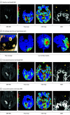
Figure 2.. First Exemplar of a Transcranial Electrical Stimulation in Stroke Early After Onset Clinical Trial Patient
Patient 1 was a male in his 70s with acute ischemic stroke due to bilateral posterior cerebral artery (PCA) occlusions. Given the symptomatic nature of the left PCA occlusion, he received 20 minutes of 1 milliamp (mA) high-definition (HD) cathodal transcranial direct current stimulation to the left occipital region only. Early alleviation of hypoperfusion, enhancement of relative cerebral blood volume, and recanalization were only observed on the stimulated side (left occipital lobe–left PCA territory). The contralateral right side, an internal control, remained ischemic due to persistent occlusion, highly suggestive of a true biological effect of stimulation. A, Acute infarctions of bilateral occipital regions (diffusion-weighted image [DWI] hyperintensities); hypoperfusion of bilateral PCA territories (time-to-maximum [Tmax] >6 seconds); symmetric bilateral relative cerebral blood volume (rCBV); bilateral PCA occlusions on MRA. B, A selection of PCA HD montage with current models predicting the e-field concentration over the ischemic region. C and D, Evolving bilateral occipital lobes acute infarcts (DWI hyperintensities); early and sustained alleviation of left occipital hypoperfusion; early and sustained rCBV enhancement of left occipital region; early and sustained left PCA partial recanalization. MRA indicates magnetic resonance angiography; MRI, magnetic resonance imaging.
Figure 3.
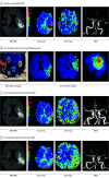
Figure 3.. Second Exemplar of a Transcranial Electrical Stimulation in Stroke Early After Onset Clinical Trial Patient
Patient 2 was a female in her 70s with acute ischemic stroke (AIS) due to left middle cerebral artery (MCA) inferior division occlusion. She received 20 minutes of 1 milliamp (mA) high-definition (HD) cathodal transcranial direct current stimulation to the left parieto-temporal region. Early alleviation of hypoperfusion region, enhancement of relative cerebral blood volume, and recanalization were observed on the stimulated side. A, Acute infarction of the left parieto-temporal region (diffusion-weighted image [DWI] hyperintensity); hypoperfusion of the parieto-temporal region (time-to-maximum [Tmax] >6 seconds); normal to low left parieto-temporal relative cerebral blood volume (rCBV); left MCA-M2 occlusion. B, A selection of MCA-inferior division HD montage with current model predicting the e-field concentration over the ischemic region. C and D, Evolving left parieto-temporal acute infarct (DWI hyperintensities); early and sustained alleviation of hypoperfusion; early and sustained rCBV enhancement of the stimulated site; early and lasting left MCA-M2 partial recanalization. MRI indicates magnetic resonance imaging.
Figure 4.
Figure 4.. Imaging Biomarkers of Neuroprotection and Collateral Enhancement Comparing Dosage Tiers
A, The enhancement of quantitative cerebral blood volume (qrCBV) in active patients early poststimulation with a dose-response pattern; B, the overall greater penumbral salvage in active vs sham patients; C, the overall greater hypoperfusion lesion (time-to-maximum >6 seconds) reduction in active vs sham patients.

References

    1. Virani SS, Alonso A, Aparicio HJ, et al. ; American Heart Association Council on Epidemiology and Prevention Statistics Committee and Stroke Statistics Subcommittee . Heart Disease and Stroke Statistics-2021 update: a report from the American Heart Association. Circulation. 2021;143(8):e254-e743. doi:10.1161/CIR.0000000000000950 - DOI - PubMed
    1. Saver JL, Goyal M, van der Lugt A, et al. ; HERMES Collaborators . Time to treatment with endovascular thrombectomy and outcomes from ischemic stroke: a meta-analysis. JAMA. 2016;316(12):1279-1288. doi:10.1001/jama.2016.13647 - DOI - PubMed
    1. Haussen DC, Nogueira RG, Elhammady MS, et al. . Infarct growth despite full reperfusion in endovascular therapy for acute ischemic stroke. J Neurointerv Surg. 2016;8(2):117-121. doi:10.1136/neurintsurg-2014-011497 - DOI - PubMed
    1. Goyal M, Menon BK, van Zwam WH, et al. ; HERMES collaborators . Endovascular thrombectomy after large-vessel ischaemic stroke: a meta-analysis of individual patient data from five randomised trials. Lancet. 2016;387(10029):1723-1731. doi:10.1016/S0140-6736(16)00163-X - DOI - PubMed
    1. Bahr Hosseini M, Hou J, Bikson M, Iacoboni M, Gornbein J, Saver JL. Central nervous system electrical stimulation for neuroprotection in acute cerebral ischemia: meta-analysis of preclinical studies. Stroke. 2019;50(10):2892-2901. doi:10.1161/STROKEAHA.119.025364 - DOI - PMC - PubMed

Publication types

Associated data