

Mechanisms of SARS-CoV-2-associated anosmia
- PMID: 37342077
- PMCID: PMC10625840
- DOI: 10.1152/physrev.00012.2023
Mechanisms of SARS-CoV-2-associated anosmia
Abstract
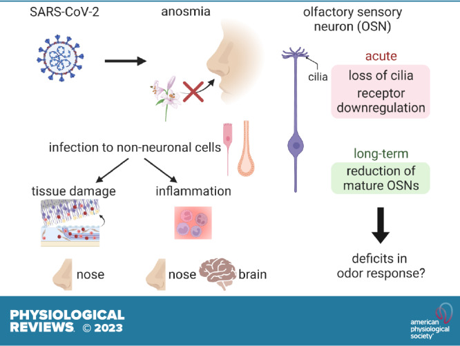
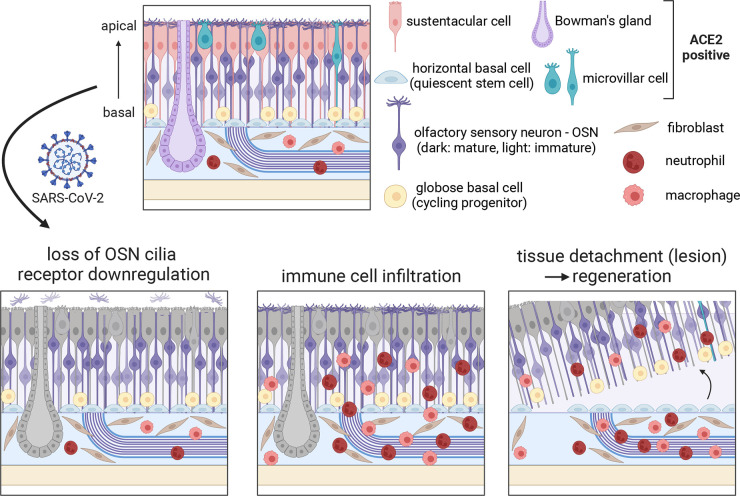
Anosmia, the loss of the sense of smell, is one of the main neurological manifestations of COVID-19. Although the SARS-CoV-2 virus targets the nasal olfactory epithelium, current evidence suggests that neuronal infection is extremely rare in both the olfactory periphery and the brain, prompting the need for mechanistic models that can explain the widespread anosmia in COVID-19 patients. Starting from work identifying the non-neuronal cell types that are infected by SARS-CoV-2 in the olfactory system, we review the effects of infection of these supportive cells in the olfactory epithelium and in the brain and posit the downstream mechanisms through which sense of smell is impaired in COVID-19 patients. We propose that indirect mechanisms contribute to altered olfactory system function in COVID-19-associated anosmia, as opposed to neuronal infection or neuroinvasion into the brain. Such indirect mechanisms include tissue damage, inflammatory responses through immune cell infiltration or systemic circulation of cytokines, and downregulation of odorant receptor genes in olfactory sensory neurons in response to local and systemic signals. We also highlight key unresolved questions raised by recent findings.
Keywords: COVID-19; SARS-CoV-2; anosmia; neuroinflammation; olfaction.
Conflict of interest statement
No conflicts of interest, financial or otherwise, are declared by the authors.
Figures

Similar articles
-
Pathophysiological relationship between COVID-19 and olfactory dysfunction: A systematic review.Braz J Otorhinolaryngol. 2022 Sep-Oct;88(5):794-802. doi: 10.1016/j.bjorl.2021.04.001. Epub 2021 Apr 25. Braz J Otorhinolaryngol. 2022. PMID: 33965353 Free PMC article.
-
Signs and symptoms to determine if a patient presenting in primary care or hospital outpatient settings has COVID-19.Cochrane Database Syst Rev. 2022 May 20;5(5):CD013665. doi: 10.1002/14651858.CD013665.pub3. Cochrane Database Syst Rev. 2022. PMID: 35593186 Free PMC article.
-
Early corticosteroid treatment enhances recovery from SARS-CoV-2 induced loss of smell in hamster.Brain Behav Immun. 2024 May;118:78-89. doi: 10.1016/j.bbi.2024.02.020. Epub 2024 Feb 15. Brain Behav Immun. 2024. PMID: 38367845
-
Designing novel "Smell-Aids" to improve olfactory function in post COVID-19 era.BMC Med. 2025 Mar 24;23(1):169. doi: 10.1186/s12916-025-03999-y. BMC Med. 2025. PMID: 40128836 Free PMC article.
-
In mice, discrete odors can selectively promote the neurogenesis of sensory neuron subtypes that they stimulate.Elife. 2025 Jun 18;13:RP96152. doi: 10.7554/eLife.96152. Elife. 2025. PMID: 40531183 Free PMC article.
Cited by
-
Vaccination against SARS-CoV-2 Does Not Protect against the Development of Anosmia in a Hamster Model.Vaccines (Basel). 2023 Oct 5;11(10):1564. doi: 10.3390/vaccines11101564. Vaccines (Basel). 2023. PMID: 37896967 Free PMC article.
-
Prevalence and Factors Associated with Olfactory Dysfunction in Individuals with COVID-19 in Brazil: A Study of 20,669 Cases from 2020 to 2021.Med Princ Pract. 2024;33(2):164-172. doi: 10.1159/000536191. Epub 2024 Jan 10. Med Princ Pract. 2024. PMID: 38198785 Free PMC article.
-
Spatiotemporal regulation by downstream genes of Prok2 in the olfactory system: from development to function.Front Cell Dev Biol. 2025 Jul 22;13:1550845. doi: 10.3389/fcell.2025.1550845. eCollection 2025. Front Cell Dev Biol. 2025. PMID: 40766783 Free PMC article.
-
Receptors Involved in COVID-19-Related Anosmia: An Update on the Pathophysiology and the Mechanistic Aspects.Int J Mol Sci. 2024 Aug 5;25(15):8527. doi: 10.3390/ijms25158527. Int J Mol Sci. 2024. PMID: 39126095 Free PMC article. Review.
-
Neurological complications caused by SARS-CoV-2.Clin Microbiol Rev. 2024 Dec 10;37(4):e0013124. doi: 10.1128/cmr.00131-24. Epub 2024 Sep 18. Clin Microbiol Rev. 2024. PMID: 39291997 Review.
References
-
- Menni C, Valdes AM, Freidin MB, Sudre CH, Nguyen LH, Drew DA, Ganesh S, Varsavsky T, Cardoso MJ, El-Sayed Moustafa JS, Visconti A, Hysi P, Bowyer RC, Mangino M, Falchi M, Wolf J, Ourselin S, Chan AT, Steves CJ, Spector TD. Real-time tracking of self-reported symptoms to predict potential COVID-19. Nat Med 26: 1037–1040, 2020. doi:10.1038/s41591-020-0916-2. - DOI - PMC - PubMed
Publication types
MeSH terms
Grants and funding
LinkOut - more resources
Full Text Sources
Medical
Miscellaneous