

Options for treatment of a small glottic gap
- PMID: 37342105
- PMCID: PMC10278110
- DOI: 10.1002/lio2.1060
Options for treatment of a small glottic gap
Abstract
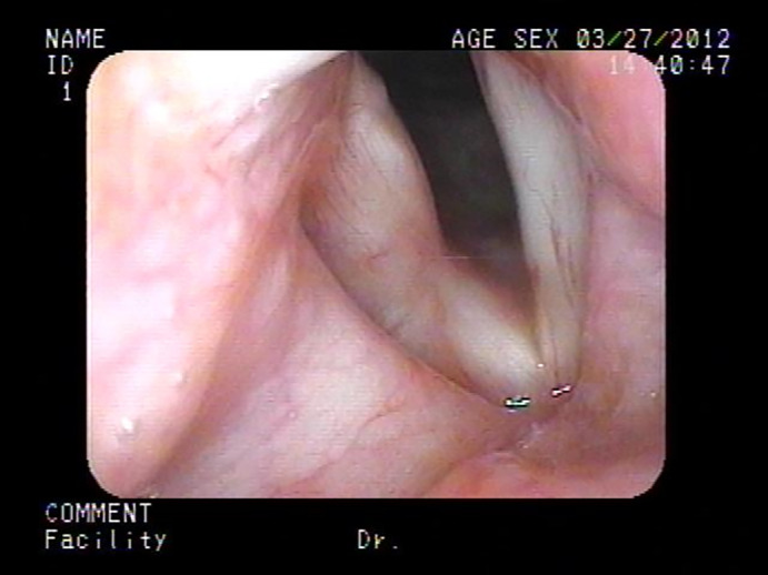
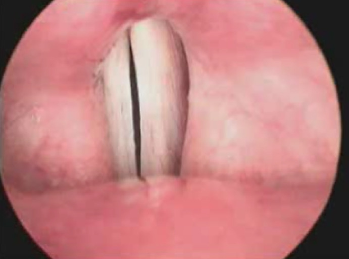
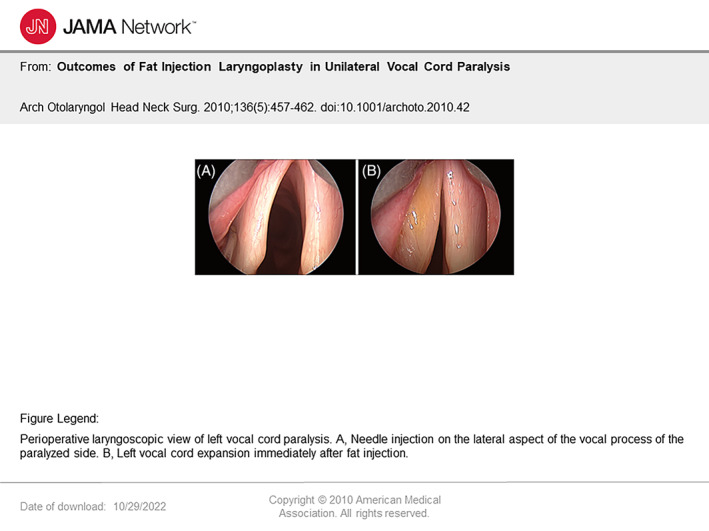
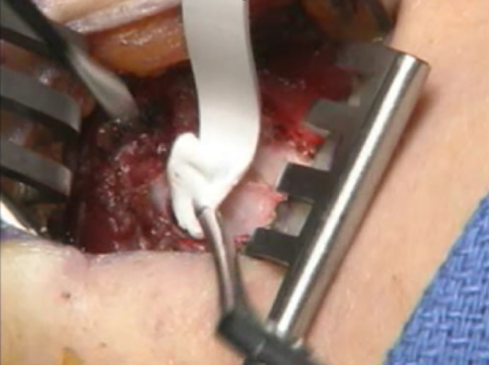
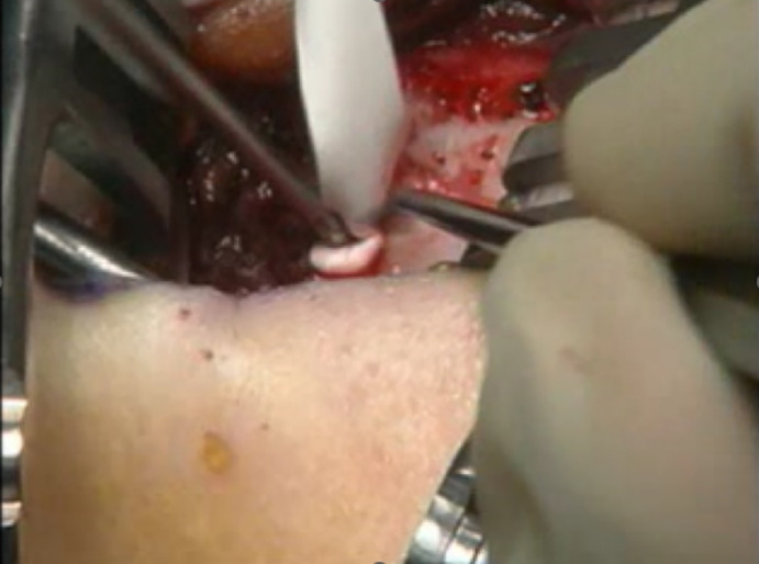
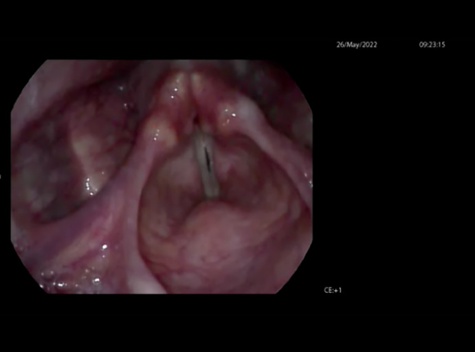
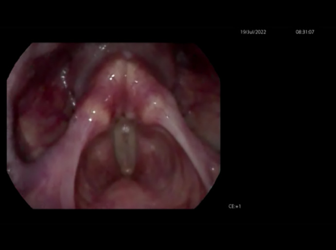
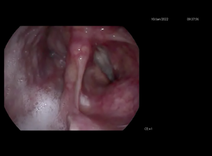
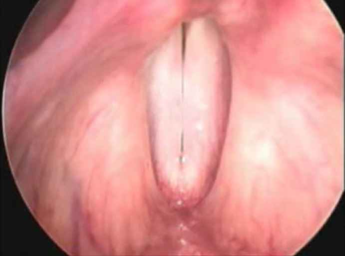
Background: Glottic insufficiency, or glottic gap as it is commonly called, is a common cause of dysphonia, producing symptoms of soft voice, decreased projection, and vocal fatigue. The etiology of glottic gap can occur from issues related to muscle atrophy, neurologic impairment, structural abnormalities, and trauma related causes. Treatment of glottic gap can include surgical and behavioral therapies or a combination of the two. When surgery is chosen, closure of the glottic gap is the primary goal. Options for surgical management include injection medialization, thyroplasty, and other methods of medializing the vocal folds.
Methods: This manuscript reviews the current literature regarding the options for treatment of glottic gap.
Discussion: This manuscript discusses options for treatment of glottic gap, including the indications for temporary and permanent treatment modalities; the differences between the available materials for injection medialization laryngoplasty and how they affect the vibratory function of the vocal folds and vocal outcome; and the evidence that supports an algorithm for treatment of glottic gap.
Level of evidence: 3a-Systematic review of case-control studies.
Keywords: glottic gap; injection laryngoplasty; thyroplasty; vocal paresis.
© 2023 The Authors. Laryngoscope Investigative Otolaryngology published by Wiley Periodicals LLC on behalf of The Triological Society.
Conflict of interest statement
Yolanda Heman‐Ackah and Chandra Ivey have nothing to disclose. Ronda Alexander discloses that she is a consultant for Smith and Nephew and receives consulting fees or honoraria as an independent contractor/speakers bureau/advisory committees/review panels.
Figures


Similar articles
-
Long-term Outcome of Autologous Lipoinjection Medialization Laryngoplasty versus Type I Thyroplasty.J Voice. 2023 Nov 6:S0892-1997(23)00321-1. doi: 10.1016/j.jvoice.2023.10.012. Online ahead of print. J Voice. 2023. PMID: 37940421
-
Medialization Thyroplasty and Arytenoid Adduction for Management of Neurological Vocal Fold Immobility.Adv Otorhinolaryngol. 2020;85:85-97. doi: 10.1159/000456686. Epub 2020 Nov 9. Adv Otorhinolaryngol. 2020. PMID: 33166967 Review.
-
Trial Vocal Fold Injection Predicts Thyroplasty Outcomes in Nonparalytic Glottic Incompetence.Ann Otol Rhinol Laryngol. 2017 Apr;126(4):279-283. doi: 10.1177/0003489416688479. Epub 2017 Jan 18. Ann Otol Rhinol Laryngol. 2017. PMID: 28100072
-
Long-term results of different treatment modalities for glottic insufficiency.Am J Otolaryngol. 2008 Jan-Feb;29(1):7-12. doi: 10.1016/j.amjoto.2006.12.001. Am J Otolaryngol. 2008. PMID: 18061825
-
Injection Laryngoplasty for Management of Neurological Vocal Fold Immobility.Adv Otorhinolaryngol. 2020;85:68-84. doi: 10.1159/000456684. Epub 2020 Nov 9. Adv Otorhinolaryngol. 2020. PMID: 33166968 Review.
Cited by
-
Vocal Fold Motion Impairment in Neurodegenerative Diseases.J Clin Med. 2024 Apr 24;13(9):2507. doi: 10.3390/jcm13092507. J Clin Med. 2024. PMID: 38731036 Free PMC article. Review.
-
Platelet Rich plasma injection of the vocal folds in benign vocal pathologies.Eur Arch Otorhinolaryngol. 2024 Oct;281(10):5419-5428. doi: 10.1007/s00405-024-08824-5. Epub 2024 Jul 17. Eur Arch Otorhinolaryngol. 2024. PMID: 39014252 Free PMC article.
-
Voice Type Component Profile Model of Glottal Gap Voice in Ex Vivo Canine Larynges.J Voice. 2024 Oct 22:S0892-1997(24)00337-0. doi: 10.1016/j.jvoice.2024.09.045. Online ahead of print. J Voice. 2024. PMID: 39443249
-
Voice Meets Swallowing: A Scoping Review of Therapeutic Connections.Am J Speech Lang Pathol. 2025 Mar 10;34(2):877-907. doi: 10.1044/2024_AJSLP-24-00194. Epub 2025 Jan 7. Am J Speech Lang Pathol. 2025. PMID: 39772835 Free PMC article.
References
-
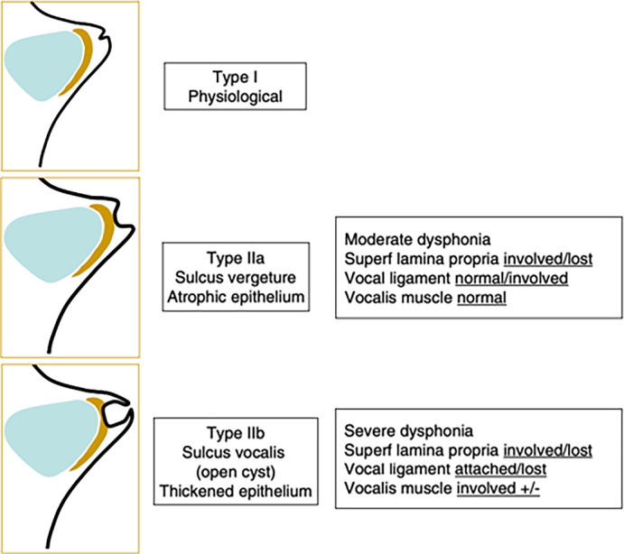
- Ford C, Inagi K, Khidr A, Bless D, Gilchrist K. Sulcus vocalis: a rational analytical approach to diagnosis and management. Ann Otol Rhinol Laryngol. 1996;105(3):189‐200. - PubMed
-
- Soni R, Dailey S. Sulcus vocalis. Otolaryngol Clin North Am. 2019;52:735‐743. - PubMed
-
- Dailey S, Spanou K, Zeitels S. The evaluation of benign glottic lesions: rigid telescopic stroboscopy versus suspension microlaryngoscopy. J Voice. 2007;21(1):112‐118. - PubMed
-
- Akbulut S, Sltintas H, Oguz H. Videolaryngostroboscopy versus microlaryngoscopy for the diagnosis of benign vocal cord lesions: a prospective clinical study. Eur Arch Oto‐Rhino‐Laryngol. 2015;272(1):131‐136. - PubMed
-
- Thibeault S, Gray S, Bless D, Chan R, Ford C. Histologic and rheologic characterization of vocal fold scarring. J Voice. 2002;16(1):96‐104. - PubMed
Publication types
LinkOut - more resources
Full Text Sources
Research Materials
Miscellaneous