The diagnostic utility of diffusion-weighted magnetic resonance imaging and high-resolution computed tomography for cholesteatoma: A meta-analysis
- PMID: 37342121
- PMCID: PMC10278117
- DOI: 10.1002/lio2.1032
The diagnostic utility of diffusion-weighted magnetic resonance imaging and high-resolution computed tomography for cholesteatoma: A meta-analysis
Abstract
Objective: The purpose of this meta-analysis was to compare the efficiency of high-resolution computed tomography (HRCT) and diffusion-weighted magnetic resonance imaging (DWI) in guiding the diagnosis of middle ear cholesteatoma in clinical practice.
Materials and methods: Cochrane Library, Medline, Embase, PubMed, and Web of Science were searched for studies that evaluated the sensitivity and specificity of HRCT or DWI in detecting middle ear cholesteatoma. A random-effects model was used to calculate and summarize the pooled estimates of sensitivity, specificity, and diagnostic odds ratios. Postoperative pathological results were considered as the diagnostic gold standard for middle ear cholesteatoma.
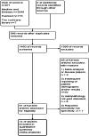
Results: Fourteen published articles (860 patients) met the inclusion criteria. The sensitivity and specificity of DWI when diagnosing cholesteatoma (regardless of type) were 0.88 (95% confidence interval [CI], 0.80-0.93) and 0.93 (95% CI, 0.86-0.97), respectively, while those of HRCT were 0.68 (95% CI, 0.57-0.77) and 0.78 (95% CI, 0.60-0.90), respectively. Notably, the sensitivity and specificity levels of DWI were similar to those of HRCT (p = .1178 for sensitivity, p = .2144 for specificity; pair-sampled t tests). The sensitivity and specificity of DWI or HRCT for the diagnosis of primary cholesteatoma were 0.78 (95% CI, 0.65-0.88) and 0.84 (95% CI, 0.69-0.93), respectively, while that for recurrent cholesteatoma were 0.93 (95% CI, 0.61-0.99) and 0.94 (95% CI, 0.82-0.98), respectively.
Conclusion: DWI and HRCT have similar levels of high sensitivity and specificity in detecting various cholesteatomas. Also, the diagnostic efficiency of HRCT or DWI for recurrent cholesteatoma is identical to that of primary cholesteatoma. Therefore, HRCT may be used in clinical settings to reduce the use of DWI and save clinical resources.
Lay summary: Data on the use of diffusion-weighted magnetic resonance imaging and high-resolution computed tomography in the diagnosis of cholesteatoma were obtained through a literature search. They were analyzed to guide the clinical diagnosis and treatment of cholesteatoma.
Level of evidence: NA.
Keywords: cholesteatoma; computed tomography; diffusion‐weighted; magnetic resonance imaging; meta‐analysis.
© 2023 The Authors. Laryngoscope Investigative Otolaryngology published by Wiley Periodicals LLC on behalf of The Triological Society.
Conflict of interest statement
The research was conducted without any commercial relationship that could be construed as a potential conflict of interest.
Figures


References
-
- Kuo CL. Etiopathogenesis of acquired cholesteatoma: prominent theories and recent advances in biomolecular research. Laryngoscope. 2015;125(1):234‐240. - PubMed
-
- Lemmerling MM, De Foer B, VandeVyver V, Vercruysse JP, Verstraete KL. Imaging of the opacified middle ear. Eur J Radiol. 2008;66(3):363‐371. - PubMed
-
- Songu M, Altay C, Onal K, et al. Correlation of computed tomography, echo‐planar diffusion‐weighted magnetic resonance imaging and surgical outcomes in middle ear cholesteatoma. Acta Otolaryngol. 2015;135(8):776‐780. - PubMed
-
- Foti G, Beltramello A, Minerva G, et al. Identification of residual‐recurrent cholesteatoma in operated ears: diagnostic accuracy of dual‐energy CT and MRI. Radiol Med. 2019;124(6):478‐486. - PubMed
Publication types
LinkOut - more resources
Full Text Sources
Research Materials
Miscellaneous
