. 2022 Sep:103:59-85.
doi: 10.3114/sim.2022.103.03. Epub 2022 Dec 12.

A review of Hyphodiscaceae

Affiliations

A review of Hyphodiscaceae

L Quijada et al. Stud Mycol. 2022 Sep.

Abstract

In a recently published classification scheme for Leotiomycetes, the new family Hyphodiscaceae was erected; unfortunately, this study was rife with phylogenetic misinterpretations and hampered by a poor understanding of this group of fungi. This manifested in the form of an undiagnostic familial description, an erroneous familial circumscription, and the redescription of the type species of an included genus as a new species in a different genus. The present work corrects these errors by incorporating new molecular data from this group into phylogenetic analyses and examining the morphological features of the included taxa. An emended description of Hyphodiscaceae is provided, notes and descriptions of the included genera are supplied, and keys to genera and species in Hyphodiscaceae are supplied. Microscypha cajaniensis is combined in Hyphodiscus, and Scolecolachnum nigricans is a taxonomic synonym of Fuscolachnum pteridis. Future work in this family should focus on increasing phylogenetic sampling outside of Eurasia and better characterising described species to help resolve outstanding issues. Citation: Quijada L, Baral HO, Johnston PR, Pärtel K, Mitchell JK, Hosoya T, Madrid H, Kosonen T, Helleman S, Rubio E, Stöckli E, Huhtinen S, Pfister DH (2022). A review of Hyphodiscaceae. Studies in Mycology 103: 59-85. doi: 10.3114/sim.2022.103.03.

Keywords: Helotiales; Leotiomycetes; keys; multi-gene phylogeny; new taxa; systematics; taxonomy.


Conflict of interest statement

The authors declare that there is no conflict of interest.

Figures

Fig. 1.
Fig. 1.
Maximum-likelihood (ML) tree based on concatenated DNA (15-gene dataset) sequences for Hyphodiscaceae, plus the taxa treated by Johnston et al. (2019) and more recently acquired sequences. Source of DNA sequence data, concatenated alignments, partitions, models and informative sites are available at https://doi.org/10.7931/b1m9-kk21. The collapsed clades represent family-level or order-level taxa accepted in Leotiomycetes. Thick branches have bootstrap support (Bps) values >95 % in black and branches with bootstrap support values >75 % are in red.
Fig. 2.
Fig. 2.
Maximum-likelihood (ML) tree based on concatenated ITS+LSU sequences for Hyphodiscaceae and related families treated by Johnston et al. (2019) and Ekanayaka et al. (2019). Lachnaceae, Solenopeziaceae and Bispora pallescens (Helotiaceae) are used as outgroups. Collection information for sequenced specimens of Hyphodiscaceae is found in Table 1. Sequences and collection information of the other taxa treated are provided in Manaaki Whenua - Landcare Research Datastore, https://doi.org/10.7931/b1m9-kk21, source of DNA sequences.xls, along with the concatenated alignment used for this analysis. Thick black branches have bootstrap support (Bps) values >95 %, whereas branches in red indicate Bps >75–95 %.
Fig. 3.
Fig. 3.
Diagrammatic representation of the sexual morphs in Hyphodiscaceae: A–E. Microscypha (M. arenula, from Hosoya & Otani 1997a: fig. 9). F–I. Hyphopeziza (H. pygmaea, from Hosoya & Otani 1997b: fig. 4). J–M. Venturiocistella (V. japonica, from Hosoya & Harada 1999: fig. 2). N–Q. Hyphodiscus (H. theiodeus, from Hosoya 2002: fig. 8). R–U. Fuscolachnum pteridis (Hosoya, this publication). D, I, J, N & R. Transverse sections showing the margin and hairs morphology. A, F, L, P & U. Asci. B, G, M, Q & S. Ascospores. C, H, K, O & T. Paraphyses. E. Multiseptate hairs of M. arenula.
Fig. 4.
Fig. 4.
Asexual morphs. A1–5. Hyphodiscus hymeniophilus culture strain TFC190382. A1. Petri dish after 17 d; A2. Tufted colonies on MEA; A3. Funiculose hyphae and phialides with collarettes and conidia in water; A4. A conidiogeneous cell with conidia, on the left a chlamydospore; A5. A conidial chain and conidia in water. B–E. A conidiogenous structure and conidia of different species of Hyphodiscus. B1, B2 = H. theiodeus; C1, C2 = H. otanii; D1, D2 = H. rhodogena; E1, E2 = H. hyaloscyphoides. Scale bars: A1 = 1 cm; A2 = 2 mm; A3–-5 = 10 μm.
Fig. 5.
Fig. 5.
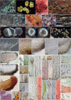
Detailed morphology of the genus Fuscolachnum. A–E. F. pteridis. A1. Fresh apothecia; A2. Dry apothecia; B1. Transverse section at margin and upper flank; B2, B5. Hairs at flanks and margin; B3, B4. Ectal excipular cells at flanks; C. Living ascospores; D. Dead paraphyses in LUG, with guttules (VBs) enhanced in contrast; E1, E3, E5. Living mature asci; E2, E6. Immature asci in LUG with euamyloid ring (dead in E6); E4. Base of asci with croziers. F–J. F. misellum. F1–3. Adult and young apothecia (fresh, rehydrated in F2); G1–8. Details of ectal excipulum and different morphologies of hairs observed; H1. Dead asci KOH-pretreated and LUG (with blue apical ring); H2 & H7. Living young and mature asci with biseriate ascospores in water; H3–5. Hemiamyloid apical ring in LUG showing blue and reddish reactions; H6. Base of asci with croziers; I1, I2. Living ascospores; J1. Dead paraphyses (in KOH); J2. Living paraphyses. K. Macrophotos of F. inopinatum showing its short and black stipe. Collections: F. pteridis = A1, B1–3, C, D, E3, E4 (ERD-4066); A2, E5, E6 (ERD-4073); B4, B5, E1, E2 (ES 201840). F. misellum (on Rubus) = F1, G4, G5, H2, H3, I1, J2 (ERD-8399); F4, G6–8, H4–7, I2 (ERD-4797); F3 (SBRH943). F. misellum (on Ribes) = F2, G1–3, H1, J1 (LE 305267). Scale bars: A1, A2, F1–4 = 200 μm; B1, G1 = 50 μm; G6 = 25 μm; B2–5, C, D, E1–4, G2–5, G7, G8, H1, H2, H7, I1, I2, J1, J2 = 10 μm; E5, E6, H3–6 = 5 μm.
Fig. 6.
Fig. 6.
Detailed morphology of the genus Hyphodiscus. A. Fresh and rehydrated apothecia showing the morphological variation in the genus. B. Transversal sections of apothecia showing hyaline vs pigmented tissues. B5. Purple reaction of tissues in KOH in H. auricolour. C. Details of ectal excipular cells at flanks, noted the thick-walled (glassy) cells embedded in gel. D. Hair morphological variation; note coloured hairs in D1 (H. luxurians) and D7 (H. auricolour) mounted in water; all hairs are ornamented with warts except D1 which has mostly smooth walls with sparse warts. E. Paraphyses. E1–3. Living paraphyses showing exudates, and cells with and without globose vacuolar bodies (guttules); E4. Dead paraphyses in CR showing septa and long apical cells. F. Morphological details of asci. F1, F2. Hemiamyloid and amyloid reaction of the ascus apex in LUG and after pretreatment with KOH; F3, F4, F7. Turgid living asci with ascospores biseriately arranged; F5, F8. Dead asci. G. Different morphologies of ascospores; noted the lipid bodies (usually two dark guttules inside each ascospore). Species in the figure: H. auricolour = A9, B4, B5, D7; H. otani = A7, E2, F6, G2; H. hyaloscyphoides = A6, B1; H. hymeniophilus = A1–4, B2, C2, C4, D3, D4, D6, E3, F1–4, F7, F8, G3–5; H; H. luxurians = A5, B3, D1, E1; H. smaragdinus = A11; H. theiodeus = A8, D2, F5, G1; Hyphodiscus sp. = A10, A12, C1, C3, D5, E4 Collections: H. auricolour = A9, B4, B5, D7 (TAAM032381). H. otanii = A7, E2, F6, G2 (ERD-7921). H. hyaloscyphoides = A6, B1 (TNS-F-13588). H. hymeniophilus = A1, C4, D6, F2, F7, F8, G4, G5 (TFCMic. 21421); A2, B2, C2, D3, D4, E3, F1, F3, F4, G3 (TFCMic. 24046); A3 (TU104853); A4 (TU104970). H. luxurians = A5, B3, D1, E1 (AP20042013). H. theiodeus = A8, D2, F5, G1 (ES 201770). H. smaragdinus = A11 (ERD-5251). H. sp. = A10, C3, D5, E4 (TFC Mic. 24497); C1 (LE235721). Scale bars: A1–7, A9, A12 = 500 μm; A8, A10, A11 = 100 μm; B1–4 = 50 μm; B5, C1–4, D2–7, E1–4, F5, F6, G1–5 = 10 μm; D1, F1–3, F7, F8 = 5 μm.
Fig. 7.
Fig. 7.
Detailed morphology of the genus Hyphopeziza. A. Fresh apothecia. B. Details of tissues. B1. Whole apothecia on top view showing long hairs in the margin; B2, B3. Ectal excipular cells at flanks and margin in CR. C. Ascospores. D. Asci. D1, D2. Living asci; D3. Hemiamyloid and amyloid reaction of the ascus apex; D4–6. Dead asci showing ascus base. E. Paraphyses. F. Hairs. F1. Ornamented hair in MLZ; F2–4. Long glassy walls of the hairs in water and CR. Collections: B1–3, C2, D4–6, E3, F4 (TAAM137705). A1 (TNS-F-17940); A2, C1, D1–3, E1, E2, F1–3 (F.G. 692); B1–3, C2, D4–6, E3, F4 (TAAM137705). Scale bars: A1, A2 = 500 μm; B1–3 = 100 μm; D1, D2, D4–6, E1–3, F1–4 = 10 μm; C1, C2, D3 = 5 μm.
Fig. 8.
Fig. 8.
Detailed morphology for Microscypha arenula. A. Fresh apothecia. B. Details of living excipular cells and hairs. C. Asci. C1. Dead asci (with croziers) and paraphyses in CR, C2, C3. Euamyloid reaction of the ascus apex (C2 dead ascus, C3 living ascus); C4. Living ascus in tap water. D. Living ascospores in tap water. Collections: A1, B3, B4, C3, C4, D2 (ERD-8386); A2, B1, B2, C1, C2, D1 (ERD-4866). Scale bars: A1, A2 = 500 μm; B1–4, C1, C4, D1, D2 = 10 μm; C2, C3 = 5 μm.
Fig. 9.
Fig. 9.
Morphological details of Scolecolachnum pteridii. A. Apothecia. B. B1. Transversal section, showing detail of the brownish flank of the ectal excipulum in the right corner; B2. Asci and paraphyses, medullary excipulum. C. Asci with ascospores. C2. Amyloid reaction of apical ring. D. Ascospores. Collection: A1–D3 (VIC 42921, from Guatimosim et al.: fig. 8 and E. Guatimosim pers. comm.). Scale bars: A1, A2 = 500 μm; B1 = ca. 100 μm; D2, B1(inset) = 50 μm; B2 = ca. 50 μm; C1–3, D1, D3 = 10 μm.
Fig. 10.
Fig. 10.
Detailed morphology of the genus Venturiocistella. A. Fresh and dry apothecia. B. B1. Median section of apothecium; B2. Squash mount showing spiny and cylindrical hairs (in polyvinyl alcohol); B3. View of lower side of apothecium; B4–6. Spiny and cylindrical hairs. C. C1–5. Living and dead asci showing arrangement of spores, hemiamyloid (C4–5, C5 KOH-pretreated) and euamyloid (C2) apex. D. Living paraphyses. Collections: V. japonica = A1, B1 (TNS-F-18030). V. diversipila = A3 (SBRH264); A4 (J.H. Petersen, JHP-99.475); B2 (HB3586b); B3, B4, C1, C2 (ES 14.10.2017). V. pini = A5 (G. Marson, 7.XII.2007). V. ulicicola = A2, B5, B6, C4, D (ERD-6422); C3, C5 (HB7648a). Scale bars: A1–A5, B3 = 100 μm; B1, B2 = 50 μm; B4–6, C1–4, D = 10 μm; C2, C5 = 5 μm.

References

    1. Albertini von JB, von Schweinitz LD. (1805). Conspectus fungorum in Lusatiae superioris agro niskiensi crescentium e methodo Persooniana. Kummerian, Germany.
    1. Allescher A. (1898). Verzeichnis in Süd-Bayern beobachteter Pilze. IV. Abteilung: Hysteriaceae, Discomycetaceae et Tuberaceae. Bericht des Botanischen Vereins in Landshut 15: 1–138.
    1. Ayel A, van Vooren N. (2005). Catalogue des Ascomycètes récoltés dans la Loire -2e partie: Leotiomycetes, Orbiliomycetes et affines (discomycètes inoperculés). Bulletin mensuel de la Société linnéenne de Lyon 74: 5–32.
    1. Baral H-O. (1992). Vital versus herbarium taxonomy: morphological differences between living and dead cells of Ascomycetes, and their taxonomic implications. Mycotaxon 44: 333–390.
    1. Baral H-O. (1993). Beiträge zür Taxonomie der Discomyceten III (Calycellina, Cyathicula, Dasyscyphella, Hyphodiscus, Olla, Trichopeziza, Venturiocistella). Zeitschrift fûr Mykologie 59: 3–22.

LinkOut - more resources