. 2022 Sep:103:25-58.
doi: 10.3114/sim.2022.103.02. Epub 2022 Oct 18.

Mortierellaceae from subalpine and alpine habitats: new species of Entomortierella, Linnemannia, Mortierella, Podila and Tyroliella gen. nov

Affiliations

Mortierellaceae from subalpine and alpine habitats: new species of Entomortierella, Linnemannia, Mortierella, Podila and Tyroliella gen. nov

A Telagathoti et al. Stud Mycol. 2022 Sep.

Abstract

Fungi are incredibly diverse, but they are unexplored, especially in the subalpine and alpine zone. Mortierellaceae are certainly one of the most abundant, species-rich, and widely distributed cultivable soil fungal families in terrestrial habitats, including subalpine and alpine zones. The phylogeny of Mortierellaceae was recently resolved based on current state of the art molecular techniques, and the paraphyletic genus Mortierella sensu lato (s.l.) was divided into 13 monophyletic genera. Our extensive sampling campaigns in the Austrian Alps resulted in 139 different Mortierellaceae pure culture isolates representing 13 new species. For the definition of taxa, we applied both classical morphological criteria, as well as modern DNA-based methods. Phylogenetic relationships were resolved based on the ribosomal DNA internal transcribed spacer (rDNA ITS), the large subunit (LSU), and the DNA-directed RNA polymerase II largest subunit 1 (RPB1). In this study, we proposed a new genus and described 13 new species belonging to the genera Entomortierella, Linnemannia, Mortierella and Podila. In addition, we proposed eight new combinations, re-defined E. jenkinii at species level, defined a neotype for M. alpina and lecto- as well as epitypes for M. fatshederae, M. jenkinii, and M. longigemmata. The rDNA ITS region is generally applied as classical barcoding gene for fungi. However, the obtained phylogenetic resolution is often too low for an accurate identification of closely related species of Mortierellaceae, especially for small sampling sizes. In such cases, unambiguous identification can be obtained based on morphological characters of pure culture isolates. Therefore, we also provide dichotomous keys for species identification within phylogenetic lineages. Taxonomic novelties: new genus: Tyroliella Telagathoti, Probst & Peintner; New species: Entomortierella galaxiae Telagathoti, M. Probst & Peintner, Linnemannia bainierella Telagathoti, M. Probst & Peintner, Linnemannia stellaris Telagathoti, M. Probst & Peintner, Linnemannia nimbosa Telagathoti, M. Probst & Peintner, Linnemannia mannui Telagathoti, M. Probst & Peintner, Linnemannia friederikiana Telagathoti, M. Probst & Peintner, Linnemannia scordiella Telagathoti, M. Probst & Peintner, Linnemannia solitaria Telagathoti, M. Probst & Peintner, Mortierella triangularis Telagathoti, M. Probst & Peintner, Mortierella lapis Telagathoti, M. Probst & Peintner, Podila himami Telagathoti, M. Probst & Peintner, Podila occulta Telagathoti, M. Probst & Peintner, Tyroliella animus-liberi Telagathoti, Probst & Peintner; New combinations: Entomortierella basiparvispora (W. Gams & Grinb.) Telagathoti, M. Probst & Peintner, Entomortierella jenkinii (A.L. Sm.) Telagathoti, M. Probst & Peintner; Entomortierella sugadairana (Y. Takash. et al.) Telagathoti, M. Probst & Peintner, Linnemannia zonata (Linnem. ex W. Gams) Telagathoti, M. Probst & Peintner, Linnemannia fluviae (Hyang B. Lee et al.) Telagathoti, M. Probst & Peintner, Linnemannia biramosa (Tiegh.) Telagathoti, M. Probst & Peintner, Linnemannia cogitans (Degawa) Telagathoti, M. Probst & Peintner, Tyroliella pseudozygospora (W. Gams & Carreiro) Telagathoti, M. Probst & Peintner; Epitypifications (basionyms): Mortierella bainieri var. jenkinii A.L. Sm., Mortierella fatshederae Linnem., Mortierella longigemmata Linnem. Neotypification (basionym): Mortierella alpina Peyronel. Citation: Telagathoti A, Probst M, Mandolini E, Peintner U (2022). Mortierellaceae from subalpine and alpine habitats: new species of Entomortierella, Linnemannia, Mortierella, Podila and Tyroliella gen. nov. Studies in Mycology 103: 25-58. doi: 10.3114/sim.2022.103.02.

Keywords: Mucoromycota systematics and taxonomy; multi-gene phylogeny; new taxa; systematics.


Conflict of interest statement

The authors declare that there is no conflict of interest.

Figures

Fig. 1.
Fig. 1.
Colony morphology of Entomortierella galaxiae grown on different media (9-cm-diam plates) at 16 °C after 7 d incubation. A. PDA. B. LCA. C. WA. Asexual morphology of Entomortierella galaxiae. D. Multi-spored sporangium attached to the sporangiophore. E. Apex of the sporangiophore after dehiscence of the peridium, with clear collarette and columella. F. Sporangiospores. G. Sporangiophores with sympodial branching pattern. H. Sporangiophores with irregular basitonous and mesotonous branching pattern. I. Sporangiophores with different size, a small with intact sporangium and a taller one where the sporangium already released the sporangiospores. All the microscopic structures of this type strain were observed on PDA. Scale bars: D–F = 10 μm; G–I = 20 μm.
Fig. 2.
Fig. 2.
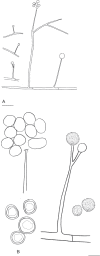
Entomortierella galaxiae. A. Sporangiophores and sporangia. B. Sporangium, sporangiophore tip and sporangiospores. Scale bars: A = 20 μm; B = 10 μm.
Fig. 3.
Fig. 3.
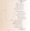
Phylogenetic relationship of the Entomortierella parvispora complex based on rDNA-ITS sequences. The Maximum Likelihood Phylogram (log likelihood -2170.60) is shown, and the branch support (Bayesian Posterior Probabilities / Parsimony Bootstrap support ≥ 70) is provided above the respective branches. Newly generated sequences are highlighted in blue, sequences generated from typus are bold and red. Gryganskiella cystojenkinii and G. fimbricystis are used as outgroup (light brown).
Fig. 4.
Fig. 4.
Colony morphology of Linnemannia bainierella grown on different media (9-cm-diam plates) at 16 °C after 7 d incubation. A. PDA. B. LCA. C. WA. Asexual morphology of Linnemannia bainierella. D. Sporangium and sporangium filled with spores. E. Cylindrical sporangiospores and sporangiophore after dehiscence of peridium, with typical collarette at the tip. F. Unbranched and branched sporangiophore. All the microscopic structures of this ex-type strain were observed on LcA medium. Scale bars: D, E = 10 μm; F = 20 μm.
Fig. 5.
Fig. 5.
Linnemannia bainierella. A. Sporangiophores. B. Sporangium, sporangiophore tip and sporangiospores. Scale bars: A = 40 μm; B = 10 μm.
Fig. 6.
Fig. 6.
Colony morphology of Linnemannia friederikiana grown on different media (9-cm-diam plates) at 16 °C after 7 d incubation. A. PDA. B. LCA. C. WA. Asexual morphology of Linnemannia friederikiana. D. Sporangium. E. Sporangiospores and tip of sporangiophore without columella and collarette. F–H. Meso, unbranched, acrotonous branching of sporangiophore. I. Chlamydospores. (D–H) Sporangium, sporangiospores, tips, branching were observed on WA, whereas the chalmydospores (I) were observed on PDA medium. Scale bars: D, E, H, I = 10 μm; F, G = 20 μm.
Fig. 7.
Fig. 7.
Linnemannia friederikiana. A. Sporangiophores; B. Sporangium, sporangiophore tip, sporangiospores, chlamydospores. Scale bars: A = 20 μm; B = 10 μm.
Fig. 8.
Fig. 8.
Colony morphology of Linnemannia mannui grown on different media (9-cm-diam plates) at 16 °C after 7 d incubation. A. PDA. B. LCA. C. WA. Asexual morphology of Linnemannia mannui. D. Cylindrical spores. E. Globose sporangium. F. Typical collarette at the tip of the sporangiophore after dehiscence of peridium, sporangium with several sporangiospores. G. Mesotonously branched sporangiophore with rhizoids at the base. H, I. Acrotonously branched sporangiophore. All the microscopic features of this ex-type strain were observed on LCA. Scale bars: D–F =10 μm; G–I = 20 μm.
Fig. 9.
Fig. 9.
Linnemannia mannui. A. Sporangiophores. B. Sporangium, sporangiophore tip with columella, sporangiospores. Scale bars: A = 40 μm; B = 10 μm.
Fig. 10.
Fig. 10.
Colony morphology of Linnemannia nimbosa grown on different media (9-cm-diam plates) at 16 °C after 7 d incubation. A. PDA. B. LCA. C. WA. Asexual morphology of Linnemannia nimbosa. D. Globose sporangium. E. Sporangium after dehiscence of peridium, with typical collarette at the tip of the sporangiophore sporangium with several sporangiospores. F. Cylindrical spores. G, H. Sporangiophore with and without the rhizoids at the base. I. Chlamydospores. All microscopic features of this ex-type strain were observed on SE agar. Scale bars: D–H, I = 10 μm; G, H = 20 μm.
Fig. 11.
Fig. 11.
Linnemannia nimbosa. A. Sporangiophores; B. Sporangiospores and chlamydospores. Scale bars: A = 20 μm, B = 10 μm.
Fig. 12.
Fig. 12.
Colony morphology of Linnemannia scordiella grown on different media (9-cm-diam plates) at 16 °C after 7 d incubation. A. PDA. B. LCA. C. WA. Asexual morphology of Linnemannia scordiella. D. Sporangia. E. Ellipsoidal sporangiospores. F. Dehiscence of peridium, with typical collarette and columella at the tip of the sporangiophore. G. Unbranched sporangiophore with several sporangiospores. H, I. Thick-walled, smooth chlamydospores. (D–G) Sporangia, sporangiospores, collarette and columella were observed on SE media whereas chlamydospores (H, I) were observed on PDA. Scale bars: D–F, H, I =10 μm; G = 20 μm.
Fig. 13.
Fig. 13.
Linnemannia scordiella. A. Sporangiophores. B. Sporangiophore tip with columella, sporangio-spores and chlamydospores. Scale bars: A = 20 μm; B = 10 μm.
Fig. 14.
Fig. 14.
Colony morphology of Linnemannia stellaris grown on different media (9-cm-diam plates) at 16 °C after 7 d incubation. A. PDA. B. LCA. C. WA. Asexual morphology of Linnemannia stellaris. D, E. Round, smooth and warted spores. F, G. Sporangiophores acrotonous branching. H. After the dehiscence of peridium, with typical collarette at the tip of the sporangiophore. I. Chlamydospores. All microscopic structures of this ex-type strain were observed on WA. Scale bars = 10 μm.
Fig. 15.
Fig. 15.
Linnemannia stellaris. A. Sporangiophores. B. Sporangiophore tip with sporangiospores, sporangiole, spiny sporangiospores, chlamydospores with oil drops. Scale bars: A = 40 μm; B = 10 μm.
Fig. 16.
Fig. 16.
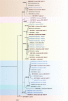
Phylogenetic relationship of Linnemannia complex based on rDNA-ITS sequences. The Maximum Likelihood Phylogram (log likelihood -2703.19) is shown, and the branch support (Bayesian Posterior Probabilities / Parsimony Bootstrap support ≥ 70) is shown above the respective branches. Newly generated sequences are highlighted in blue, sequences generated from typus are bold and dark red. Mortierella antarctica and Linnemannia amoeboidea are used as an outgroup (light brown).
Fig. 17.
Fig. 17.
Colony morphology of Mortierella lapis grown on different media (9-cm-diam plates) at 16 °C after 7 d incubation. A. PDA. B. LCA. C. WA. Asexual morphology of Mortierella lapis. D, E. Intact sporangia and the matured sporangia leaving sporangiospores. F. After the dehiscence of peridium, with typical collarette at the tip of the sporangiophore sporangium with several sporangiospores. G, H. Sporangiophores with swollen and wide base. I. Septated hyphae. All the microscopic structures for this ex-type strain were observed on SE media. Scale bars = 10 μm.
Fig. 18.
Fig. 18.
Colony morphology of Mortierella triangularis grown on different media (9-cm-diam plates) at 16 °C after for 7 d incubation. A. PDA. B. LCA. C. WA. Asexual morphology of Mortierella triangularis. D, E. Sporangiophore with wide and sporangiophore with a swollen base. F. After dehiscence of peridium, with typical collarette at the tip of the sporangiophore, sporangium and several sporangiospores. G, H. Globose and triangular shaped sporangia. I. Hyphal side branches reminding of gametangia, forming anastomoses. All microscopic structures of this ex-type strain were observed on WA. Scale bars = 10 μm.
Fig. 19.
Fig. 19.
Mortierella lapis and Mortierella triangularis. A. Mortierella lapis sporangiophore, sporangia and sporangiospores. B. Mortierella triangularis sporangiophore, sporangia and sporangiospores. Scale bars = 10 μm.
Fig. 20.
Fig. 20.
Phylogenetic relationships of Mortierella sensu stricto based on rDNA-ITS sequences. The Maximum Likelihood Phylogram (log likelihood -1815.59) is shown, and the branch support (Bayesian Posterior Probabilities / Parsimony Bootstrap support ≥ 70) is shown above the respective branches. Sequences generated from the typus are highlighted in red. Mortierella antarctica and Linnemannia amoeboidea are used as an outgroup (light brown).
Fig. 21.
Fig. 21.
Colony morphology of Podila himami grown on different media (9-cm-diam plates) at 16 °C after 7 d incubation. A. PDA. B. LCA. C. WA. Asexual morphology of Podila himami. D–F. Single sporangiophore, with sporangiospores, columella, narrow tip, and a wider base. G. Sporangium. H. Sporangiophore tip with columella and sporangiospores. I. Sporangiospores with different size. (D–G) Single sporangiophore, sporangiospores, sporangia were observed on SE media whereas (H, I) the sporangiophore tip and sporangiospores were observed on LCA. Scale bars: D = 20 μm; E–I = 10 μm.
Fig. 22.
Fig. 22.
Podila himami. A. Sporangiophores and sporangia. B. Sporangiophore tip with columella, sporangiospores. Scale bars: A = 20 μm; B = 10 μm.
Fig. 23.
Fig. 23.
Colony morphology of Podila occulta grown on different media (9-cm-diam plates) at 16 °C after 7 d incubation. A. PDA. B. LCA C. WA. Asexual morphology of Podila occulta. D–F. Branched sporangiophores. G. Sporangium with several sporangiospores. H. Sporangium after dehiscence of peridium, with typical collarette at the tip of the sporangiophore. I. Hyphal coils. All microscopic structures were observed on SE media except for the hyphal coils, which were observed on PDA media. Scale bars: D–F = 20 μm; G–I = 10 μm.
Fig. 24.
Fig. 24.
Podila occulta. A. Sporangiophores on aerial hyphae. B. Sporangium, sporangiophore tip, sporangiospores and chlamydospores. Scale bars: A = 20 μm; B = 10 μm.
Fig. 25.
Fig. 25.
Phylogenetic relationship of Podila species based on rDNA-ITS sequences. The Maximum Likelihood Phylogram (log likelihood -1604.45) is shown, and the branch support (Bayesian Posterior Probabilities / Parsimony Bootstrap support ≥ 70) is shown above the respective branches. Newly generated sequences are highlighted in blue, sequences generated from typus are bold and dark red. Necromortierella dichotoma is used as an outgroup (light brown).
Fig. 26.
Fig. 26.
Colony morphology of Tyroliella animus-liberi grown on different media (9-cm-diam plates) at 16 °C after 7 d incubation. A. PDA. B. LCA. C. WA. Asexual morphology of T. animus-liberi. D. Unbranched sporangiophore with slightly enlarged basal part. E. Sporangiospores after dehiscence or peridial wall. F. Typical, brownish pigmented chlamydospore with hyphal outgrowings. All the microscopic structures for this ex-type strain were observed on SE media. Scale bars = 20 μm.
Fig. 27.
Fig. 27.
Tyroliella animus-liberi. A. Sporangiophore with sporangium. B. Sporangiophore tip, sporangiospores and chlamydospore. Scale bars: A = 40 μm; B = 10 μm.
Fig. 28.
Fig. 28.
Phylogenetic relationship of Tyroliella animus-liberi based on rDNA-ITS sequences. The Maximum Likelihood Phylogram (log likelihood -2339.17) is shown, and the branch support (Bayesian Posterior Probabilities / Parsimony Bootstrap support ≥ 70) is shown above the respective branches. Newly generated sequences are highlighted in blue, sequences generated from typus are bold and dark red. Actinomortierella is used as an outgroup (background light brown).
Fig. 29.
Fig. 29.
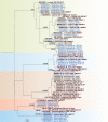
Maximum Likelihood phylogram (log likelihood -4069.81) of isolated Mortierellaceae species based on rDNA-ITS region. For each clade, one or two isolate sequences were randomly picked. Our isolates were grouped into five genera (main colours) and nine well-supported lineages representing species groups (shaded colours). The branch support (Bayesian Posterior Probabilities / Parsimony Bootstrap support ≥ 70) is shown above the respective branches. Newly generated sequences are highlighted in blue, sequences generated from typus are bold and dark red.
Fig. 30.
Fig. 30.
Maximum Likelihood phylogram (loglikelihood -9496.62) of Mortierellaceae species based on RPB1 sequences. Lineages with support of 100 % Bayesian Posterior Probability (BPP) are in bold in the phylogram. The branch support (Bayesian Posterior Probabilities / Parsimony Bootstrap support ≥ 70) is shown above the respective branches. Sequences generated from typus are highlighted in red. The colour-indicated lineages representing the genera Podila, Entomortierella, Linnemannia, Mortierella, and the new genus Tyroliella are well supported.

References

    1. Bonfante P, Desiro A. (2017). Who lives in a fungus? The diversity, origins and functions of fungal endobacteria living in Mucoromycota. ISME Journal 11: 1727–1735. - PMC - PubMed
    1. Boonmee S, Wanasinghe DN, Calabon MS, et al. (2021). Fungal diversity notes 1387–1511: taxonomic and phylogenetic contributions on genera and species of fungal taxa. Fungal Diversity 111: 1–335. - PMC - PubMed
    1. Büttner H, Niehs SP, Vandelannoote K, et al. (2021). Bacterial endosymbionts protect beneficial soil fungus from nematode attack. Proceedings of the National Academy of Sciences of the USA 118: e2110669118. - PMC - PubMed
    1. Coemans E. (1863). Quelques Hyphomycètes nouveaux. 1. notice: Mortierella polycephala et Martinella pectinata. Bulletin de l’Académie Royale des Sciences de Belgique Classe des Sciences 15: 536-540.
    1. Degawa Y, Gams W. (2004). A new species of Mortierella, and an associated sporangiiferous mycoparasite in a new genus, Nothadelphia. Studies in Mycology 50: 567–572.

LinkOut - more resources