. 2023 Jun 5:13:1165756.
doi: 10.3389/fcimb.2023.1165756. eCollection 2023.

Identification of natural killer markers associated with fatal outcome in COVID-19 patients

Affiliations

Identification of natural killer markers associated with fatal outcome in COVID-19 patients

Nadine Tarantino et al. Front Cell Infect Microbiol. .

Abstract

Introduction: Increasing evidence has shown that coronavirus disease 19 (COVID-19) severity is driven by a dysregulated immunological response. Previous studies have demonstrated that natural killer (NK) cell dysfunction underpins severe illness in COVID-19 patients, but have lacked an in-depth analysis of NK cell markers as a driver of death in the most critically ill patients.

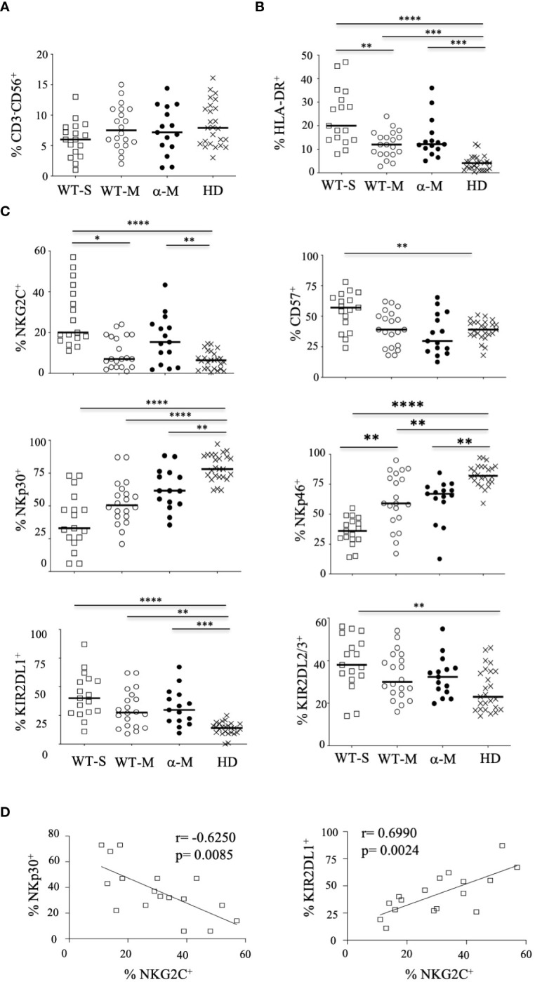
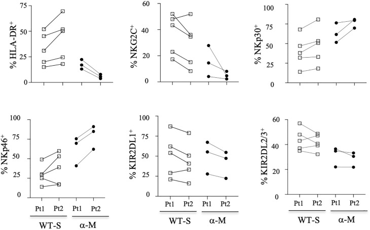
Methods: We enrolled 50 non-vaccinated hospitalized patients infected with the initial virus or the alpha variant of SARS-CoV-2 with moderate or severe illness, to evaluate phenotypic and functional features of NK cells.

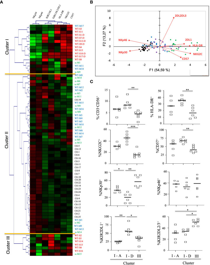
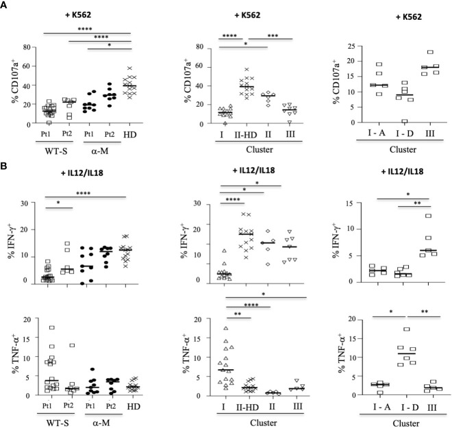
Results: Here, we show that, consistent with previous studies, evolution NK cells from COVID-19 patients are more activated, with the decreased activation of natural cytotoxicity receptors and impaired cytotoxicity and IFN-γ production, in association with disease regardless of the SARS-CoV-2 strain. Fatality was observed in 6 of 17 patients with severe disease; NK cells from all of these patients displayed a peculiar phenotype of an activated memory-like phenotype associated with massive TNF-α production.

Discussion: These data suggest that fatal COVID-19 infection is driven by an uncoordinated inflammatory response in part mediated by a specific subset of activated NK cells.

Keywords: COVID-19; SARS-CoV-2 infection; fatal outcome; natural killer (Nk) cell; tNF-alpha.


Conflict of interest statement

The authors declare that the research was conducted in the absence of any commercial or financial relationships that could be construed as a potential conflict of interest.

Figures

Figure 1
Figure 1
Phenotypic characteristics of NK cells from COVID-19 patients. (A) Percentage of CD3-CD56+ NK cells within the lymphocyte gate. (B) Percentage of HLA-DR+ activated cells among CD3-CD56+ NK cells. (C) Percentage of cells expressing cell-surface markers among CD3-CD56+ NK cells. Data are shown at the first time point (Pt1) for healthy donors (HD, n=25), patients with severe (WT-S, n=17) or moderate COVID-19 (WT-M, n=18) infected with the WT virus, or patients with moderate COVID-19 infected with the Alpha variant (α-M, n=15). Black lines represent the median. *p<0.05; **p<0.001; ***p<0.0005, ****p<0.0001. (D) Correlation between NKp30 or KIR2DL1 and NKG2C in NK cells from WT-S COVID-19 patients.
Figure 2
Figure 2
Kinetic study of NK-cell markers from COVID-19 patients. Data from some patients are shown at the two-time point (Pt1 and Pt 2) for healthy donors (HD), patients with severe (WT-S) or moderate COVID-19 (WT-M) infected with the WT virus, or patients with moderate COVID-19 infected with the Alpha variant (α-M). Black lines represent the median.
Figure 3
Figure 3
Hierarchical clustering of NK cell markers from COVID-19 patients. (A) Clustering analysis of the seven significant cell-surface markers is shown at the first time point (Pt1) for the samples of healthy donors (HD, n=25), patients with severe (WT-S, n=17) or moderate COVID-19 (WT-M, n=18) infected with the WT virus and patients with moderate COVID-19 infected with the Alpha variant (α-M, n=15). Patients with fatal outcomes are indicated by a “D”. Each vertical line is dedicated to a definite NK marker, with the color of each square reflecting the percentage of expression of the corresponding marker in each NK cell sample. The values measured for samples were color displayed and rank ordered considering the healthy donors’ median as a reference: green indicates inferior to the median and red indicates superior to the median. Analysis was performed using the Genesis program (www.genome.tugraz.at). The three different clusters (Cluster I, II and III) are separated by yellow lines. (B) Principal component analysis graphically shows the statistical proximity between the different variables that were tested, as well as the distribution of healthy donors (black circles; n=25), patients with severe (green circles, n=17) or moderate COVID-19 (blue circles, n=18) infected with the WT virus and patients with a moderate COVID-19 infected with the Alpha variant (purple circles, n=15). (C) Percentage of CD3-CD56+ and other cell-surface markers among NK cells at the first time point (Pt1) in samples of WT-S patients from clusters I and III. In cluster I, data from surviving (I-A) and deceased (I-D) WT-S patients are presented in two different columns. Black lines represent the median. *p<0.05; **p<0.001; ***p<0.0005.
Figure 4
Figure 4
Functional activity of NK cells from COVID-19 patients. (A) Degranulation of NK cells measured by the cell-surface expression of CD107a in CD3-CD56+ NK cells, tested in the presence of the standard K562 target cells (ratio 1:1). Data are taken at two time points (Pt1 and Pt2). (B) Intracellular production of IFN-γ or TNF-α in CD3-CD56+ NK cells after IL-12+IL-18 overnight stimulation (+IL12/IL18). In the left panels, data are shown for the two time points (Pt1 and Pt2) in severe COVID-19+ patients infected with WT (WT-S, n=12), or patients with moderate COVID-19 infected by the Alpha variant (α-M, n=9), compared to healthy donors (HD, n=13). In the middle panels, data are shown at the first time point (Pt1) for WT-S and α-M COVID-19 patients from clusters I (n=12), II (n=7) and III (n=8), compared to healthy donors in cluster II (II-HD, n=13). In the right panels, data are shown for WT-S patients from clusters I (n=11) and III (n=5). In cluster I, data from surviving (I-A, n=5) and deceased (I-D, n=6) WT-S patients are presented in two different columns. Black lines represent the median. *p<0.05; **p<0.001; ***p<0.0005, ****p<0.0001.
Figure 5
Figure 5
Functional correlation with cell-surface markers associated with fatality. Principal component analysis shows the statistical proximity between the different variables that were tested, as well as the distribution of each patient according to the differential expression of these variables. Blue circles: severely affected patients with a fatal outcome; Green circles: surviving patients with severe COVID-19.

Similar articles

Cited by

References

    1. Abdul-Careem M. F., Mian M. F., Yue G., Gillgrass A., Chenoweth M. J., Barra N. G., et al. . (2012). Critical role of natural killer cells in lung immunopathology during influenza infection in mice. J. Infect. Dis. 206 (2), 167–177. doi: 10.1093/infdis/jis340 - DOI - PubMed
    1. Béziat V., Liu L. L., Malmberg J. A., Ivarsson M. A., Sohlberg E., Björklund A. T., et al. . (2013). NK cell responses to cytomegalovirus infection lead to stable imprints in the human KIR repertoire and involve activating KIRs. Blood 121 (14), 2678–2688. doi: 10.1182/blood-2012-10-459545 - DOI - PMC - PubMed
    1. Bi J. (2022). NK cell dysfunction in patients with COVID-19. Cell. Mol. Immunol. 19 (2), 127–129. doi: 10.1038/s41423-021-00825-2 - DOI - PMC - PubMed
    1. Bordoni V., Sacchi A., Cimini E., Notari S., Grassi G., Tartaglia E., et al. . (2020). An inflammatory profile correlates with decreased frequency of cytotoxic cells in coronavirus disease. Clin. Infect. Dis. 71 (16), 2272–2275. doi: 10.1093/cid/ciaa577 - DOI - PMC - PubMed
    1. Carbonnel M., Daclin C., Tarantino N., Groiseau O., Morin V., Rousseau A., et al. . (2022). Plasticity of natural killer cells in pregnant patients infected with SARS-CoV-2 and their neonates during childbirth. Front. Immunol. 13. doi: 10.3389/fimmu.2022.893450 - DOI - PMC - PubMed

Publication types

MeSH terms

Substances