. 2023 Jun 5:14:1184105.
doi: 10.3389/fimmu.2023.1184105. eCollection 2023.

Free heme exacerbates colonic injury induced by anti-cancer therapy

Affiliations

Free heme exacerbates colonic injury induced by anti-cancer therapy

Philippa Seika et al. Front Immunol. .

Abstract

Gastrointestinal inflammation and bleeding are commonly induced by cancer radiotherapy and chemotherapy but mechanisms are unclear. We demonstrated an increased number of infiltrating heme oxygenase-1 positive (HO-1+) macrophages (Mø, CD68+) and the levels of hemopexin (Hx) in human colonic biopsies from patients treated with radiation or chemoradiation versus non-irradiated controls or in the ischemic intestine compared to matched normal tissues. The presence of rectal bleeding in these patients was also correlated with higher HO-1+ cell infiltration. To functionally assess the role of free heme released in the gut, we employed myeloid-specific HO-1 knockout (LysM-Cre : Hmox1flfl), hemopexin knockout (Hx-/-) and control mice. Using LysM-Cre : Hmox1flfl conditional knockout (KO) mice, we showed that a deficiency of HO-1 in myeloid cells led to high levels of DNA damage and proliferation in colonic epithelial cells in response to phenylhydrazine (PHZ)-induced hemolysis. We found higher levels of free heme in plasma, epithelial DNA damage, inflammation, and low epithelial cell proliferation in Hx-/- mice after PHZ treatment compared to wild-type mice. Colonic damage was partially attenuated by recombinant Hx administration. Deficiency in Hx or Hmox1 did not alter the response to doxorubicin. Interestingly, the lack of Hx augmented abdominal radiation-mediated hemolysis and DNA damage in the colon. Mechanistically, we found an altered growth of human colonic epithelial cells (HCoEpiC) treated with heme, corresponding to an increase in Hmox1 mRNA levels and heme:G-quadruplex complexes-regulated genes such as c-MYC, CCNF, and HDAC6. Heme-treated HCoEpiC cells exhibited growth advantage in the absence or presence of doxorubicin, in contrast to poor survival of heme-stimulated RAW247.6 Mø. In summary, our data indicate that accumulation of heme in the colon following hemolysis and/or exposure to genotoxic stress amplifies DNA damage, abnormal proliferation of epithelial cells, and inflammation as a potential etiology for gastrointestinal syndrome (GIS).

Keywords: free heme; gastrointestinal syndrome; heme oxygenase-1; hemopexin; radiation enteritis.


Conflict of interest statement

The authors declare that the research was conducted in the absence of any commercial or financial relationships that could be construed as a potential conflict of interest.

Figures

Figure 1
Figure 1
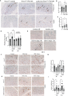
Expression of HO-1, Hx, and γH2AX in human intestinal biopsies from patients treated with radiation or suffering from intestinal ischemia. (A) Representative immunostainings of HO-1 in colonic biopsies from cancer patients treated with radiotherapy (“Radiation”) or diagnosed with colonic ischemia (ischemic and matched non-ischemic control tissue regions are shown). Arrows indicate HO-1+ Mø. Staining from two patients with ischemic colons and matched normal mucosa are shown. (B–E) Analyses of the HO-1 staining intensities in the following groups: n=11 control (untreated), n=12 irradiated, and n=11 with intestinal ischemia. Mean values +/- SD are shown. *p<0.05, **p<0.01, ***p<0.001. (F, G) Analyses of samples based on the presence of colonic bleeding. (H–J) Immunostaining of colonic biopsies as in (A) with antibodies against Hx. The representative staining is shown in (H) Analysis of n=11-12 cases is shown in (I, J). ****p<0.001, **p<0.01. Mean values +/- SD are shown. Analyses of samples based on the presence of colonic bleeding is shown in (K). (L–N) Immunostaining of colonic biopsies as in (A) with antibodies against DNA damage marker γH2AX. The representative staining is shown in (L). Analysis of n=11-12 cases is shown in (M, N) *p<0.05. Mean values +/- SD are shown. Analyses of samples based on the presence of colonic bleeding is shown in (O).
Figure 2
Figure 2
Expression of HO-1 and γH2AX in human colonic biopsies from patients treated with CRT. (A–C) HO-1 immunostainings of CRT treated and normal mucosa from n=8 patients with rectal tumors. Representative staining is shown in (A). Evaluation of HO-1 staining in Mø is shown in (B) and qualitative evaluation of the staining based on the rectal bleeding status is shown in (C). Mean values +/- SD are shown. *p<0.05. ns= not statistically significant. (D–F) Immunostainings of γH2AX of chemotherapy and normal mucosa from n=8 patients with rectal tumors treated with radiation therapy. Representative staining is shown in (D). Evaluation of γH2AX staining is shown in (E) and statistical analysis of the staining based on clinically diagnosed rectal bleeding is shown in (F). Ns, not significant.
Figure 3
Figure 3
The role of Mø-expressed HO-1 in modulating DNA damage and proliferation of colonic epithelial cells in in vivo model of hemolysis and doxorubicin. (A–C) Immunohistochemical analyses of P-HH3 (a marker of proliferation) (A, B) and γH2AX (a marker of DNA damage) (A, C) in colon samples isolated from LysM-Cre : Hmox1flfl and Hmox1flfl mice treated with PHZ (150 mg/kg, i.p.) and harvested at 48 hours after treatment. A p-value of <0.05 was considered significant. *p<0.05, **p<0.01. n=4-7 mice per group. Multiple FOVs were evaluated for quantification of the number of positive cells per crypt. (D) Heme levels were measured in the colon lysates from mice treated as above. Relative heme levels (μmol) per protein concentration are shown **p<0.01. (E) Immunohistochemical analysis of HO-1 expression in the colons of mice treated as in (D) Representative pictures are shown. (F–I) Immunohistochemical analyses of P-HH3 (a marker of proliferation) (F, G) and γH2AX (a marker of DNA damage) (H, I) in colon samples as in (D) Arrows indicate positive staining. The quantification and statistical analysis are shown in G and (H) Mean values +/- SD are shown. *p<0.05, **p <0.01, ***p<0.0001.
Figure 4
Figure 4
Lack of Hx leads to colonic inflammation and DNA damage in vivo. (A) Geo profiles of relative expression of Hx in the mouse intestinal tract based on the Affymetrix MuU74v2 Gene chip set (24). N=3 per group. (B, C) Hx-/- and Hx+/+ mice were treated with PHZ (100 mg/kg, i.p.) for 48 h. Hemolysis was measured in the plasma by absorbance at 420 nm, detecting primarily free heme (B). Mean values +/- SD are shown. n=3-7 mice per group. *p<0.05, **p<0.001. Free heme was measured in the same samples by BioVision kit and the data are shown in (C). n=7-13 mice per group. (D–G) Immunohistochemical staining of P-HH3 (a marker of proliferation) (D) and γH2AX (a marker of DNA damage) (E) of the colonic tissues from Hx-/- and WT mice treated with PHZ (100 mg/kg, i.p.) and harvested at day 5. 200x magnification. (F, G) The quantifications of the number of cells positive for P-HH3 (F) or γH2AX (G) in the colons of mice treated as described in (B) n=3 male mice per group; 2-3 sections were analyzed in (B) FOV-field of view. Mean values +/- SD are shown. **p<0.01; ***p<0.001. (H, I) Immunohistochemical staining of γH2AX (a marker of DNA damage) of the colonic tissues from Hx-/- and WT mice treated with PHZ (100 mg/kg, i.p.) ± recombinant Hx (rHx, 2.5 mg/kg, i.v.) and harvested at day 2. Mean values +/- SD are shown. N=3-8 mice per group. *p<0.05.
Figure 5
Figure 5
Deficiency in Hx results in altered DNA damage and proliferation in response to Dox treatment in the colon. (A, B) Immunohistochemical staining of HO-1 of the colonic tissues from Hx-/- and WT (Hx+/+ ) mice treated with doxorubicin (Dox, 8 mg/kg, i.v.) and harvested at day 2. Representative pictures are shown in A and quantification is shown in (B). (C–E) Differential gene expression pattern of Hmox1 (C), C-MYC (D), and IL-10 (E) in the colon tissues upon Dox treatment was compared between the WT and Hx-/- mice by RT-PCR analysis. β-Actin was used as the housekeeping gene. One-way ANOVA or/and t-tests were used to determine statistical significance. Mean values +/- SD are shown. n=6-14 mice per group. *p<0.05. (F–I) Immunohistochemical staining of γH2AX (a marker of DNA damage) (F) or P-HH3 (a marker of proliferation) (H) and of the colonic tissues from Hx-/- and WT (Hx+/+ ) mice treated with Dox and harvested at day 2. 200x magnification. Quantification is shown in G, (I) n=7 (Hx+/+ , Control), n=6 (Hx+/+ , Dox), n=13 (Hx-/- , Control) and n=14 (Hx-/- , Dox), each evaluated in triplicates. Mean values +/- SD are shown. ***p<0.001, *p<0.05.
Figure 6
Figure 6
Abdominal radiation induces hemolysis and increased DNA damage in Hx-/- mice. (A) A scheme showing the abdominal radiation (shielded IR) procedure in mice. The upper body is shielded, and only the lower part of the body is irradiated. (B) Plasma levels of free heme were measured in WT and Hx-/- mice treated with abdominal irradiation (one dose of 12 Gy) and harvested at 48 hours. n=3 control mice per group, n=6 irradiated mice per group. Mean values +/- SD are shown. *p<0.05. (C–F) Immunohistochemical staining of γH2AX (a marker of DNA damage) (C) and P-HH3 (a marker of proliferation) (E) of the colonic tissues from Hx-/- and WT mice treated with radiation as above and harvested at day 2. 200x magnification. Quantification of the number of cells positive for γH2AX (D) or P-HH3 (F) or in the colons of mice treated as described in (B) Mean values +/- SD are shown. N=3 control mice per group, n=6 irradiated mice per group. 2-3 sections were analyzed. *p<0.05; ***p<0.001. ns, not significant.
Figure 7
Figure 7
Heme prevents doxorubicin-induced cell death in human colonic epithelial cells but not in Mø. (A) HCoEpiC cells were co-treated with Dox (1 μM) and heme (5-50 μM) and assayed for viability after 24 hours of treatment by crystal violet staining. The graph illustrates the percentage change in viability as compared to untreated controls. *p<0.05, ***p<0.001. (B) RAW267.4 Mø were co-treated with Dox (0.5-10 μM) and heme (0-100 μM) and assayed for viability after 24 hours of treatment by crystal violet staining. Mean values +/- SD from n=5 replicates are shown. n=12. Data from 2 independent experiments are shown. (C) RAW267.4 Mø stably transfected with shRNA against Hmox1 or scrambled shRNA were co-treated with Dox (0.5-10 μM) and heme (0-100 μM) assayed for viability after 24 hours of treatment by crystal violet staining. Mean values +/- SD from n=5 replicates are shown. Data represenatative for 2 independent experiments are shown. (D) Differential gene expression pattern of heme-modulated genes in HCoEpiC cells treated with heme and analyzed by RT-PCR. β-Actin was used as the housekeeping gene. One-way ANOVA or/and t-tests were used to determine statistical significance. Mean values +/- SD; n=6. Data representative for 2 independent experiments are shown. *p<0.05, **p<0.01. (E) Scheme depicting the role of free heme released in response to chemotherapy, radiation, or hemolysis in the gut. Lack of HO-1 in macrophage or Hx may result in heme-induced DNA damage and altered proliferation.

References

    1. Sohail M, Sun Z, Li Y, Gu X, Xu H. Research progress in strategies to improve the efficacy and safety of doxorubicin for cancer chemotherapy. Expert Rev Anti Cancer Ther (2021) 21(12):1385–98. doi: 10.1080/14737140.2021.1991316 - DOI - PubMed
    1. Liu YQ, Wang XL, He DH, Cheng YX. Protection against chemotherapy- and radiotherapy-induced side effects: a review based on the mechanisms and therapeutic opportunities of phytochemicals. Phytomedicine (2021) 80:153402. doi: 10.1016/j.phymed.2020.153402 - DOI - PubMed
    1. Morales Chamorro R, Serrano Blanch R, Méndez Vidal MJ, Gómez España MA, Rubio Pérez MJ, de la Haba Rodríguez JR, et al. . Pseudomembranous colitis associated with chemotherapy with 5-fluorouracil. Clin Transl Oncol (2005) 7(6):258–61. doi: 10.1007/BF02710173 - DOI - PubMed
    1. Rodrigues FG, Dasilva G, Wexner SD. Neutropenic enterocolitis. World J Gastroenterol (2017) 23(1):42–7. doi: 10.3748/wjg.v23.i1.42 - DOI - PMC - PubMed
    1. Andreyev J. Gastrointestinal complications of pelvic radiotherapy: are they of any importance? Gut (2005) 54(8):1051–4. doi: 10.1136/gut.2004.062596 - DOI - PMC - PubMed

Publication types