. 2023 May 16;14(6):2689-2708.
doi: 10.1364/BOE.488630. eCollection 2023 Jun 1.

Primary assessment of medicines for expected migrastatic potential with holographic incoherent quantitative phase imaging

Affiliations

Primary assessment of medicines for expected migrastatic potential with holographic incoherent quantitative phase imaging

Markéta Šuráňová et al. Biomed Opt Express. .

Abstract

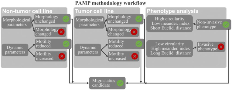
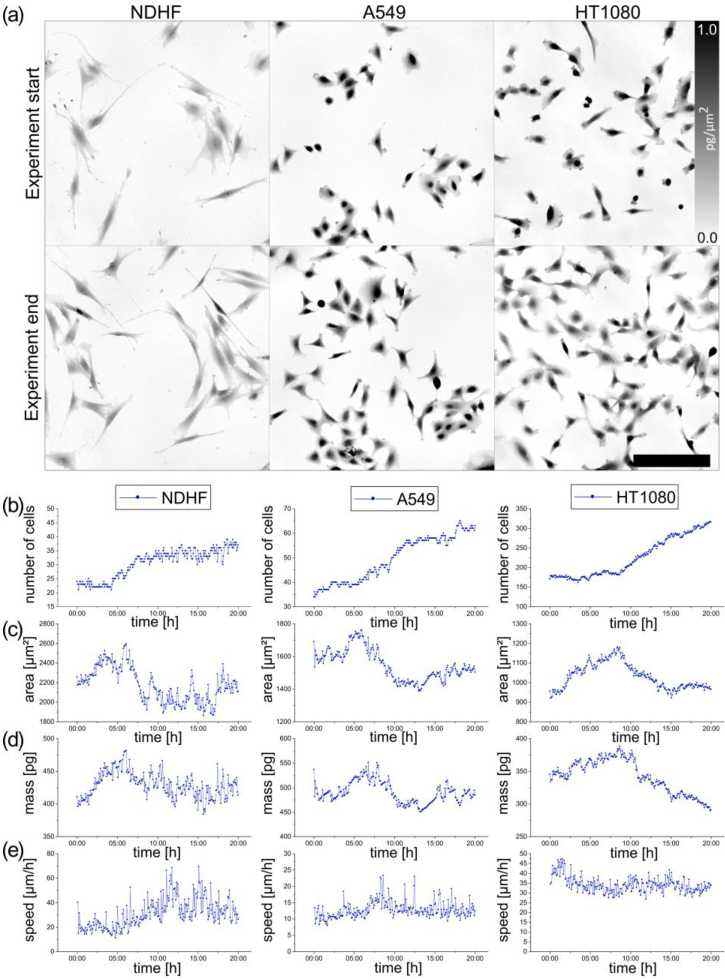
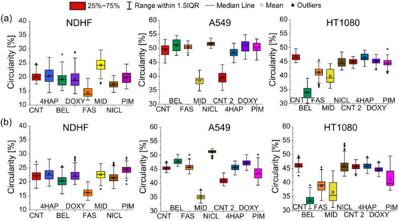
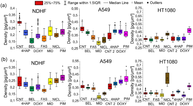
Solid tumor metastases cause most cancer-related deaths. The prevention of their occurrence misses suitable anti-metastases medicines newly labeled as migrastatics. The first indication of migrastatics potential is based on an inhibition of in vitro enhanced migration of tumor cell lines. Therefore, we decided to develop a rapid test for qualifying the expected migrastatic potential of some drugs for repurposing. The chosen Q-PHASE holographic microscope provides reliable multifield time-lapse recording and simultaneous analysis of the cell morphology, migration, and growth. The results of the pilot assessment of the migrastatic potential exerted by the chosen medicines on selected cell lines are presented.


Conflict of interest statement

R.C. is a co-author of patents covering Q-Phase (EA 018804 B1, US 8526003 B2, JP 5510676 B2, CN102279555A, EP 2378244 B1, and CZ302491) and a recipient of related royalties from Telight.

Figures

Fig. 1.
Fig. 1.
Optical setup of the coherence-controlled holographic microscope: S, light source; IF, interference filter; L, relay lens; BS, beam splitters; M, mirrors; Mm, movable mirrors; C, condensers; O, objective lenses; TL, tube lenses; DG, diffraction grating; OL, output lenses; IP, interference plane.
Fig. 2.
Fig. 2.
Diagram showing the PAMP methodology procedure. This concept consists of three parts (evaluation of non-tumor cells, tumor, and invasive phenotype). Subsequently, these parts are broken down, and a possible candidate for migrastatics is determined.
Fig. 3.
Fig. 3.
Example of the first step of the PAMP method ― the evaluation of non-tumor NDHF cells and A549 and HT1080 tumor cell lines in the control sample. (a) Images of QPI of NDHF, A549 and HT1080 cells in the control sample. Color bar in pg/µm2. Time-lapse QPI at the beginning and end of the measurement. Objective lens 10x/0.3. Scale bar 200 µm. (b) Time graph of number of cells. (c) Time graph of area. This parameter is calculated in µm2. Average values over time are shown. (d) Time graph of mass. This parameter is calculated in pg. Average values over time are shown. (e) Time graph of NDHF cell movement speed in [µm/h].
Fig. 4.
Fig. 4.
Sample of evaluating invasive phenotype of individual A549 tumor cells in the control sample. (a) Sample of QPI time-lapse of one scanned field of view at the start and end of the experiment. Objective lens 10x/0.3. Scale bar 200 µm. The color scale in pg/µm2. (b) Sample of feature plot of the circularity per mass at the start and end of the experiment. (c) Example of QPI images with trajectories of individual cells at the end of measurement. Trajectories are in µm. (d) Feature plot for the invasive phenotype. Graph of the ratio of Euclidean distance (x) and meandering index (y) with the division of cells into four quadrants. (e) The table of all parameters of individual groups of cells evaluates the state of the cells at the end of the measurement. (f) Time plot of the average perimeter values showing the time course of given groups of cells. The perimeter is in µm. (g) Migration rose plot showing individual cell trajectories. Individual tracks were shifted to the common origin. Axes are in µm.
Fig. 5.
Fig. 5.
Boxplots of circularity illustrating responses of NDHF non-tumor cell line and A549 and HT1080 tumor cell lines to selected migrastatics. The line represents the medians, and the square represents the mean. (a) Box plots in the first 10 hours of measurement (00:00–10:00). (b) Box plots in the second 10 hours (10:05–20:00) of the measurement. Circularity is calculated in %. Each migrastatic has its own color in all graphs.
Fig. 6.
Fig. 6.
Boxplots of density illustrating responses of non-tumor NDHF cell line and A549 and HT1080 tumor cell lines to selected migrastatics. The line represents the medians, and the square represents the mean. (a) Box plots in the first 10 hours of measurement (00:00-10:00). (b) Box plots in the second 10 hours (10:05-20:00) of the measurement. The density is calculated in pg/µm2 as the mass per area of cells.
Fig. 7.
Fig. 7.
Examples of QPI images picked up from time-lapse recordings of NDHF, A549 and HT1080 cells show morphological changes induced by niclosamide. In magnified areas from the red squares the red arrows indicate cytoplasmic vacuolation suggesting apparently un endoplasmic reticulum stress. Objective lens 10x/0.3. Scale bar 200 µm.

References

    1. Fares J., Fares M. Y., Khachfe H. H., Salhab H. A., Fares Y., “Molecular principles of metastasis: a hallmark of cancer revisited,” Signal Transduction Targeted Ther. 5(1), 28 (2020).10.1038/s41392-020-0134-x - DOI - PMC - PubMed
    1. Sleeman J., Steeg P. S., “Cancer metastasis as a therapeutic target,” Eur. J. Cancer 46(7), 1177–1180 (2010).10.1016/j.ejca.2010.02.039 - DOI - PMC - PubMed
    1. Gandalovičová A., Rosel D., Fernandes M., Veselý P., Heneberg P., Čermák V., Petruželka L., Kumar S., Sanz-Moreno V., Brábek J., “Migrastatics-Anti-metastatic and Anti-invasion Drugs: Promises and Challenges,” Trends Cancer 3(6), 391–406 (2017).10.1016/j.trecan.2017.04.008 - DOI - PMC - PubMed
    1. Fares J.D., Kanojia A., Rashidi I., Ulasov M.S., Lesniak, “Landscape of combination therapy trials in breast cancer brain metastasis,” Int. J. Cancer 147, 1939–1952 (2020).10.1002/ijc.32937 - DOI - PMC - PubMed
    1. Solomon J., Rašková M., Rösel D., Brábek J., Gil-Henn H., “Are We Ready for Migrastatics?” Cell 10(8), 2021 (1845).10.3390/cells10081845 - DOI - PMC - PubMed

LinkOut - more resources