. 2023 May 2;14(6):2432-2448.
doi: 10.1364/BOE.486363. eCollection 2023 Jun 1.

Non-invasive diffuse optical monitoring of cerebral physiology in an adult swine-model of impact traumatic brain injury

Affiliations

Non-invasive diffuse optical monitoring of cerebral physiology in an adult swine-model of impact traumatic brain injury

Rodrigo M Forti et al. Biomed Opt Express. .

Abstract

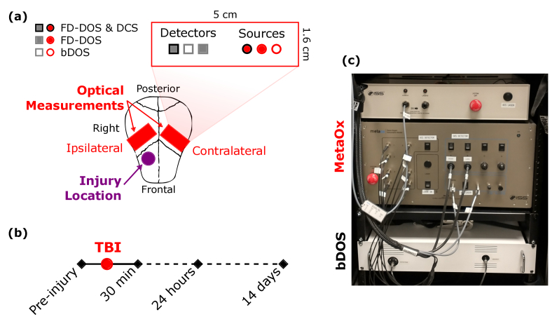
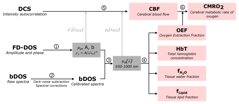
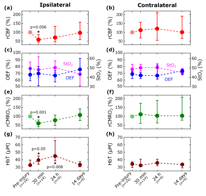
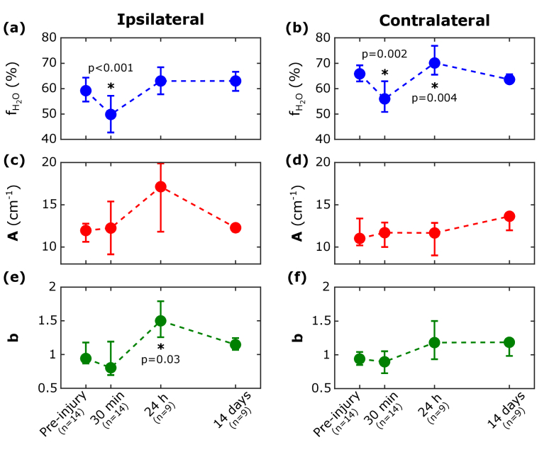
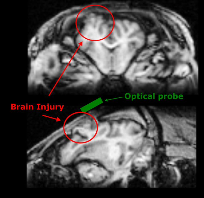
In this study, we used diffuse optics to address the need for non-invasive, continuous monitoring of cerebral physiology following traumatic brain injury (TBI). We combined frequency-domain and broadband diffuse optical spectroscopy with diffuse correlation spectroscopy to monitor cerebral oxygen metabolism, cerebral blood volume, and cerebral water content in an established adult swine-model of impact TBI. Cerebral physiology was monitored before and after TBI (up to 14 days post injury). Overall, our results suggest that non-invasive optical monitoring can assess cerebral physiologic impairments post-TBI, including an initial reduction in oxygen metabolism, development of cerebral hemorrhage/hematoma, and brain swelling.


Conflict of interest statement

The authors disclose partial ownership of the following patents. Pending: WO2021/091961 [TSK, DJL, WBB, AGY, TJK], 63/257685 [WBB, DJL, TSK, TJK, RMF], WO2013/090658Al [AGY], PCT/US2012/069626 [AGY], PCT/US2015/017286 [AGY], PCT/US2015/017277 [AGY]. US8082015 [AGY], US10064554 [AGY], US10342488 [WBB and AGY], US10827976 [WBB, DJL, AGY]. No author currently receives royalties or payments from these patents.

Figures

Fig. 1.
Fig. 1.
(a) Schematic of the optical probe and the locations of the bilateral optical measurements. The optical probe comprised 4 source-detector separations (SDS) for frequency-domain diffuse optical specotrscopy (FD-DOS, 1.5, 2.0, 2.5, 3.0 cm), a single SDS for diffuse correlation spectorscopy (DCS, 2.5 cm), and a single SDS for broadband DOS (bDOS, 3.0 cm). Mild-to-moderate focal contusion traumatic brain injury (TBI) was induced using a controlled cortical impact device in adult-aged swine (the skull and dura were permanently removed at the injury location). (b) Timeline of the study’s pre- and post-injury bilateral optical measurements. (c) Image of the instrument showing the commercial MetaOx (which combines FD-DOS and DCS), and our lab built bDOS instrument.
Fig. 2.
Fig. 2.
Flow chart of the analysis algorithm for the integrated diffuse correlation spectroscopy (DCS), frequency-domain diffuse optical spectroscopy (FD-DOS), and broadband diffuse optical spectroscopy (bDOS) data. The tissue absorption and reduced scattering coefficients at wavelength λ are μa(λ) and μs(λ) , A is the scattering amplitude, and b is the scattering power (from Eq. (1), i.e., μs(λ)=A(λ/λ0)b , with λ0=500nm ).
Fig. 3.
Fig. 3.
Pre- and post-injury diffuse optical measurements of relative cerebral blood (rCBF100×CBF/CBF0) , oxygen extraction fraction (OEF), oxygen saturation (StO2), relative cerebral metabolic rate of oxygen (rCMRO2100×CMRO2/CMRO2,0) , and tissue hemoglobin concentration (HbT) for the ipsilateral (first column) and contralateral (second column) hemispheres. rCBF and rCMRO2 are normalized to their pre-injury levels, i.e., CBF0 and CMRO2,0 . The medians (circles) and interquartile ranges (error bars) across subjects for each measurement timepoint are shown. P values indicate whether the median pre-injury to post-injury change was different from zero.
Fig. 4.
Fig. 4.
Pre-injury and post-injury diffuse optical measurements of cerebral tissue water volume fraction (fH2O) , scattering amplitude (A), and scattering power (b) for the ipsilateral and contralateral hemispheres (A and b are defined by Eq. (1)). The medians (circles) and interquartile ranges across subjects for each measurement timepoint are shown. P values indicate whether the median pre-injury to post-injury change was different from zero.
Fig. 5.
Fig. 5.
– An exemplar structural MRI image collected at 24 hours post injury, revealing significant brain contusion and injury at the injury location (red circle). Here, we also represent the approximate location of the optical measurements (in green); to avoid the cranial incision we always placed our optical probes in a region posterior to the injury site.

References

    1. Daugherty J., “Differences in State Traumatic Brain Injury–Related Deaths, by Principal Mechanism of Injury, Intent, and Percentage of Population Living in Rural Areas: United States, 2016–2018,” MMWR Morb. Mortal. Wkly. Rep. 70(41), 1447–1452 (2021).10.15585/mmwr.mm7041a3 - DOI - PMC - PubMed
    1. Daugherty J., “Traumatic Brain Injury–Related Deaths by Race/Ethnicity, Sex, Intent, and Mechanism of Injury — United States, 2000–2017,” MMWR Morb. Mortal. Wkly. Rep. 68(46), 1050–1056 (2019).10.15585/mmwr.mm6846a2 - DOI - PMC - PubMed
    1. Masel B. E., DeWitt D. S., “Traumatic brain injury: a disease process, not an event,” Journal of Neurotrauma 27(8), 1529–1540 (2010).10.1089/neu.2010.1358 - DOI - PubMed
    1. Werner C., Engelhard K., “Pathophysiology of traumatic brain injury,” Br. J. Anaesth. 99(1), 4–9 (2007).10.1093/bja/aem131 - DOI - PubMed
    1. Carney N., Totten A. M., O’Reilly C., Ullman J. S., Hawryluk G. W. J., Bell M. J., Bratton S. L., Chesnut R., Harris O. A., Kissoon N., Rubiano A. M., Shutter L., Tasker R. C., Vavilala M. S., Wilberger J., Wright D. W., Ghajar J., “Guidelines for the Management of Severe Traumatic Brain Injury, Fourth Edition,” Neurosurgery 80(1), 6–15 (2017).10.1227/NEU.0000000000001432 - DOI - PubMed

LinkOut - more resources