. 2023 Aug 3;31(8):987-1004.e8.
doi: 10.1016/j.str.2023.05.017. Epub 2023 Jun 20.

How aberrant N-glycosylation can alter protein functionality and ligand binding: An atomistic view

Affiliations

How aberrant N-glycosylation can alter protein functionality and ligand binding: An atomistic view

Matteo Castelli et al. Structure. .

Abstract

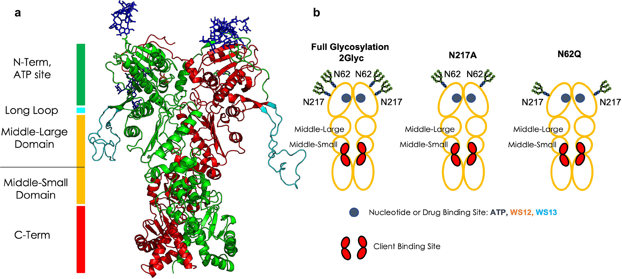
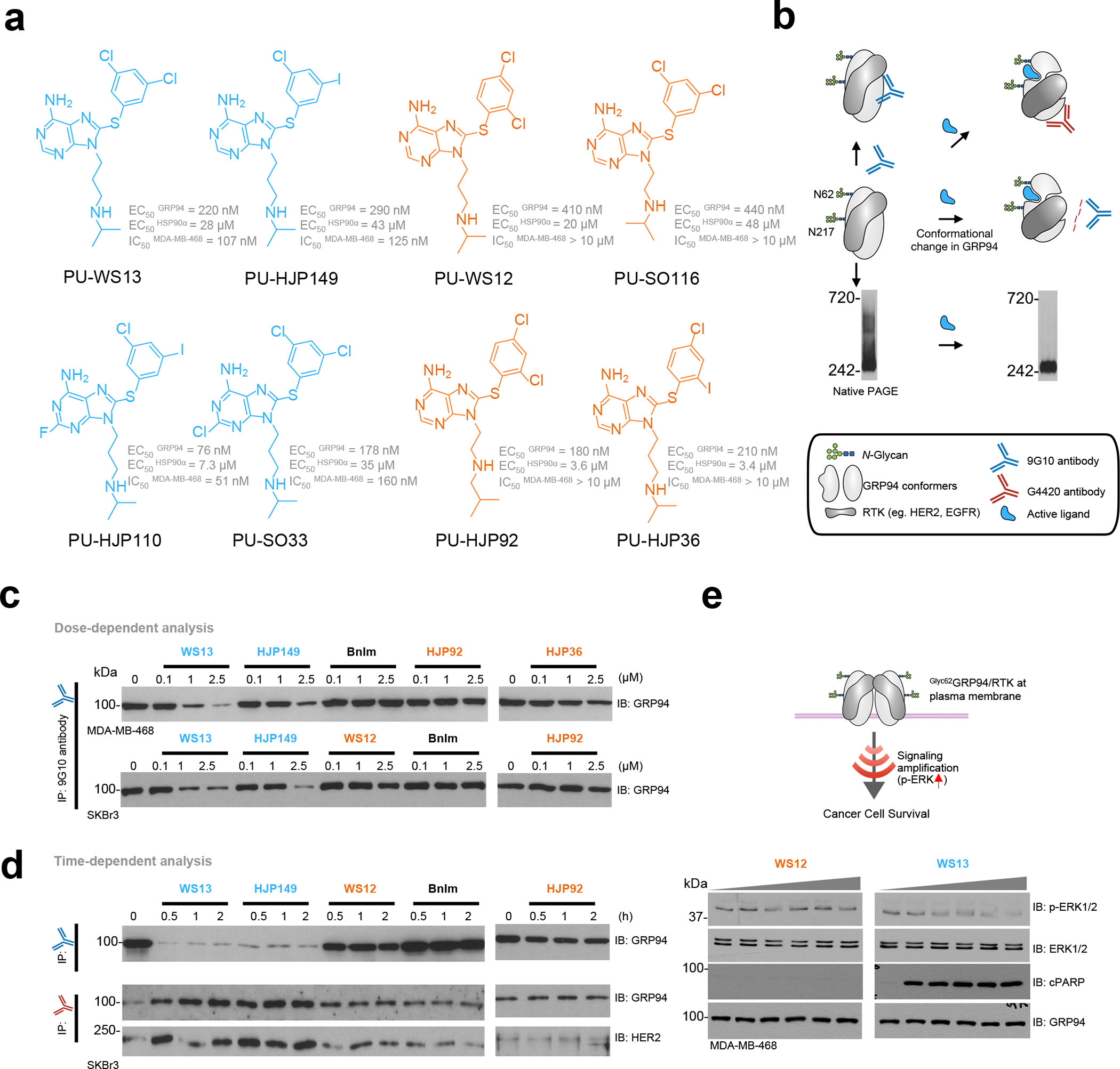
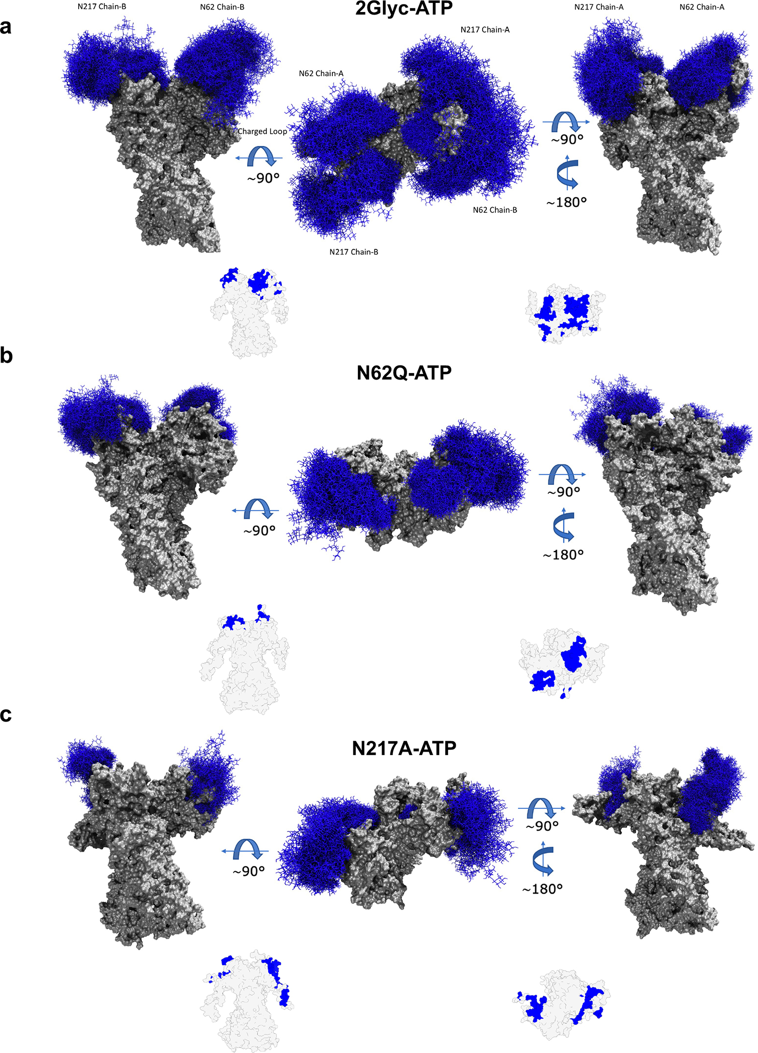
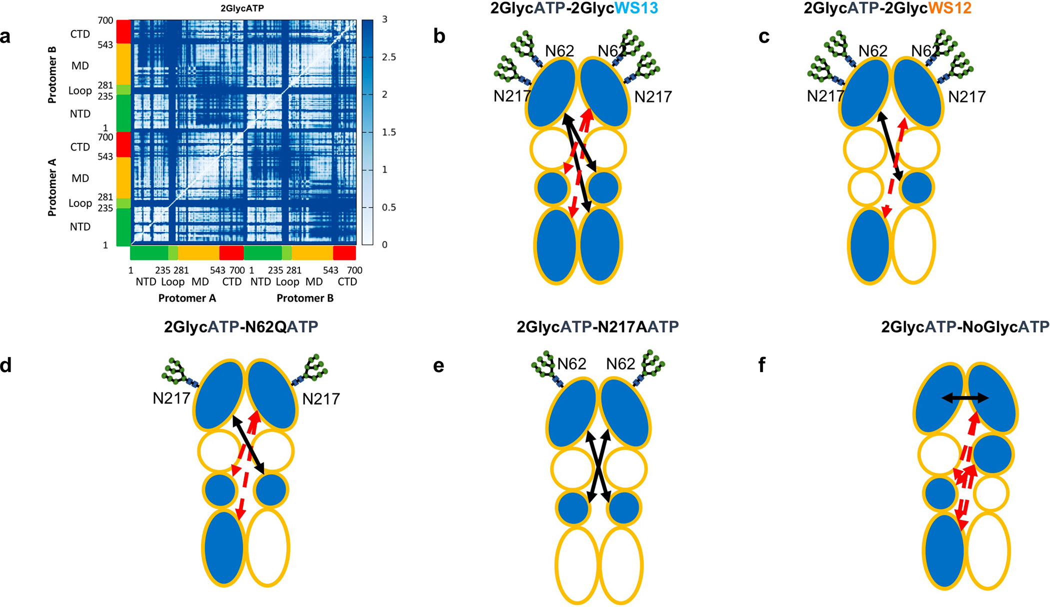
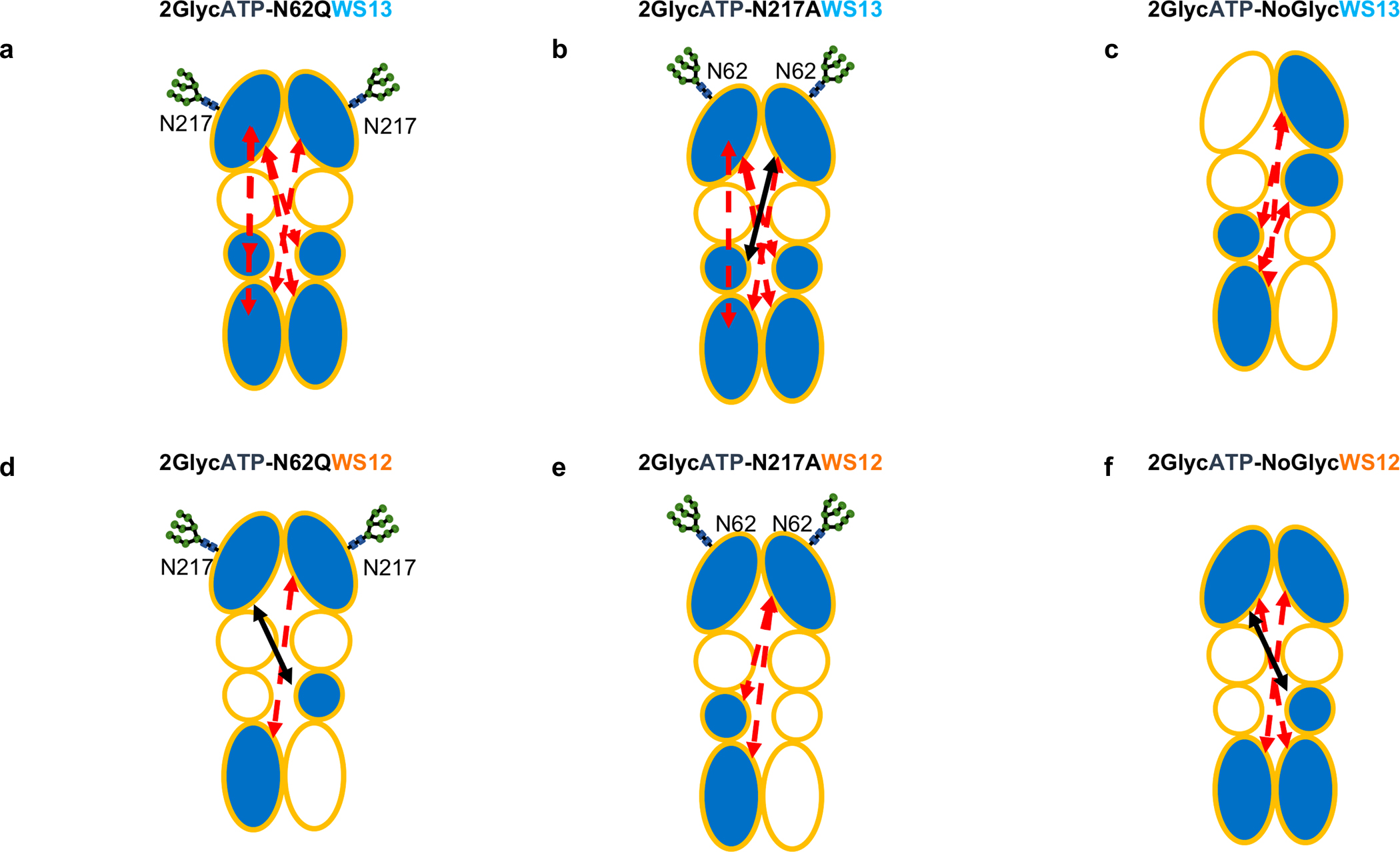
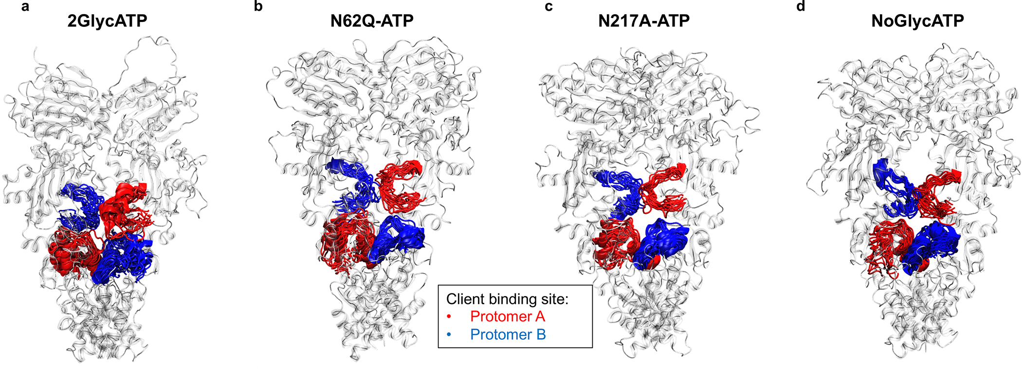
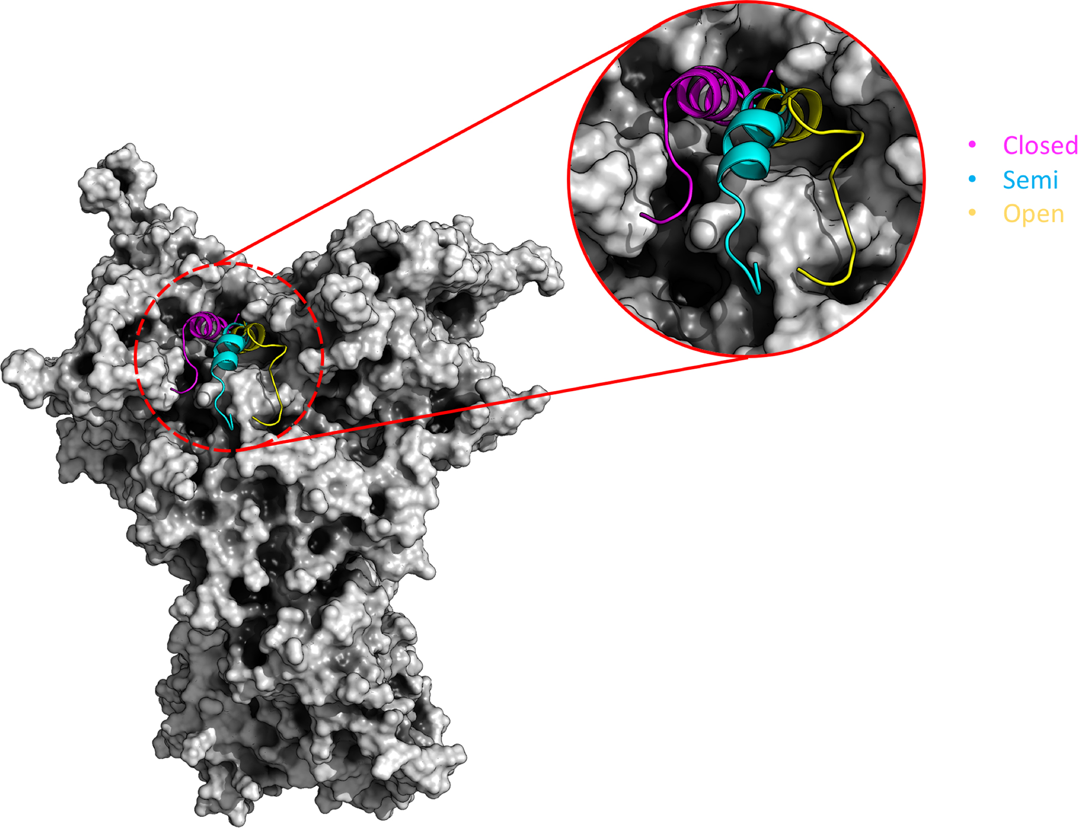
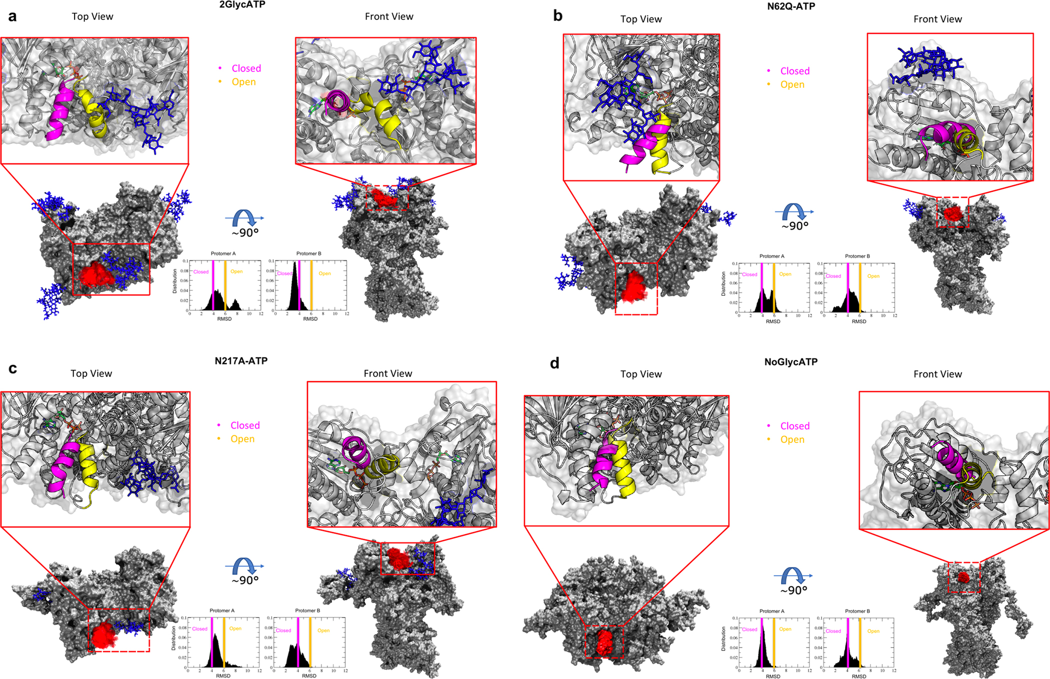
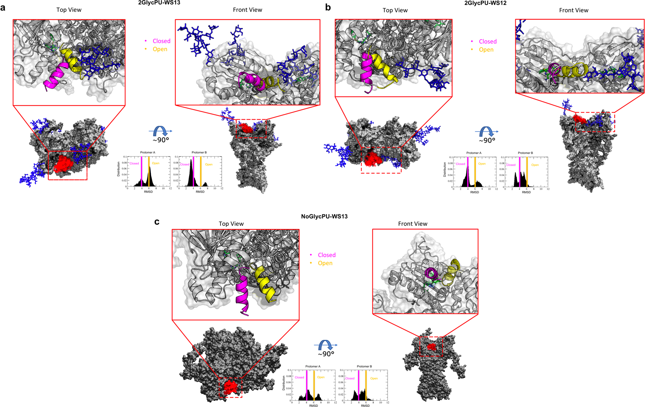
Protein-assembly defects due to an enrichment of aberrant conformational protein variants are emerging as a new frontier in therapeutics design. Understanding the structural elements that rewire the conformational dynamics of proteins and pathologically perturb functionally oriented ensembles is important for inhibitor development. Chaperones are hub proteins for the assembly of multiprotein complexes and an enrichment of aberrant conformers can affect the cellular proteome, and in turn, phenotypes. Here, we integrate computational and experimental tools to investigte how N-glycosylation of specific residues in glucose-regulated protein 94 (GRP94) modulates internal dynamics and alters the conformational fitness of regions fundamental for the interaction with ATP and synthetic ligands and impacts substructures important for the recognition of interacting proteins. N-glycosylation plays an active role in modulating the energy landscape of GRP94, and we provide support for leveraging the knowledge on distinct glycosylation variants to design molecules targeting GRP94 disease-associated conformational states and assemblies.

Keywords: GRP94; aberrant protein conformations; allostery; disease; drug design; drug selectivity; epichaperomes; functional dynamics; glycosylation; post-translational modifications; protein assembly mutations.


Conflict of interest statement

Declaration of interests G. Chiosis, A.R., C.S.D., and P.Y. are inventors on patents covering PU-WS13 and associated composition of matter. G. Chiosis is a founder of Samus Therapeutics.

Figures

Figure 1.
Figure 1.. Structural and domain organization of GRP94.
(a) The 3D structure of 2Glyc Grp94. The two protomers are colored in green and red respectively, while the glycosyl PTMs are shown in sticks. (b) Schematic of the domains, binding sites, glycosylation sites, and glycosylation states, as they are referred to throughout the paper. The red areas indicate the client binding site regions.
Figure 2.
Figure 2.. Biochemical and functional evaluation of GRP94 ligands for biological activity via Glyc62GRP94.
(a) Chemical structure of GRP94 small molecule ligands. EC50GRP94 and EC50HSP90, relative binding affinity constant to recombinant GRP94 and HSP90α, respectively. IC50MDA-MB-468, half maximal inhibitory concentration determined in the MDA-MB-468 EGFR-overexpressing and Glyc62GRP94-expressing breast cancer cells (ATP quantitation assay). Values, mean of three independent experiments. (b) Schematic showing the conformational changes induced in GRP94 expected from small molecule ligands that act on Glyc62GRP94. (c,d) Immuno-capture as in (b) of SKBr3 HER2-overexpressing or MDA-MB-468 EGFR-overexpressing (both Glyc62GRP94-expressing) breast cancer cells treated with the indicated ligands in a dose- (for 4 h) and time-dependent (at 2.5 μM) manner. Experiments were repeated trice with similar results. (e) Western blot analysis for inhibition of Glyc62GRP94 function in MDA-MB-468 cancer cells. Downstream signaling (p-ERK/ERK) and induction of apoptosis (cleaved PARP, cPARP) were analyzed for MDA-MB-468 cancer cells treated for 24 h with 0, 0.5, 1, 2.5, 5 and 10 μM of the indicated inhibitors. GRP94, loading control. Gels are representative of three independent results. See also Figure S1.
Figure 3.
Figure 3.. Glycan dynamics on the surfaces of GRP94.
(a) Molecular representation of the 2GlycATP system. Glycans at several frames (namely, 300 frames, one every 30 ns from one replica) are represented with blue lines, while the protein is shown as a gray surface. (b) Same as a), for the N62Q mutant in complex with ATP; c) Same as a), for the N217A mutant in complex with ATP. Insets: Analysis of accessible area for more than 70% of the simulation time; gray shading indicates the areas that are accessible for more than 70% of the simulation time; in blue the ones that are shielded by carbohydrates.
Figure 4.
Figure 4.. Residue-Pair Distance Fluctuations and Domain Cross-Talk for GRP94 in Different Glycosylation and Ligand Conditions.
The internal dynamics of GRP94 characterized from the various trajectories. (a) The original Distance Fluctuation matrix is reported for the fully-glycosylated ATP-bound state. The axes report the residue numbering and domain organisation, as depicted also in Figure 2. In this view, lighter pixels correspond to highly coordinated residue pairs, while darker ones report on low coordination pairs. The single original matrices for all 9 conditions, together with the difference matrices obtained by subtracting the DF matrix of a certain state from the reference one pertaining to 2GlycATP are reported in Supplementary Information. (b-f) Domain based representations of the modulation of internal flexibility as a function of ligand-state and/or glycosylation state. This representation is a simplified graphical translation of the difference matrices reported in the Supplementary Information (Figures S8–S9). A certain domain is colored in light-blue if its coordination with other domains changes as a function of the condition. The variation in internal dynamics is evaluated as a difference for the DF of the system under exam from that of the fully glycosylated ATP-bound state, 2GlycATP. A black arrow indicates increased coordination (rigidity) between two domains with respect to the fully glycosylated ATP-bound state, 2GlycATP. A red, broken arrow indicates decreased coordination (flexibility) between two domains with respect to the fully glycosylated ATP-bound state, 2GlycATP. The various ligand states are reported in each subfigure.
Figure 5.
Figure 5.. Residue-pair Distance Fluctuations and Domain Cross-Talk for GRP94 in Different Glycosylation and Ligand Conditions.
Same as Figure 4(b–f), for systems with different degrees of glycosylation.
Figure 6.
Figure 6.. Conformational dynamics of GRP94 Client-Binding Site.
The client binding site is depicted as red (Protomer A) or blue (protomer B) ribbons, whereas the rest of the protein is depicted as transparent cartoons. (a) 2GlycATP system. (b) Same as A, for the N62Q mutant in complex with ATP. (c) Same as A, for the N217A mutant in complex with ATP. (d) Same as A, for the completely unglycosylated NoGlyc state in complex with ATP.
Figure 7.
Figure 7.. Simplified representation of the structural ensembles of GRP94 ATP-lid.
The cartoon shows the whole GRP94 represented as a surface. The zoom on the lid structure shows the three main conformational families of the lid covering the active site of Grp94 in the closed (magenta), semi-open (cyan), open (yellow).
Figure 8.
Figure 8.. Conformational dynamics of the ATP-lid covering the active site of Grp94 and its interactions with sugar chains in the open or closed forms.
Two orientations are depicted. ATP-lid and glycans at several frames (300 frames) are represented with red and blue lines, respectively. The insets show the distributions of RMSD values from the conformation presented in crystal structure PDB 5ULS.pdb. The blue and yellow vertical lines show the RMSD threshold for the closed and open states, respectively. (a) 2GlycATP. (b) N62Q mutant in complex with ATP. (c) N217A mutant in complex with ATP. (d) NoGlyc system in complex with ATP.
Figure 9.
Figure 9.. The effect of ligands on the conformational dynamics of the ATP-lid covering the active site of GRP94 and its interactions with sugar chains in the open or closed forms.
Same as Figure 8. (a) 2Glyc system in complex with PU-WS13. (b) 2Glyc system in complex with PU-WS12. (c) NoGlyc system in complex with PU-WS13.

References

    1. Nussinov R, Ma B, and Tsai CJ (2014). Multiple conformational selection and induced fit events take place in allosteric propagation. Biophys. Chem. 186, 22. - PMC - PubMed
    1. Wei G, Xi W, Nussinov R, and Ma B (2016). Protein Ensembles: How Does Nature Harness Thermodynamic Fluctuations for Life? The Diverse Functional Roles of Conformational Ensembles in the Cell. Chem. Rev. 116, 6516–6551. - PMC - PubMed
    1. Nussinov R, Tsai C-J, and Jang H (2019). Protein ensembles link genotype to phenotype. PLoS computational biology 15, e1006648–e1006648. 10.1371/journal.pcbi.1006648. - DOI - PMC - PubMed
    1. Ferreiro DU, Hegler JA, Komives EA, and Wolynes PG (2011). On the role of frustration in the energy landscapes of allosteric proteins. Proc. Natl. Acad. Sci. U. S. A. 108, 3499. - PMC - PubMed
    1. Gfeller D, Michielin O, and Zoete V (2013). Shaping the interaction landscape of bioactive molecules. Bioinformatics 29, 3073–3079. - PubMed

Publication types

LinkOut - more resources