. 2023 Oct;44(10):2075-2090.
doi: 10.1038/s41401-023-01113-7. Epub 2023 Jun 21.

Pregnane X receptor activation alleviates renal fibrosis in mice via interacting with p53 and inhibiting the Wnt7a/β-catenin signaling

Affiliations

Pregnane X receptor activation alleviates renal fibrosis in mice via interacting with p53 and inhibiting the Wnt7a/β-catenin signaling

Wen-Hua Ming et al. Acta Pharmacol Sin. 2023 Oct.

Abstract

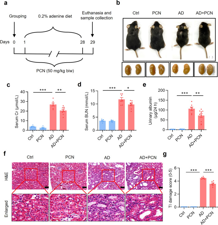
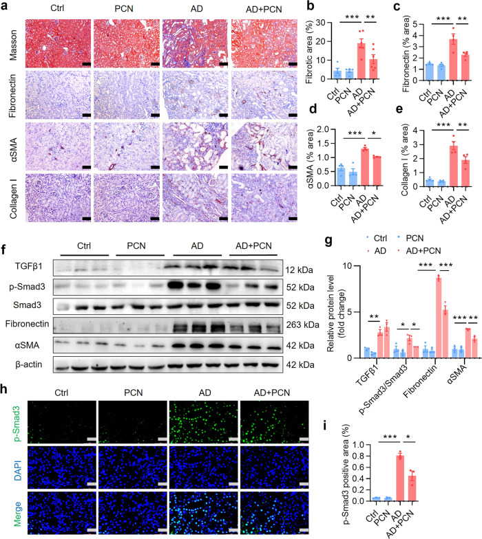
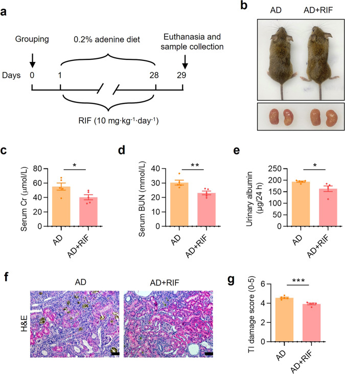
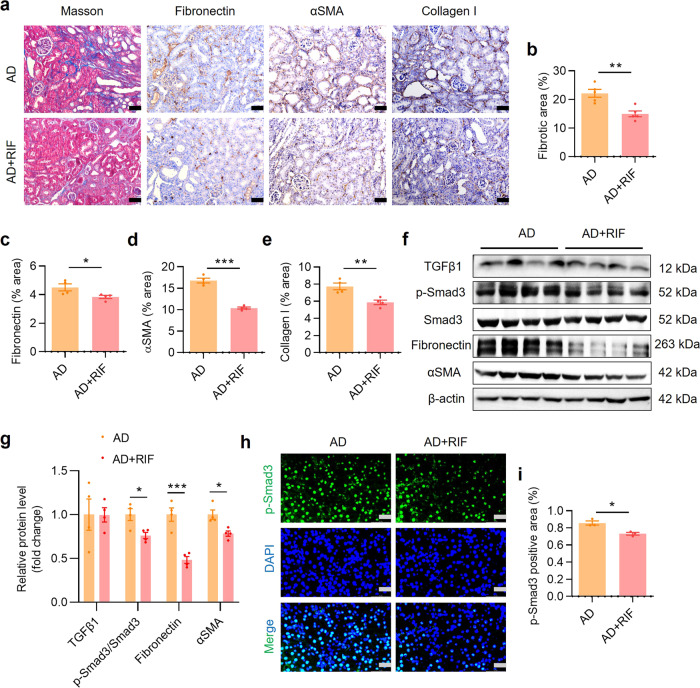
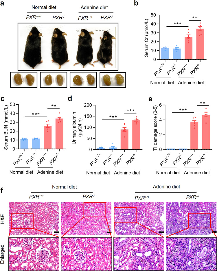
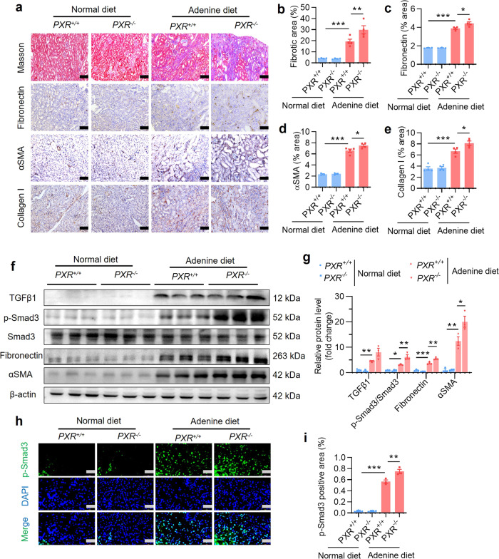
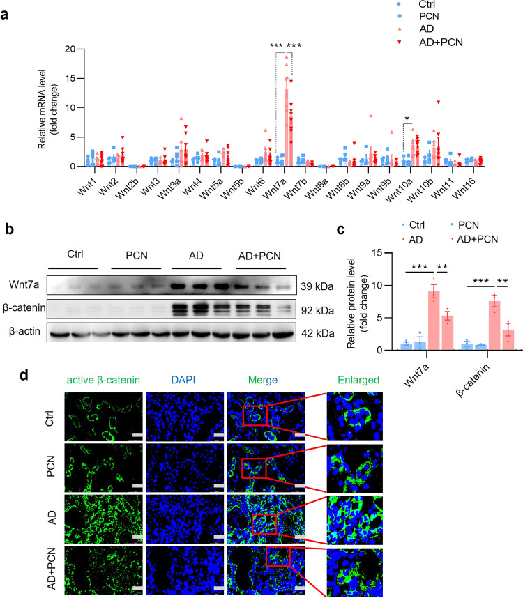
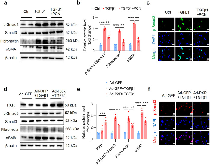
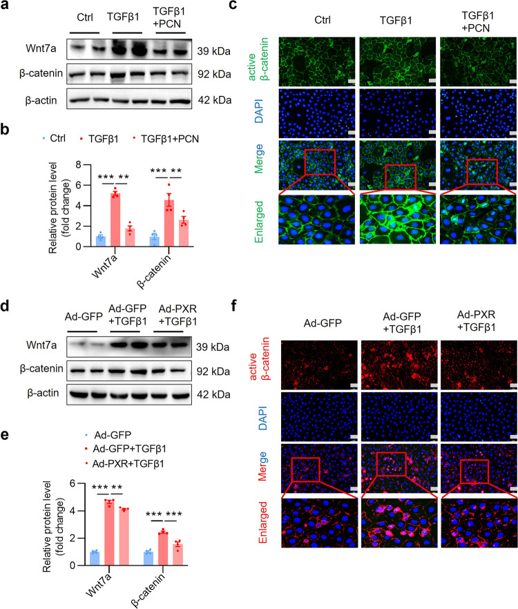
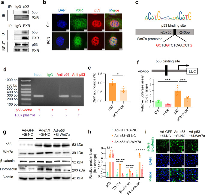
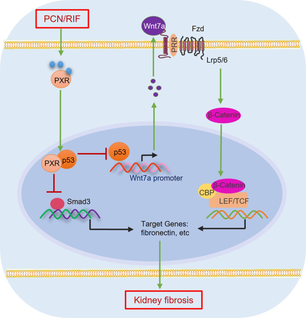
Renal fibrosis is a common pathological feature of chronic kidney disease (CKD) with various etiologies, which seriously affects the structure and function of the kidney. Pregnane X receptor (PXR) is a member of the nuclear receptor superfamily and plays a critical role in regulating the genes related to xenobiotic and endobiotic metabolism in mammals. Previous studies show that PXR is expressed in the kidney and has protective effect against acute kidney injury (AKI). In this study, we investigated the role of PXR in CKD. Adenine diet-induced CKD (AD) model was established in wild-type and PXR humanized (hPXR) mice, respectively, which were treated with pregnenolone-16α-carbonitrile (PCN, 50 mg/kg, twice a week for 4 weeks) or rifampicin (RIF, 10 mg·kg-1·d-1, for 4 weeks). We showed that both PCN and RIF, which activated mouse and human PXR, respectively, improved renal function and attenuated renal fibrosis in the two types of AD mice. In addition, PCN treatment also alleviated renal fibrosis in unilateral ureter obstruction (UUO) mice. On the contrary, PXR gene deficiency exacerbated renal dysfunction and fibrosis in both adenine- and UUO-induced CKD mice. We found that PCN treatment suppressed the expression of the profibrotic Wnt7a and β-catenin in AD mice and in cultured mouse renal tubular epithelial cells treated with TGFβ1 in vitro. We demonstrated that PXR was colocalized and interacted with p53 in the nuclei of tubular epithelial cells. Overexpression of p53 increased the expression of Wnt7a, β-catenin and its downstream gene fibronectin. We further revealed that p53 bound to the promoter of Wnt7a gene to increase its transcription and β-catenin activation, leading to increased expression of the downstream profibrotic genes, which was inhibited by PXR. Taken together, PXR activation alleviates renal fibrosis in mice via interacting with p53 and inhibiting the Wnt7a/β-catenin signaling pathway.

Keywords: PXR; Wnt7a; chronic kidney disease; p53; renal fibrosis; β-catenin.


Conflict of interest statement

The authors declare no competing interests.

Figures

Fig. 1
Fig. 1. PXR activation by PCN alleviates mouse renal damage induced by adenine.
a Schematic diagram of adenine-induced CKD with PCN treatment. b The gross view of the mice and the macroscopic appearance of the kidneys were photographed at the end of 4-week treatment. PCN treatment significantly improved adenine-induced body size reduction and granular appearance of the kidneys. ce PCN treatment significantly attenuated adenine-induced increase in serum creatinine and BUN levels and urinary albumin excretion (n = 5–9). f Representative images of H&E staining. PCN significantly alleviated adenine-induced renal tubular atrophy, dilatation with crystal deposition and cellular casts, and tubulointerstitial fibrosis. Scale bar: 100 μm. g Tubulointerstitial (TI) damage score of histopathologic features in H&E staining (n = 5–6). Results are expressed as mean ± SEM. *P < 0.05, **P < 0.01, ***P < 0.001.
Fig. 2
Fig. 2. PXR activation by PCN attenuates renal fibrosis induced by adenine.
a Masson trichrome staining and immunostaining showing that PCN treatment significantly attenuated renal extracellular matrix protein deposition and the protein expression of fibronectin, αSMA and collagen I. Scale bar: 100 μm. be Semiquantitative analysis of a (n = 4–6). f Western blot analysis demonstrating that PCN treatment markedly reduced adenine-induced expression of fibrosis-related proteins including p-Smad3, fibronectin and αSMA (n = 3). g Semiquantitative measurement for the protein levels in f. h Immunofluorescence assay showing that PCN treatment significantly inhibited adenine-induced activation of p-Smad3. Scale bar: 50 μm. i Semiquantitative measurement for the protein levels in h (n = 3). Data represents the mean ± SEM. *P < 0.05, **P < 0.01, ***P < 0.001.
Fig. 3
Fig. 3. PXR activation by rifampicin alleviates adenine-induced renal damage in PXR-humanized mice.
a Schematic diagram of adenine-induced CKD with or without rifampicin (RIF) treatment. Adenine diet-fed PXR-humanized (hPXR) mice were treated with or without daily intragastric administration of RIF (10 mg/kg) for 28 days. b The gross view of the mice and the macroscopic appearance of the kidneys were photographed on day 29. RIF treatment reduced adenine-induced renal atrophy. RIF treatment significantly reduced the levels of sCr (c), BUN (d) and urinary albumin excretion (e) (n = 5). f H&E staining showed that RIF treatment mitigated renal tubular atrophy, dilatation with crystal deposition and cellular casts, and tubulointerstitial fibrosis. Scale bar: 100 μm. g The TI damage score of histopathologic features in f (n = 5). Results are expressed as mean ± SEM. *P < 0.05, **P < 0.01, ***P < 0.001.
Fig. 4
Fig. 4. PXR activation by rifampicin alleviates renal fibrosis induced by adenine in PXR-humanized mice.
a Masson trichrome staining and immunostaining showing that rifampicin (RIF) treatment significantly alleviated renal extracellular matrix protein deposition and reduced the protein expression of fibronectin, αSMA and collagen I. Scale bar: 100 μm. Semiquantitative analysis of fibrotic area (b), fibronectin (c), αSMA (d) and collagen I (e) positive staining area (n = 4–5). f Western blot assay demonstrating that RIF treatment inhibited protein expression of p-Smad3, fibronectin and αSMA. g Semiquantitative measurement for the protein levels in f (n = 4). h Immunofluorescence analysis showed that RIF treatment markedly reduced p-Smad3 expression. Scale bar: 50 μm. i Semiquantitative measurement of p-Smad3 levels in h (n = 3). Data represents the mean ± SEM. *P < 0.05, **P < 0.01, ***P < 0.001.
Fig. 5
Fig. 5. PXR gene ablation exacerbates adenine-induced renal damages.
a Gross view of the mice and the macroscopic appearance of the kidneys of mice treated with adenine or vehicle for 4 weeks. PXR gene knockout exacerbated adenine-induced body size reduction and granular appearance of the kidneys. PXR gene deficiency aggravated adenine-induced increase in sCr (b), BUN (c) and urinary albumin excretion (d) (n = 5–6). e The TI damage score of histopathologic features (n = 4–6). f H&E staining showing that PXR gene knockout aggravated renal tubular atrophy, dilatation with crystal deposition and cellular casts, and tubulointerstitial fibrosis. Scale bar: 100 μm. Results are expressed as mean ± SEM. **P < 0.01, ***P < 0.001.
Fig. 6
Fig. 6. PXR gene ablation exacerbates adenine-induced renal fibrosis.
a Masson trichrome staining and immunostaining demonstrating that PXR gene knockout increased adenine-induced extracellular matrix protein deposition and renal protein expression of fibronectin, αSMA and collagen I. Scale bar: 100 μm. be Semiquantitative analysis of fibrotic area and protein expression levels in a (n = 4). f Western blot analysis demonstrating that PXR gene knockout aggravated adenine-induced p-Smad3, fibronectin and αSMA expression. g Semiquantitative measurement for the protein levels in f (n = 3). h Immunofluorescence analysis showing that PXR gene knockout increased adenine-induced p-Smad3 activation. Scale bar: 50 μm. i Semiquantitative measurement of p-Smad3 in h (n = 3). Data represents the mean ± SEM. *P < 0.05, **P < 0.01, ***P < 0.001.
Fig. 7
Fig. 7. PXR activation inhibits the Wnt7a/β-catenin signaling in the kidneys of adenine-treated mice.
a Real-time PCR analysis demonstrating the effect of PCN treatment on the expression of all 19 Wnt members in mice treated with adenine for 4 weeks. Adenine induced ~13-fold increase in Wnt7a expression, which was significantly attenuated by PCN treatment (n = 4–8). b, c Western blot assay and semiquantitative analysis indicate that PCN treatment significantly decreased adenine-induced protein levels of Wnt7a and its downstream effector β-catenin (n = 3). d Immunofluorescence assay showed that PCN treatment inhibited adenine-induced activation of β-catenin. Scale bar: 50 μm. Results are expressed as mean ± SEM. *P < 0.05, **P < 0.01, ***P < 0.001.
Fig. 8
Fig. 8. PXR activation and overexpression inhibit TGFβ1-induced fibrotic gene expression in cultured MTEC.
a Western blot analysis demonstrating that PXR activation decreased TGFβ1-induced upregulation of p-Smad3, fibronectin and αSMA protein expression in MTEC. b Semiquantitative analysis of the protein levels in a (n = 4). c Immunofluorescence assay showed that PXR activation attenuated TGFβ1-induced activation of p-Smad3. Scale bar: 25 μm. d Western blot analysis demonstrated that adenovirus-mediated PXR overexpression decreased TGFβ1-induced p-Smad3, fibronectin and αSMA expression in MTEC. e Semiquantitative analysis of the protein levels in d (n = 4). f Immunofluorescence assay showed that PXR overexpression abolished the activation of p-Smad3 induced by TGFβ1. Scale bar: 25 μm. Results are expressed as mean ± SEM. **P < 0.01, ***P < 0.001.
Fig. 9
Fig. 9. PXR activation and overexpression inhibit the Wnt7a/β-catenin signaling in cultured MTEC.
a Western blot analysis demonstrating that PXR activation inhibited Wnt7a and β-catenin expression after TGFβ1 treatment in MTEC. b Semiquantitative analysis of the protein levels in a (n = 4). c Immunofluorescence assay showed that PXR activation inhibited the activation of β-catenin induced by TGFβ1 (5 ng/mL) for 24 h. Scale bar: 25 μm. d Western blot analysis demonstrated that PXR overexpression inhibited Wnt7a and β-catenin expression after TGFβ1 treatment in MTEC. e Semiquantitative analysis of the protein levels in d (n = 4). f Immunofluorescence assay showed that PXR activation inhibited the activation of β-catenin induced by TGFβ1 (5 ng/mL) for 24 h. Scale bar: 25 μm. Results are expressed as mean ± SEM. **P < 0.01, ***P < 0.001.
Fig. 10
Fig. 10. PXR physically interacts with p53 protein and inhibits p53-induced Wnt7a gene transcription and β-catenin activation.
a Co-IP assay showing protein-protein interaction between PXR and p53 in HEK293T cells co-transfected with both mouse PXR and human p53 expression vector. b Immunofluorescence assay demonstrating that PCN treatment increased nuclear co-localization of p53 and PXR protein. Scale bar: 5 μm. c Identification of a putative p53 binding element using the JASPAR website. The predicted p53 binding site in mouse Wnt7a promoter is located between −257 bp and −243 bp. ChIP assay (d) and quantitative analysis (e) revealed that PXR markedly blocked the binding of p53 to mouse Wnt7a gene promoter. Mouse IgG served as a negative control (n = 3). f PXR overexpression significantly inhibited p53-induced Wnt7a promoter-driven luciferase reporter activity in HEK293T cells (n = 4–7). g Western blot analysis demonstrated that adenovirus-mediated p53 overexpression increased expression of Wnt7a, β-catenin and fibronectin. Wnt7a siRNA treatment eliminated p53-induced β-catenin activation and fibronectin expression in MTEC. h Quantitative analysis of the protein levels in g (n = 4). i Immunofluorescence assay showed that p53 overexpression increased the expression and activation of β-catenin, which was abolished by Wnt7a siRNA. Scale bar: 25 μm Results are expressed as mean ± SEM. *P < 0.05, **P < 0.01, ***P < 0.001.
Fig. 11
Fig. 11. Proposed mechanisms by which PXR alleviates kidney fibrosis through interacting with p53.
Fibrotic kidney exhibits increased expression of p53, which promotes the assembly of the p53/p-Smad3 complex to induce profibrotic gene expression. In addition, p53 can bind to the promoter region of Wnt7a to increase Wnt7a transcription and β-catenin activation, leading to increased expression of the downstream profibrotic genes. By physically interacting with p53, PXR reduces the binding of p53 to p-Smad3 protein and Wnt7a gene promoter to suppress both the p-Smad3 and β-catenin signaling, thereby attenuating renal fibrosis. Fzd Frizzled, LRP5/6 low density lipoprotein receptor related protein-5 or −6, PRR prorenin receptor, CBP cAMP response element binding protein (CREB)-binding protein, TCF T-cell factor, LEF lymphoid enhancer-binding factor.

Similar articles

Cited by

References

    1. Stenvinkel P, Chertow GM, Devarajan P, Levin A, Andreoli SP, Bangalore S, et al. Chronic inflammation in chronic kidney disease progression: role of Nrf2. Kidney Int Rep. 2021;6:1775–87. doi: 10.1016/j.ekir.2021.04.023. - DOI - PMC - PubMed
    1. Neovius M, Jacobson SH, Eriksson JK, Elinder CG, Hylander B. Mortality in chronic kidney disease and renal replacement therapy: a population-based cohort study. BMJ Open. 2014;4:e004251. doi: 10.1136/bmjopen-2013-004251. - DOI - PMC - PubMed
    1. Jha V, Garcia-Garcia G, Iseki K, Li Z, Naicker S, Plattner B, et al. Chronic kidney disease: global dimension and perspectives. Lancet. 2013;382:260–72. doi: 10.1016/s0140-6736(13)60687-x. - DOI - PubMed
    1. Tonelli M, Wiebe N, Culleton B, House A, Rabbat C, Fok M, et al. Chronic kidney disease and mortality risk: a systematic review. J Am Soc Nephrol. 2006;17:2034–47. doi: 10.1681/asn.2005101085. - DOI - PubMed
    1. Reidy K, Kang HM, Hostetter T, Susztak K. Molecular mechanisms of diabetic kidney disease. J Clin Invest. 2014;124:2333–40. doi: 10.1172/jci72271. - DOI - PMC - PubMed