. 2023 Jun 21;11(1):102.
doi: 10.1186/s40478-023-01593-y.

DHCR24 reverses Alzheimer's disease-related pathology and cognitive impairment via increasing hippocampal cholesterol levels in 5xFAD mice

Affiliations

DHCR24 reverses Alzheimer's disease-related pathology and cognitive impairment via increasing hippocampal cholesterol levels in 5xFAD mice

Wen-Bin Zhang et al. Acta Neuropathol Commun. .

Abstract

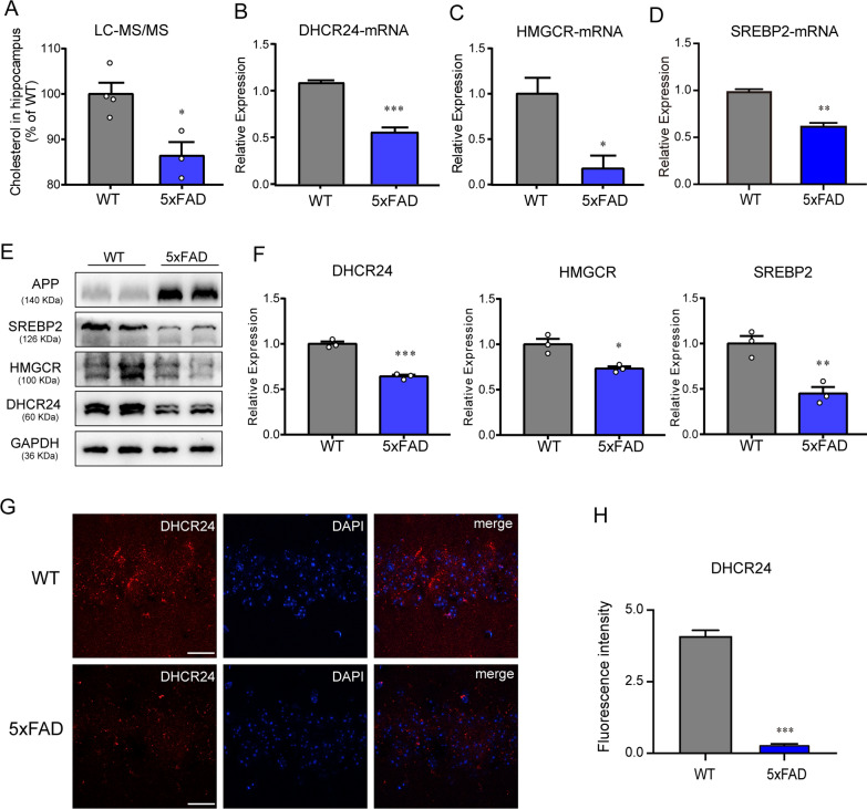
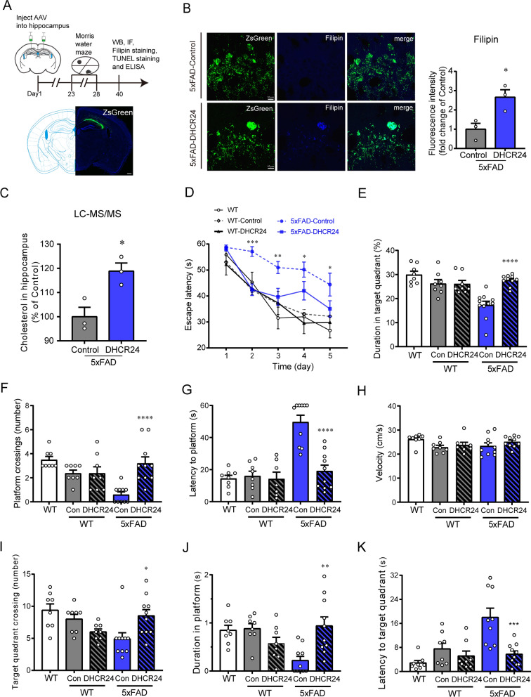
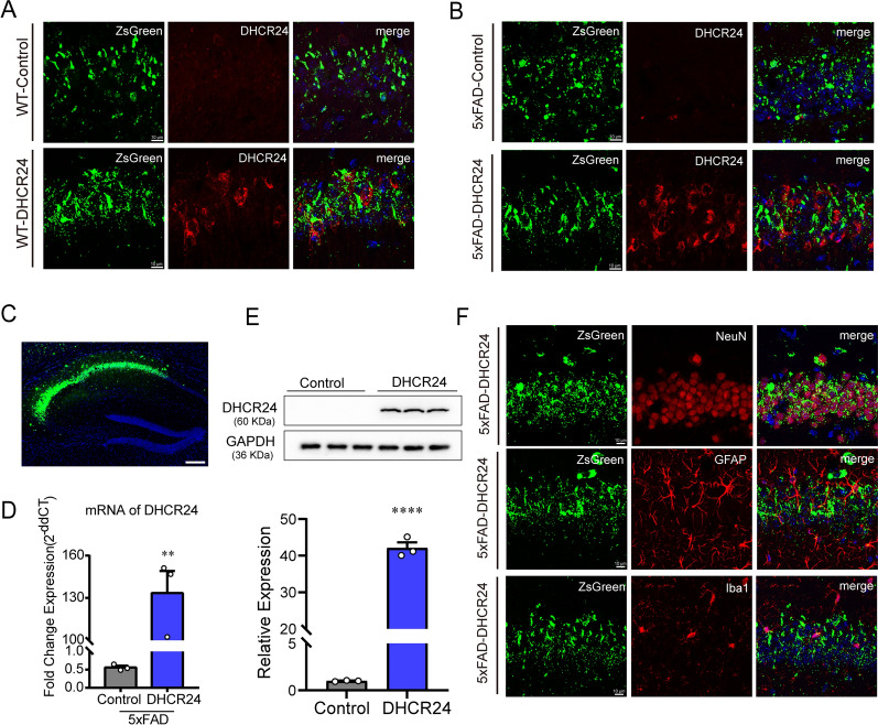
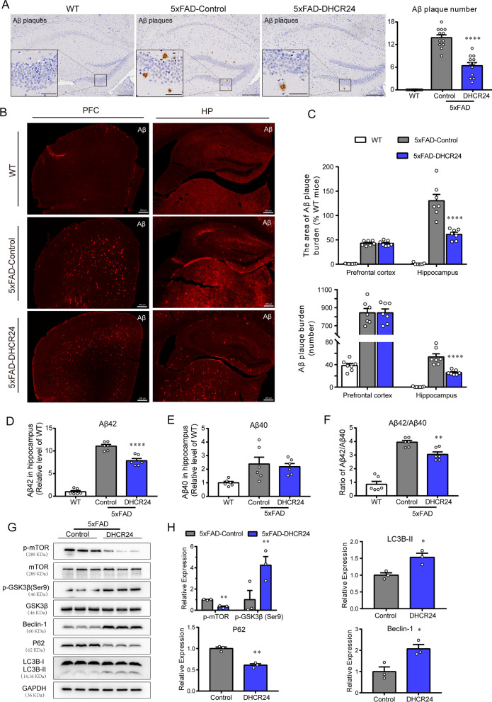
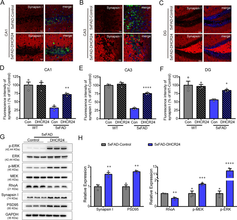
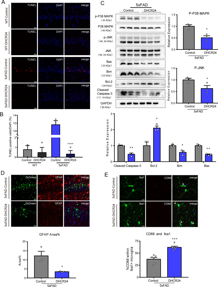
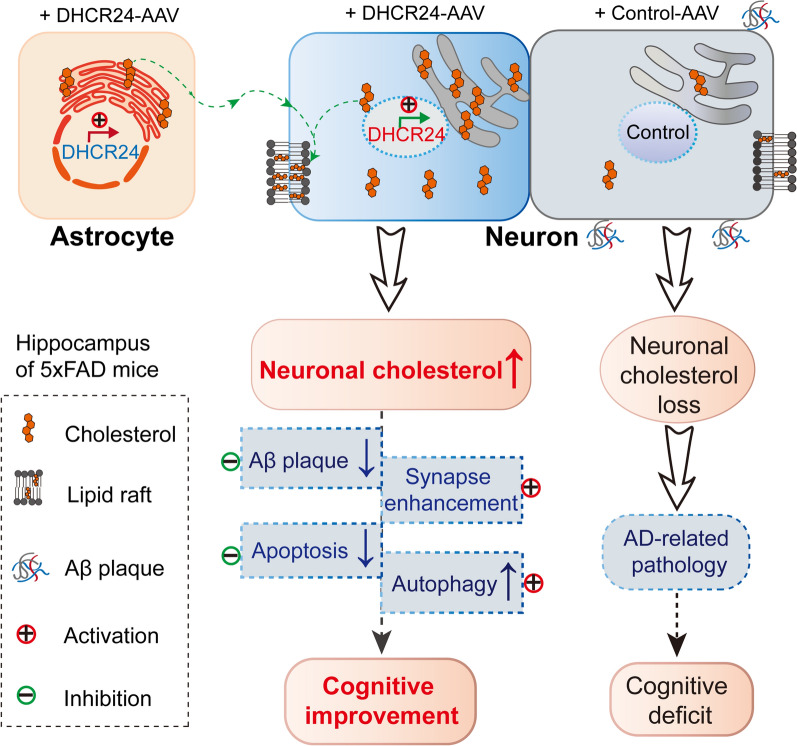
Accumulating evidences reveal that cellular cholesterol deficiency could trigger the onset of Alzheimer's disease (AD). As a key regulator, 24-dehydrocholesterol reductase (DHCR24) controls cellular cholesterol homeostasis, which was found to be downregulated in AD vulnerable regions and involved in AD-related pathological activities. However, DHCR24 as a potential therapeutic target for AD remains to be identified. In present study, we demonstrated the role of DHCR24 in AD by employing delivery of adeno-associated virus carrying DHCR24 gene into the hippocampus of 5xFAD mice. Here, we found that 5xFAD mice had lower levels of cholesterol and DHCR24 expression, and the cholesterol loss was alleviated by DHCR24 overexpression. Surprisingly, the cognitive impairment of 5xFAD mice was significantly reversed after DHCR24-based gene therapy. Moreover, we revealed that DHCR24 knock-in successfully prevented or reversed AD-related pathology in 5xFAD mice, including amyloid-β deposition, synaptic injuries, autophagy, reactive astrocytosis, microglial phagocytosis and apoptosis. In conclusion, our results firstly demonstrated that the potential value of DHCR24-mediated regulation of cellular cholesterol level as a promising treatment for AD.

Keywords: 24-dehydrocholesterol reductase (DHCR24); Alzheimer’s disease; Cholesterol; Gene therapy; Neurodegeneration; Neuroprotection; Pathogenesis.


Conflict of interest statement

The authors have declared that no conflict of interest exists.

Figures

Fig. 1
Fig. 1
The significant decrease of hippocampal cholesterol level in 5xFAD mice. A Quantification of cholesterol level in hippocampus of WT and 5xFAD mice (3 month) by LC–MS/MS analysis. n = 3–4 sample/group, every sample was included 2–3 mice randomly. BD The fold change expression of mRNA of DHCR24, HMGCR and SREBP2 in hippocampus of WT and 5xFAD mice (3 month) by RT-PCR. E The immunoblotting bands of DHCR24, HMGCR, SREBP2 in hippocampus of 3-month-old WT mice and 5xFAD mice. F Analysis of western blot with mean gray value which all were quantification on the ratio of target proteins against GAPDH. G The fluorescence images of DHCR24 (red) in hippocampus of 3-month-old WT and 5xFAD mice (scale bar, 20 μm). H Mean fluorescence intensity of DHCR24. n = 3 mice per group in [B-H]. Data expressed as mean ± SEM, statistical analysis was performed using unpaired two-tailed Student’s t-test. *P < 0.05; **P < 0.01; ***P < 0.001; compared with aged-matched WT group
Fig. 2
Fig. 2
DHCR24 knock-in markedly improves cognitive ability of 5xFAD mice. A The timeline of the experiment and fluorescence images of ZsGreen (green) one month after AAV injection (scale bar, 200 μm). B Fluorescence images of Filipin staining (blue) in the CA1 region of hippocampus (scale bar, 10 μm) and the analysis results of mean fluorescence intensity which were normalized with 5xFAD-Control group. C Quantification of cholesterol level in hippocampus two groups by LC–MS/MS analysis which were normalized with 5xFAD-Control group. n = 3 sample/group, every sample was included 2–4 mice randomly. D Escape latency during the 5 days acquisition phase of Morris water maze (MWM) test. E The percentage of time spent in target quadrant in MWM test. F The number of platform crossings in MWM test. G The latency to platform in MWM test. H The swim speed of mice in MWM test. I The number of target quadrant crossings in MWM test. J The time spent in platform in MWM test. K The latency to the target quadrant in MWM test. n = 7–10 mice per group in [G], n = 8–10 mice per group in [DF and HK]. Data expressed as mean ± SEM, statistical analysis between the five groups was analyzed by one-way ANOVA with Tukey’s post hoc test, except for escape latency was analyzed by three-way repeated measures ANOVA with LSD post hoc test. *P < 0.05; **P < 0.01; ***P < 0.001; compared with aged-matched 5xFAD-Control group. The images of MWM trials were showed in Additional file 1: Fig. S1
Fig. 3
Fig. 3
The over-expression of DHCR24 in the hippocampus of 5xFAD mice. A, B The fluorescence images of ZsGreen (green) and DHCR24 (red) in hippocampus (scale bar, 10 μm). C Fluorescence image of ZsGreen (green) in hippocampus (scale bar, 200 μm). D The fold change expression of mRNA of DHCR24 in hippocampus by RT-PCR. E The immunoblotting bands of DHCR24 protein and the analysis results of western blot with mean gray value which were quantification on the ratio of target proteins against GAPDH. n = 3 mice per group. F Co-staining of ZsGreen (green) with NeuN (red) and GFAP (red) or Iba1 (red) in hippocampus (scale bar, 10 μm). Data expressed as mean ± SEM, statistical analysis between the two groups was analyzed by unpaired two-tailed Student’s t-test. *P < 0.05; **P < 0.01; ***P < 0.001; compared with aged-matched 5xFAD-Control group
Fig. 4
Fig. 4
DHCR24 knock-in alleviates Aβ pathology and activates autophagy flux in hippocampus of 5xFAD mice. A The immunohistochemistry images of Aβ plagues using the antibody MOAB-2 in the hippocampus (Scale bars, 200 μm). Insets show a higher magnification of Aβ plagues. Inset scale bar, 50 μm. The right is analysis of number of Aβ plagues in hippocampus. n = 12 slices from 4 mice per group. B Fluorescence images of Aβ (red) in prefrontal cortex and whole hippocampus (scale bar, 200 μm). C The top half is the analysis of the area of Aβ plagues, and the bottom half is the analysis of number of Aβ plagues in prefrontal cortex and hippocampus. n = 7 slices from 4 mice per group. D, E The relative level of Aβ42 and Aβ40 in hippocampus by ELISA which normalized with WT mice. n = 6 mice per group. F The ratio of Aβ42/Aβ40 in hippocampus which normalized with WT mice. G The immunoblotting bands of p-mTOR, p-GSK3β (ser9), Beclin-1, P62 and LC3B in the hippocampus of 5xFAD-Control group and 5xFAD-DHCR24 group. H Analysis of western blot with mean gray value which all were quantification on the ratio of target proteins against GAPDH except p-mTOR and p-GSK3β (ser9) was against total mTOR and GSK3β, n = 3 mice per group. Data expressed as mean ± SEM, statistical analysis between two groups was analyzed by unpaired two-tailed Student’s t-test, between three groups was analyzed by one-way ANOVA with Tukey’s post hoc test. *P < 0.05; **P < 0.01; ***P < 0.001; compared with aged-matched 5xFAD-Control group
Fig. 5
Fig. 5
DHCR24 knock-in improves synaptic function in 5xFAD mice. A, B Fluorescence images of Synapsin I in the CA1 and CA3 region of hippocampus (scale bar, 20 μm). C Fluorescence images of Synapsin I in the DG region of hippocampus (scale bar, 50 μm). DF Mean fluorescence intensity of Synapsin I in the CA1, CA3, and DG region. n = 3 mice/group. G The immunoblotting bands of PSD95, Synapsin I, RhoA, p-MEK and p-ERK in the hippocampus of 5xFAD-Control group and 5xFAD-DHCR24 group. H Analysis of western blot with mean gray value which all were quantification on the ratio of target proteins against GAPDH except p-MEK and p-ERK were the ratio against total MEK and ERK. n = 3 mice/group. Data expressed as mean ± SEM, statistical analysis between the two groups was analyzed by unpaired two-tailed Student’s t-test, between the four groups was analyzed by one-way ANOVA with Tukey’s post hoc test. *P < 0.05; **P < 0.01; ***P < 0.001; compared with aged-matched 5xFAD-Control group. The images of Synapsin I in CA1, CA3 and DG regions of WT-Control group and WT-DHCR24 group were showed in Additional file 2: Fig. S2
Fig. 6
Fig. 6
DHCR24 knock-in inhibits apoptosis or reactive astrocytosis and promotes microglial phagocytosis in 5xFAD mice. A Fluorescence images of TdT-mediated dUTP Nick-End Labeling (TUNEL) staining (red) in the CA1 region of hippocampus (scale bar, 20 μm). B The ratio of TUNEL-positive cells against DAPI. C Representative immunoblotting bands of Cleaved caspase3, Bcl-2, Bim, Bax, p-JNK and p-P38 MAPK and analysis of western blot with mean gray value which all were quantification on the ratio of target proteins against GAPDH except p-JNK and p-P38 MAPK were against total JNK and P38 MAPK. D Co-staining of ZsGreen (green) with GFAP (red) in hippocampus and analyzing with the percentage of fluorescence area of GFAP (scale bar, 20 μm). E Co-staining of Iba1 (green) with CD68 (red) in hippocampus and analyzing with the percentage of CD68 + positive within Iba1 + microglia (scale bar, 10 μm). The arrows indicate CD68 + and Iba1 + microglia. n = 3 mice/group in [A-D], n = 6 brain slices from 3 mice/group in [E]. Data expressed as mean ± SEM, statistical analysis between the two groups was analyzed by unpaired two-tailed Student’s t-test, between the four groups was analyzed by one-way ANOVA with Tukey’s post hoc test. *P < 0.05; **P < 0.01; ***P < 0.001; compared with aged-matched 5xFAD-Control group
Fig. 7
Fig. 7
Schematic summary of DHCR24 knock-in reverses AD-related pathology and enhances cognitive ability of 5xFAD mice. To demonstrate the contribution of DHCR24 in improving the cognitive ability of AD, we employed delivery of adeno-associated virus (AAV) carrying DHCR24 gene into the hippocampus of 5xFAD mice. After DHCR24 transfection, DHCR24 is universally co-expressed in neurons and astrocytes, resulting in the increase of neuronal cholesterol level. By enhancing neuronal cholesterol level, DHCR24 knock-in successfully prevented or reversed AD-related pathology, including amyloid-β deposition, synaptic injuries, inhibition of autophagy, and apoptosis. Finally, DHCR24 knock-in obviously improved the cognitive ability of 5xFAD mice

Similar articles

Cited by

References

    1. Abad-Rodriguez J, Ledesma MD, Craessaerts K, Perga S, Medina M, Delacourte A, Dingwall C, De Strooper B, Dotti CG. Neuronal membrane cholesterol loss enhances amyloid peptide generation. J Cell Biol. 2004;167:953–960. doi: 10.1083/jcb.200404149. - DOI - PMC - PubMed
    1. Allen LB, Genaro-Mattos TC, Porter NA, Mirnics K, Korade Z. Desmosterolosis and desmosterol homeostasis in the developing mouse brain. J Inherit Metab Dis. 2019;42:934–943. doi: 10.1002/jimd.12088. - DOI - PMC - PubMed
    1. Anchisi L, Dessi S, Pani A, Mandas A. Cholesterol homeostasis: a key to prevent or slow down neurodegeneration. Front Physiol. 2012;3:486. doi: 10.3389/fphys.2012.00486. - DOI - PMC - PubMed
    1. Andersson HC, Kratz L, Kelley R. Desmosterolosis presenting with multiple congenital anomalies and profound developmental delay. Am J Med Genet. 2002;113:315–319. doi: 10.1002/ajmg.b.10873. - DOI - PubMed
    1. Attems J, Jellinger KA. Amyloid and tau: neither chicken nor egg but two partners in crime! Acta Neuropathol. 2013;126:619–621. doi: 10.1007/s00401-013-1167-9. - DOI - PubMed

Publication types

Substances