

Androgen Receptor Is Expressed in the Majority of Breast Cancer Brain Metastases and Is Subtype-Dependent
- PMID: 37345085
- PMCID: PMC10216529
- DOI: 10.3390/cancers15102748
Androgen Receptor Is Expressed in the Majority of Breast Cancer Brain Metastases and Is Subtype-Dependent
Abstract
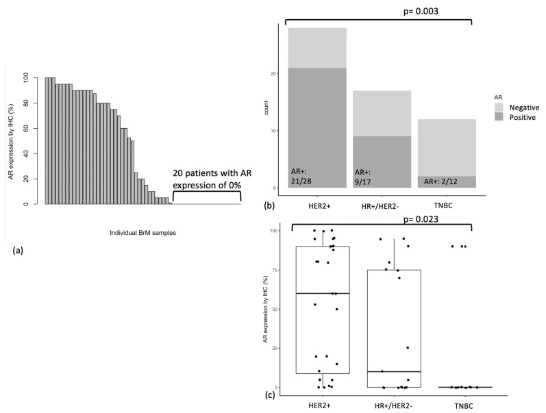
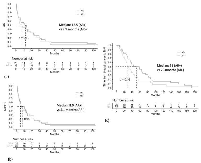
We aimed to evaluate the expression of the "targetable" androgen receptor (AR) in breast cancer brain metastases (BrM). An established, retrospective 57-patient cohort with metastatic breast cancer who underwent surgery for BrM at the Sunnybrook Odette Cancer Centre between 1999-2013 was studied. AR expression in BrM samples was assessed in triplicate using immunohistochemistry (IHC). AR positive status was defined as nuclear AR expression ≥ 10% by IHC using the SP107 antibody. The median age of patients was 52 years (range 32-85 years). 28 (49%) of BrM were HER2+, 17 (30%) were hormone receptor positive (HR+)/HER2-, and 12 (21%) were triple negative breast cancers (TNBCs). 56% (n = 32/57) of BrM were AR positive, and median AR expression was 20% (CI 1.6-38.3%). AR expression was different across breast cancer subtypes; AR was most frequently expressed in HER2+ (n = 21/28), followed by HR+/HER2- (n = 9/17), and lowest in TNBC (n = 2/12) BrM (p = 0.003). Patients with AR positive versus AR negative BrM had similar overall survival (12.5 vs. 7.9 months, p = 0.6), brain-specific progression-free survival (8.0 vs. 5.1 months, p = 0.95), and time from breast cancer diagnosis to BrM diagnosis (51 vs. 29 months, p = 0.16). AR is expressed in the majority of breast cancer BrM and represents a potential therapeutic target.
Keywords: androgen receptor; brain metastasis; breast cancer; immunohistochemistry.
Conflict of interest statement
K.J.J. has served as a consultant and/or advisory board member for: Amgen, AstraZeneca, Apo Biologix, Eli Lilly, Esai, Genomic Health, Gilead Sciences, Knight Therapeutics, Merck, Myriad Genetics Inc., Pfizer, Roche, Seagen, Novartis, and Viatris. K.J.J has also received research funding from AstraZeneca, Eli Lilly and Seagen (paid to the institution). We did not receive any funding for this study.
Figures
References
-
- Gerratana L., Basile D., Buono G., De Placido S., Giuliano M., Minichillo S., Coinu A., Martorana F., De Santo I., Del Mastro L., et al. Androgen Receptor in Triple Negative Breast Cancer: A Potential Target for the Targetless Subtype. Cancer Treat. Rev. 2018;68:102–110. doi: 10.1016/j.ctrv.2018.06.005. - DOI - PubMed
LinkOut - more resources
Full Text Sources
Research Materials
Miscellaneous
