. 2023 Jun 6:14:1178817.
doi: 10.3389/fimmu.2023.1178817. eCollection 2023.

Disruption of the sialic acid/Siglec-9 axis improves antibody-mediated neutrophil cytotoxicity towards tumor cells

Affiliations

Disruption of the sialic acid/Siglec-9 axis improves antibody-mediated neutrophil cytotoxicity towards tumor cells

Marta Lustig et al. Front Immunol. .

Abstract

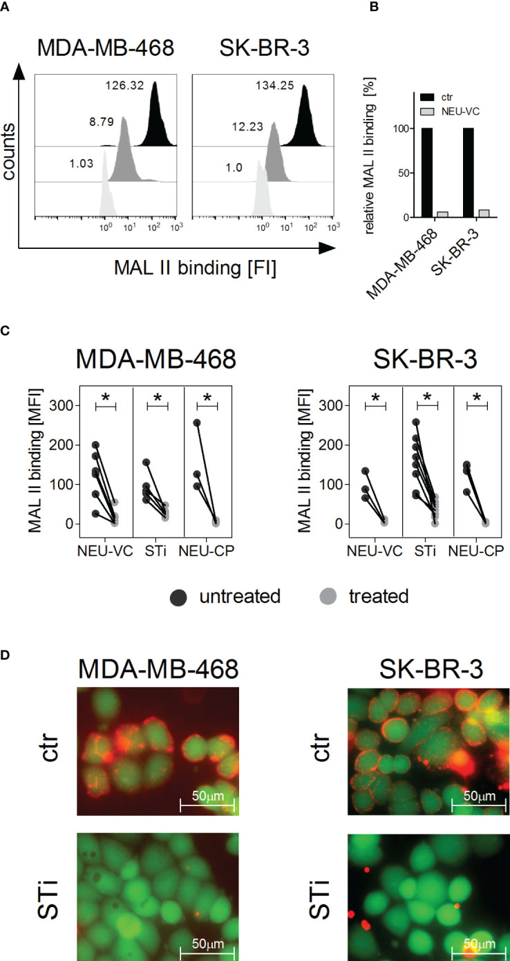
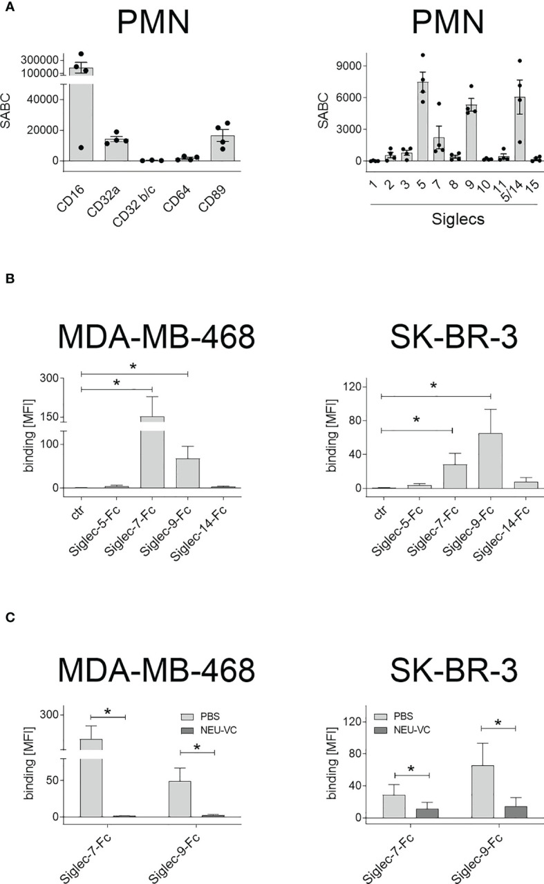
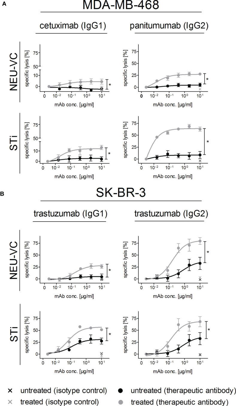
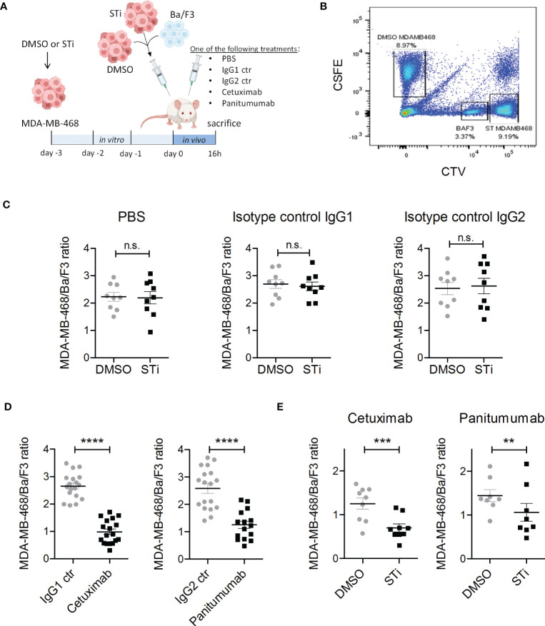
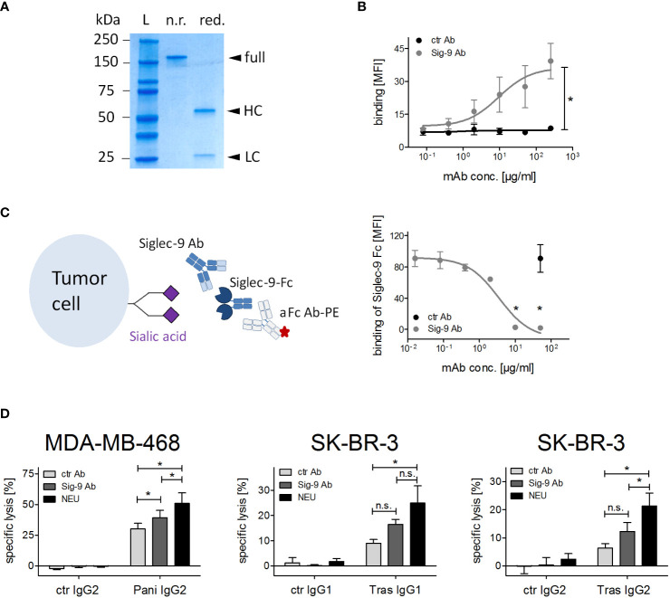
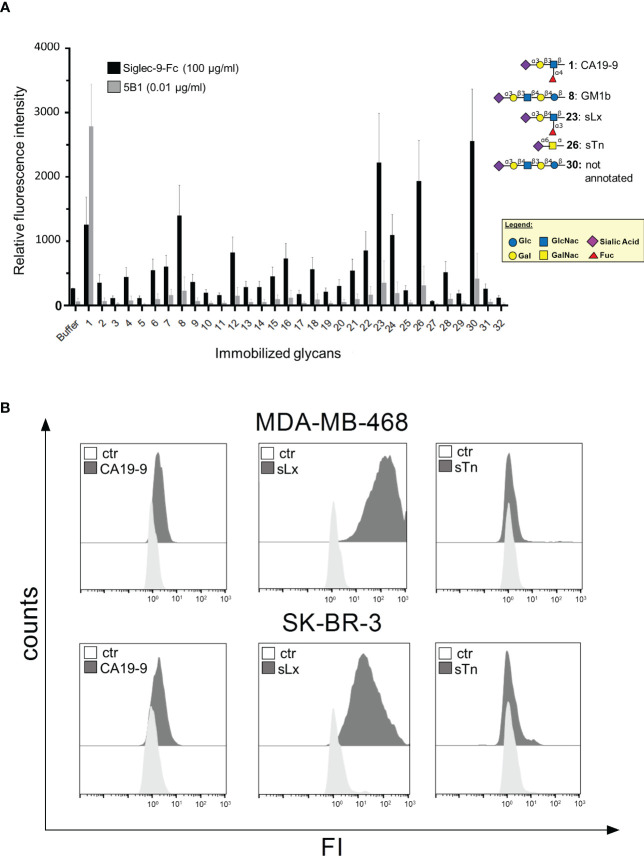
Upregulation of surface expressed sialoglycans on tumor cells is one of the mechanisms which promote tumor growth and progression. Specifically, the interactions of sialic acids with sialic acid-binding immunoglobulin-like lectins (Siglecs) on lymphoid or myeloid cells transmit inhibitory signals and lead to suppression of anti-tumor responses. Here, we show that neutrophils express among others Siglec-9, and that EGFR and HER2 positive breast tumor cells express ligands for Siglec-9. Treatment of tumor cells with neuraminidases or a sialyl transferase inhibitor significantly reduced binding of a soluble recombinant Siglec-9-Fc fusion protein, while EGFR and HER2 expression remained unchanged. Importantly, the cytotoxic activity of neutrophils driven by therapeutic EGFR or HER2 antibodies in vitro was increased by blocking the sialic acid/Siglec interaction, either by reducing tumor cell sialylation or by a Siglec-9 blocking antibody containing an effector silenced Fc domain. In vivo a short-term xenograft mouse model confirmed the improved therapeutic efficacy of EGFR antibodies against sialic acid depleted, by a sialyltransferase inhibitor, tumor cells compared to untreated cells. Our studies demonstrate that sialic acid/Siglec interactions between tumor cells and myeloid cells can impair antibody dependent tumor cell killing, and that Siglec-9 on polymorphonuclear cells (PMN) is critically involved. Considering that PMN are often a highly abundant cell population in the tumor microenvironment, Siglec-9 constitutes a promising target for myeloid checkpoint blockade to improve antibody-based tumor immunotherapy.

Keywords: ADCC - antibody dependent cellular cytotoxicity; Siglec-9; glycans; myeloid cells; neutrophils (PMNs); sialic acid; siglecs.


Conflict of interest statement

MB and PS are employed by Tacalyx GmbH. JHWL is scientific founder and shareholder of TigaTx. All other authors declare that the research was conducted in the absence of any commercial or financial relationships that could be construed as a potential conflict of interest.

Figures

Figure 1
Figure 1
α2,3 linked sialic acid on tumor cells can be reduced by neuraminidase treatment or by sialyltransferase inhibition. (A, B) Binding of Maackia amurensis leukagglutinin (MAL) II (5 µg/ml), detected with streptavidin-PE, on MDA-MB-468 and SK-BR-3 cells is reduced after treatment with neuraminidase of Vibrio cholerae (NEU-VC) (0.1 U/ml). Black, untreated cells; grey, treated cells; white, PBS was used as control for MAL II on untreated cells. Shown are representative histograms (A) and MAL II binding normalized to the control (ctr) (B). (C) MAL II binding on MDA-MB-468 and SK-BR-3 cells untreated (black dots) or treated (grey dots) with NEU-VC (0.1 U/ml), NEU-Clostridium perfringens (CP) (1 U/ml), or a sialyltransferase inhibitor (STi) (100 µM). Dot plots show MAL II binding as mean fluorescence intensity (MFI) values. * depicts significant differences (p < 0.05) between treated and untreated cells (two-way ANOVA). FI, fluorescence intensity. (D) Overlay of microscopic images of live (green fluorescent, excitation at 495nm) and sialic acid positive (red fluorescent, excitation at 532 nm) MDA-MB-468 and SK-BR-3 cells. Untreated (ctr) or STi treated cells (STi) were stained with Calcein-AM (green fluorescent) and MAL II-streptavidin-PE (red fluorescent).
Figure 2
Figure 2
Expression of FcR and Siglecs on PMN, and soluble Siglec binding to tumor cells. (A) Expression of Fc receptors (FcR) (left panel) and Siglecs (right panel) on GM-CSF stimulated PMN stained with mouse monoclonal antibodies (5 µg/ml) and detected with FITC-conjugated goat anti-mouse Fcγ-specific F(ab)2 fragments. Antigen expression levels are depicted as mean specific antibody binding capacities (SABC) ± SEM of at least 3 different donors. (B) Siglec-binding epitopes on tumor cells were assessed by binding of soluble Siglec-Fc proteins (10 µg/ml) and detected with PE-conjugated goat anti-human Fcγ-specific F(ab)2 fragments. (C) Siglec-7-Fc and Siglec-9-Fc binding (10 µg/ml) on MDA-MB-468 and SK-BR-3 cells was reduced after treatment with neuraminidase (NEU-VC). The mean fluorescence intensity (MFI) values of more than three independent replicates are shown. * indicates significant differences (p < 0.05) compared to the controls (non-parametric one-tailed paired t-test).
Figure 3
Figure 3
Reduced α2,3 linked sialic acid on tumor cells improves antibody-dependent PMN mediated ADCC. MDA-MB-468 (A) and SK-BR-3 (B) served as targets in [51Cr] release assays with GM-CSF (50 U/ml) stimulated PMN at an E:T cell ratio of 40:1. Tumor cells were treated (grey curves) with neuraminidase (NEU-VC) (0.1 U/ml) or with the sialyltransferase inhibitor (STi) (100 µM). IgG1 or IgG2 antibodies against EGFR or HER2 were used at indicated concentrations and control antibodies at 10 µg/ml. Shown are the mean values ± SEM as % specific lysis of at least 3 independent experiments with cells from different donors. Data were analysed by two-way ANOVA, and significant differences between treated and non-treated cells (*) are indicated.
Figure 4
Figure 4
Sialyltransferase inhibition in tumor cells improves EGFR antibody therapeutic efficacy in a xenograft tumor model. (A) MDA-MB-468 tumor cells were treated for 3 days in vitro with 100 μM sialyltransferase inhibitor (STi) (MDA-MB-468 STi) or DMSO (MDA-MB-468 DMSO) prior to intraperitoneal (IP) injection into SCID mice. EGFR-negative murine Ba/F3 cells served as a recovery control. MDA-MB-468 DMSO, MDA-MB-468 STi and Ba/F3 cells were injected together at an initial ratio of 5:5:1. After injection, mice were separated into five groups receiving different treatments: PBS, cetuximab, panitumumab, IgG1 or IgG2 isotype controls (10 μg antibodies), respectively. After 16 h, mice were sacrificed and the numbers of residual tumor cells in the peritoneal fluid were evaluated by flow cytometry. (B) Gating strategy to discriminate MDA-MB-468 DMSO, MDA-MB-468 STi and Ba/F3 cells in flow cytometry. MDA-MB-468 DMSO cells were labelled with CSFE, MDA-MB-468 STi with CTV high and Ba/F3 with CTV low. (C) Effect of STi treatment on tumor cell recovery compared to DMSO in groups treated with PBS or isotype controls. (D) Reduction of MDA-MB-468 cells recovery from mice treated with EGFR antibodies with respect to their isotype. (E) Tumor cell recovery of STi treated cells compared to DMSO treated tumor cells in mice treated with EGFR antibodies. Results are presented as the mean ratio ± SEM of total MBA-MD-468 to Ba/F3 cells (at least 8 mice per group). Significant differences (*) were analysed by two-tailed, paired t-test (ns, not significant). **p < 0.01, ***p < 0.001, ****p < 0.0001. Figure 4A was created with BioRender.com.
Figure 5
Figure 5
Siglec-9 blockade on PMN improves ADCC by therapeutic IgG antibodies. (A) SDS-PAGE under non-reducing (n.r.) and reducing (red.) conditions of the Siglec-9 antibody. HC, heavy chain; LC, light chain; L, protein ladder. (B) Binding of Siglec-9 antibody on PMN. A non-binding IgG2σ antibody served as control, anti-human kappa-FITC was used for detection. Mean of the mean fluorescence intensity (MFI) values ± SEM of 3 independent experiments with PMN from 3 different donors are shown. Data were analysed by two-way ANOVA, and * indicates a significant difference. (C) Binding of Siglec-9-Fc fusion protein (5 µg/ml) on tumor cells is blocked with increasing concentrations of the Siglec-9 antibody. The non-binding IgG2σ antibody was used as control (50 μg/ml). The Siglec-9-Fc fusion protein was detected by a goat anti-human IgG PE-conjugated antibody. Mean MFI values ± SEM of 3 independent experiments are shown. Data were analysed by t-student test and significant blocking is depicted by *. (D) PMN ADCC with MDA-MB-468 or SK-BR-3 was performed in the presence of panitumumab IgG2 (10 µg/ml), trastuzumab (10 µg/ml), or trastuzumab IgG2 (2 µg/ml) using the Siglec-9 blocking antibody (20 µg/ml). The effect of the Siglec-9 blocking antibody was also compared to neuraminidase treatment (0.1 U/ml). At least 3 independent experiments with different donors, are displayed as mean values ± SEM (% specific lysis). Data were analysed by two-way ANOVA with Bonferroni post-test correction. Significant differences (*) are indicated. n.s., not significant.
Figure 6
Figure 6
Siglec-9-Fc binds to tumor associated carbohydrate antigens. (A) Tumor associated carbohydrate antigens (TACAs), with O-glycosylation structure bound to an amino-pentanol linker, were analysed in a glycan array by their ability to bind soluble Siglec-9-Fc (100 µg/ml). Reference antibody 5B1 (0,01 µg/ml) binds only to CA19-9. Shown are mean values +/- SEM of the 3 individual experiments. In each experiment, six data points for each sugar were collected, up to 2 were excluded if the background was too high. (B) Expression of CA19-9, sLewisX (sLx) and sTn on MDA-MB-468 and SK-BR-3 cells as analysed by indirect flow cytometry. Binding of primary antibodies (mIgG1 or mIgM, 10 μg/ml) (grey) were detected with FITC-conjugated goat anti-mouse IgG and IgM-specific F(ab)2 fragments. Representative histograms of 3 replicates are shown.

Similar articles

Cited by

References

    1. Varki A. Biological roles of glycans. Glycobiology (2017) 27(1):3–49. doi: 10.1093/glycob/cww086 - DOI - PMC - PubMed
    1. Gagneux P, Hennet T, Varki A. Biological functions of glycans. In: Varki A, Cummings RD, Esko JD, Stanley P, Hart GW, Aebi M, et al., editors. Essentials of glycobiology. NY: Cold Spring Harbor; (2022). p. 79–92.
    1. Smith BAH, Bertozzi CR. The clinical impact of glycobiology: targeting selectins, siglecs and mammalian glycans. Nat Rev Drug Discovery (2021) 20(3):217–43. doi: 10.1038/s41573-020-00093-1 - DOI - PMC - PubMed
    1. Läubli H, Nalle SC, Maslyar D. Targeting the siglec-sialic acid immune axis in cancer: current and future approaches. Cancer Immunol Res (2022) 10(12):1423–32. doi: 10.1158/2326-6066.CIR-22-0366 - DOI - PMC - PubMed
    1. Varki A, Kannagi R, Toole B, Stanley P. Glycosylation changes in cancer. In: Varki A, Cummings RD, Esko JD, Stanley P, Hart GW, et al., editors. Essentials of glycobiology. NY: Cold Spring Harbor; (2015). p. 597–609.

Publication types