

Computed tomography findings of intersigmoid hernia
- PMID: 37346424
- PMCID: PMC10280369
- DOI: 10.5114/pjr.2023.127559
Computed tomography findings of intersigmoid hernia
Abstract
Purpose: To evaluate the computed tomography findings of intersigmoid hernias.
Material and methods: Between April 2010 and March 2018, 7 patients who were surgically diagnosed with intersigmoid hernia in 3 institutions were enrolled in this study. Two radiologists evaluated imaging findings for the herniated small bowel, the distance between the occlusion point and bifurcation of the left common iliac artery, and the anatomic relationship with adjacent organs.
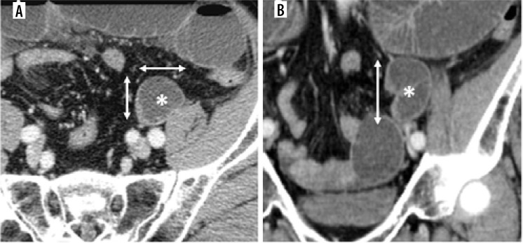
Results: All patients were male, and their mean age (standard deviation, range) was 61.0 (13.5, 36-85) years. The mean size of the bowel loops was 5.2 (1.3, 4.0-8.3) cm in the caudal direction, 3.6 (0.8, 2.5-5.1) cm in the lateral, and 3.4 (0.6, 2.5-4.7) cm in the anterior-posterior direction. The volume was 37.9 (27.8, 15.6-103.0) cm3 approximated by an ellipse, and 24.0 (17.7, 9.9-65.6) cm3 approximated by a truncated cone. The obstruction point was located 3.6 (0.6, 2.8-4.7) cm inferior to the bifurcation of the left common iliac artery. In all cases, the small bowel ran under the point at which the inferior mesenteric vessels bifurcated to the superior rectal vessels and the sigmoid vessels and formed a sac-like appearance between the left psoas muscle and the sigmoid colon. The ureter ran dorsal to the point of the bowel stenosis, and the left gonadal vein ran outside the small bowel loops.
Conclusions: All cases showed common imaging findings, which may be characteristic of men's intersigmoid hernia. In addition, the fossa's position was lower, and the size was larger than in the previous study, which may be a risk factor.
Keywords: internal hernia; intersigmoid fossa; intersigmoid hernia.
© Pol J Radiol 2023.
Conflict of interest statement
The authors report no conflict of interest.
Figures


References
-
- Meyers MA, Charnsangavej C, Oliphant M. Internal abdominal hernias. In: Meyers MA, Charnsangavej C, Oliphant M (eds.). Meyers’ Dynamic Radiology of the Abdomen. 6th ed. New York: Springer; 2011, pp. 381-409.
-
- Akyildiz H, Artis T, Karahan I , et al. . Internal hernia: complex diagnostic and therapeutic problem. Int J Surg 2009; 7: 334-337. - PubMed
-
- Somé OR, Ndoye JM, Chaffanjon P, et al. . An anatomical study of the intersigmoid fossa and applications for internal hernia surgery. Surg Radiol Anat 2017; 39: 243-248. - PubMed
LinkOut - more resources
Full Text Sources
Miscellaneous