. 2023 Nov;261(11):3149-3158.
doi: 10.1007/s00417-023-06147-5. Epub 2023 Jun 22.

Optical coherence tomography angiography-guided vs indocyanine green angiography-guided half-dose photodynamic therapy for acute central serous chorioretinopathy: 6-month randomized trial results

Affiliations

Optical coherence tomography angiography-guided vs indocyanine green angiography-guided half-dose photodynamic therapy for acute central serous chorioretinopathy: 6-month randomized trial results

Siying Li et al. Graefes Arch Clin Exp Ophthalmol. 2023 Nov.

Abstract

Purpose: This study aimed to compare the anatomic and functional results of optical coherence tomography angiography (OCTA)-guided half-dose photodynamic therapy (PDT) versus indocyanine green angiography (ICGA)-guided PDT in eyes with acute central serous chorioretinopathy (CSC).

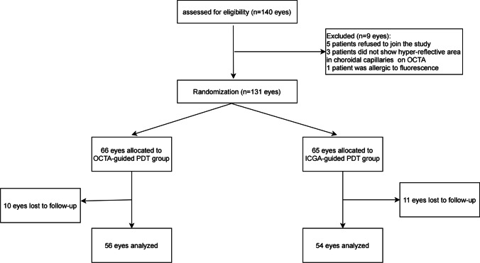
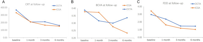
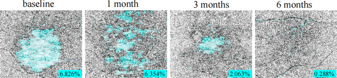
Methods: One hundred and thirty-one eyes of 131 patients with acute central serous chorioretinopathy (CSC) were recruited, and randomly assigned to the OCTA-guided group and ICGA-guided group. The primary outcome measures were the rates of complete subretinal fluid (SRF) resolution at 1 month, 3 months, and 6 months. The secondary outcomes included best-corrected visual acuity (BCVA), central retinal thickness (CRT), choroidal capillary flow deficit density at each scheduled visit, and recurrence rate of SRF at 3 months and 6 months.

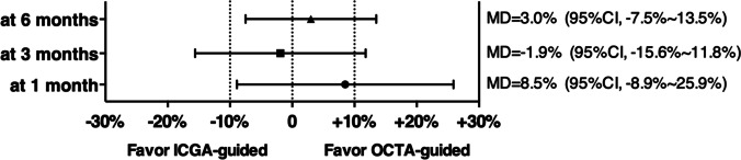
Results: There were 110 eyes that finished the follow-up, with 56 eyes in the OCTA-guided group and 54 eyes in the ICGA guided group. OCTA-guided PDT was demonstrated to be noninferior to ICGA-guided PDT for SRF resolution rate at 1 months and 6 months (P = 0.021 and P = 0.037), but not at 3 months for acute CSC (P = 0.247). The average CRT of the ICGA-guided group was significantly lower than that of the OCTA-guided group at 3-month visit (P = 0.046), but no significant difference was found between them at the 1-month and 6-month visits (P = 0.891 and 0.527). There was no significant difference between the two groups for BCVA (P = 0.359, 0.700, and 0.143, respectively) and the deficit area on CC (P = 0.537, 0.744,and 0.604, respectively) at 1, 3, and 6 months.

Conclusion: OCTA may replace ICGA to guide PDT for the treatment of acute CSC and their follow-up.

Keywords: Acute CSC; ICGA-guided; OCTA-guided; PDT.


Conflict of interest statement

The authors declare no competing interests.

Figures

Fig. 1
Fig. 1
Binarizing of choriocapillaris flow imaging and measurement of flow deficit density (FDD) by using Image J
Fig. 2
Fig. 2
Flowchart showing the distribution of the study population
Fig. 3
Fig. 3
Non-inferiority test of OCTA-guided PDT to ICGA-guided PDT for complete SRF resolution rate at 1, 3, and 6 months after treatment
Fig. 4
Fig. 4
Images of a 23-year-old man with acute CSC treated with ICGA-guided PDT. The patient companied of blurred vision of the left eye for 1 week. CFP, ICGA, and FFA images, 6 × 6 mm en face OCTA image at the level of the choriocapillaris, B scan of the structural OCT before PDT (A1–E1), 1 month after PDT (A2–E2), 3 months after PDT (A3–E3), and 6 months after PDT (A4–E4). Red circles indicate the PDT spot area. Note the SRF was completely resolved at 1 month after PDT (CSC, central serous chorioretinopathy; OCTA, optical coherence tomography angiography; PDT, photodynamic therapy; CFP, color fundus photograph; ICGA, indocyanine green angiography; FFA, fundus fluorescein angiography; SRF, subretinal fluid)
Fig. 5
Fig. 5
Images of a 42-year-old man with acute CSC treated with OCTA-guided PDT. CFP, ICGA, and FFA images, 6 × 6 mm en face OCTA image at the level of the choriocapillaris, B scan of the structural OCT before PDT (A1–E1), 1 month after PDT (A2–E2), 3 months after PDT (A3–E3), and 6 months after PDT (A4–E4). Red circles indicate the PDT spot area. Note the SRF was partially resolved at 1 and 3 months after PDT and completely resolved with a small serous PED appeared at 6 months after PDT. The leakage on FFA and ICGA partially resolved at 1 and 3 months after PDT. The en face OCTA image at the level of the choriocapillaris hyper-reflective area on OCTA also gradually disappeared during the 6 months of follow-up (CSC, central serous chorioretinopathy; OCTA, optical coherence tomography angiography; PDT, photodynamic therapy; CFP, color fundus photograph; ICGA, indocyanine green angiography; FFA, fundus fluorescein angiography; SRF, subretinal fluid)
Fig. 6
Fig. 6
A Comparison of CRT at each visit between two groups. B Comparison of Log MAR BCVA at each visit between two groups. C Comparison of FDD at CC level on 6 × 6 mm en face OCTA image at each visit between two groups
Fig. 7
Fig. 7
The change in choroicapillaris flow deficit density (FDD) after half dose PDT calculated by using binarized OCTA image at CC level throughout follow-up period

References

    1. Eandi CM, Ober M, Iranmanesh R, Peiretti E, Yannuzzi LA. Acute central serous chorioretinopathy and fundus autofluorescence. Retina (Philadelphia, Pa) 2005;25:989–993. doi: 10.1097/00006982-200512000-00006. - DOI - PubMed
    1. Daruich A, Matet A, Dirani A, Bousquet E, Zhao M, Farman N, Jaisser F, Behar-Cohen F. Central serous chorioretinopathy: recent findings and new physiopathology hypothesis. Prog Retin Eye Res. 2015;48:82–118. doi: 10.1016/j.preteyeres.2015.05.003. - DOI - PubMed
    1. Loo RH, Scott IU, Flynn HW, Jr, Gass JD, Murray TG, Lewis ML, Rosenfeld PJ, Smiddy WE. Factors associated with reduced visual acuity during long-term follow-up of patients with idiopathic central serous chorioretinopathy. Retina (Philadelphia, Pa) 2002;22:19–24. doi: 10.1097/00006982-200202000-00004. - DOI - PubMed
    1. Ober MD, Yannuzzi LA, Do DV, Spaide RF, Bressler NM, Jampol LM, Angelilli A, Eandi CM, Lyon AT. Photodynamic therapy for focal retinal pigment epithelial leaks secondary to central serous chorioretinopathy. Ophthalmology. 2005;112:2088–2094. doi: 10.1016/j.ophtha.2005.06.026. - DOI - PubMed
    1. Chan WM, Lai TY, Lai RY, Liu DT, Lam DS. Half-dose verteporfin photodynamic therapy for acute central serous chorioretinopathy: one-year results of a randomized controlled trial. Ophthalmology. 2008;115:1756–1765. doi: 10.1016/j.ophtha.2008.04.014. - DOI - PubMed