. 2023 Sep 4;220(9):e20230661.
doi: 10.1084/jem.20230661. Epub 2023 Jun 22.

Autoantibodies neutralizing type I IFNs underlie West Nile virus encephalitis in ∼40% of patients

Adrian Gervais #  1   2 Francesca Rovida #  3   4 Maria Antonietta Avanzini #  5 Stefania Croce #  6 Astrid Marchal #  1   2 Shih-Ching Lin #  7 Alessandro Ferrari #  4 Christian W Thorball  8   9 Orianne Constant  10 Tom Le Voyer  1   2 Quentin Philippot  1   2 Jérémie Rosain  1   2   11 Micol Angelini  12 Malena Pérez Lorenzo  1 Lucy Bizien  1   2 Cristian Achille  12 Francesca Trespidi  12 Elisa Burdino  13 Irene Cassaniti  4 Daniele Lilleri  4 Chiara Fornara  4 José Camilla Sammartino  4 Danilo Cereda  14 Chiara Marrocu  15 Antonio Piralla  4 Chiara Valsecchi  5 Stefano Ricagno  16   17 Paola Cogo  18 Olaf Neth  19   20 Inés Marín-Cruz  20 Monia Pacenti  21 Alessandro Sinigaglia  22 Marta Trevisan  22 Andrea Volpe  22 Antonio Marzollo  23 Francesca Conti  24 Tiziana Lazzarotto  25   26 Andrea Pession  24 Pierluigi Viale  27 Jacques Fellay  8   9 Stefano Ghirardello  12 Mélodie Aubart  1   2   28 Valeria Ghisetti  13 Alessandro Aiuti  29   30   31 Emmanuelle Jouanguy  1   2   32 Paul Bastard  1   2   32   33 Elena Percivalle  4 Fausto Baldanti  3   4 Anne Puel  1   2   32 Margaret R MacDonald #  34 Charles M Rice #  34 Giada Rossini #  25 Kristy O Murray #  35 Yannick Simonin #  10 Anna Nagy #  36 Luisa Barzon #  21   22 Laurent Abel #  1   2   32 Michael S Diamond #  7 Aurélie Cobat #  1   2   32 Shen-Ying Zhang #  1   2   32 Jean-Laurent Casanova #  1   2   32   37   38 Alessandro Borghesi #  9   12
Affiliations

Autoantibodies neutralizing type I IFNs underlie West Nile virus encephalitis in ∼40% of patients

Adrian Gervais et al. J Exp Med. .

Abstract

Mosquito-borne West Nile virus (WNV) infection is benign in most individuals but can cause encephalitis in <1% of infected individuals. We show that ∼35% of patients hospitalized for WNV disease (WNVD) in six independent cohorts from the EU and USA carry auto-Abs neutralizing IFN-α and/or -ω. The prevalence of these antibodies is highest in patients with encephalitis (∼40%), and that in individuals with silent WNV infection is as low as that in the general population. The odds ratios for WNVD in individuals with these auto-Abs relative to those without them in the general population range from 19.0 (95% CI 15.0-24.0, P value <10-15) for auto-Abs neutralizing only 100 pg/ml IFN-α and/or IFN-ω to 127.4 (CI 87.1-186.4, P value <10-15) for auto-Abs neutralizing both IFN-α and IFN-ω at a concentration of 10 ng/ml. These antibodies block the protective effect of IFN-α in Vero cells infected with WNV in vitro. Auto-Abs neutralizing IFN-α and/or IFN-ω underlie ∼40% of cases of WNV encephalitis.


Conflict of interest statement

Disclosures: Q. Philippot reported grants from Assistance Publique Hopitaux de Paris, Fondation Bettencourt Schueller, ARS Ile de France, and personal fees from Gilead outside the submitted work. M.S. Diamond reported personal fees from Topspin Therapeutics outside the submitted work. J.-L. Casanova reported a patent to PCT/US2021/042741 pending. No other disclosures were reported.

Figures

Figure 1.
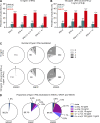
Figure 1.
Auto-Abs neutralizing type I IFNs in individuals infected with WNV. (A) Age and sex distribution of individuals in the WNVIC, WNVF, and WNVD groups. (B) Detection of auto-Abs against IFN-α2, IFN-β, and IFN-ω by ELISA. An OD > 0.5 (dotted line) was considered to correspond to positive samples based on the signal typically observed for serum/plasma from healthy donors. Each sample was tested once. (C) Luciferase-based neutralization assay to detect auto-Abs neutralizing 10 ng/ml IFN-α2, IFN-ω, or IFN-β (left panel) and 100 pg/ml IFN-α2 or IFN-ω, or 1 ng/ml IFN-β (right panel). Plasma samples from WNVIC (black), patients with WNVF (dark cyan), and patients with WNVD (red) were diluted 1:10. HEK293T cells were transfected with (1) a plasmid containing the firefly luciferase gene under the control of an IFN-sensitive response element (ISRE)-containing promotor and (2) a plasmid containing the Renilla luciferase gene. The cells were then treated with type I IFNs, and relative luciferase activity (RLA) was calculated by normalizing firefly luciferase activity against Renilla luciferase activity. An RLA <15% of the value for the mock treatment was considered to correspond to neutralizing activity (dotted line; Bastard et al., 2021a). Each sample was tested once.
Figure S1.
Figure S1.
Detailed demographic characteristics of the individuals enrolled in the study. (A) Number of individuals of each WNV group enrolled at each center. (B) Age distribution of the individuals enrolled, by the WNV group. (C) Age distribution of the individuals enrolled, by the center. (D) Sex distribution of the individuals enrolled, by the WNV group. (E) Sex distribution of the individuals enrolled, by the center. WNND: WNV neuroinvasive disease, a subgroup of WNVD.
Figure S2.
Figure S2.
Detection of auto-Abs against type I IFNs by ELISA and Gyros. (A) Detection, by ELISA, of auto-Abs against IFN-α2, IFN-β, and IFN-ω for the WNVD group and the two subgroups of WNVD patients: non-neuroinvasive WNVD and neuroinvasive WNVD (WNND). (B) Detection, by ELISA, of auto-Abs against IFN-α2, IFN-β, and IFN-ω in subgroups of WNND patients: WNV encephalitis (WNE), WNV meningitis (WNM), acute flaccid paralysis (AFP), and unspecified neurological syndrome (UNS). (C) Number of auto-Abs detected by ELISA for the WNVIC, WNVF, and WNVD groups. (D) Detection, by Gyros, of auto-Abs against IFN-α2 for the WNVIC, WNVF, and WNVD groups and for patients hospitalized with WNND and without neuroinvasive disease, the two main subgroups of WNVD. (E) Correlation between the detection of auto-Abs against IFN-α2 by ELISA and by Gyros. In all the experiments each sample was tested once.
Figure 2.
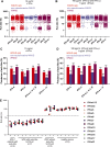
Figure 2.
Proportions of patients with auto-Abs neutralizing type I IFNs. (A and B) Proportions of individuals with auto-Abs neutralizing type I IFNs at a concentration of 10 ng/ml (A) or 100 pg/ml (B) in the three groups of WNV-infected individuals (WNVIC, WNVF, and WNVD), as determined with the luciferase-based neutralization assay. IFN-α, auto-Abs neutralizing IFN-α2 (regardless of their effects on other IFNs); IFN-ω, auto-Abs neutralizing IFN-ω (regardless of their effects on other IFNs); IFN-α ± ω ± β, auto-Abs neutralizing IFN-α2 and/or IFN-ω and/or IFN-β; IFN-α + ω, auto-Abs neutralizing both IFN-α2 and IFN-ω; *P < 0.05, **P < 0.01, ***P < 0.001, ****P < 0.0001. (C) The number of type I IFNs neutralized in the three groups of WNV-infected individuals (WNVIC, WNVF, and WNVD) as determined with the luciferase-based neutralization assay. (D) Proportion of type I IFNs neutralized in the three groups of WNV-infected individuals (WNVIC, WNVF, and WNVD) according to the nature and combination of auto-Abs.
Figure S3.
Figure S3.
Detection of auto-Abs neutralizing type I IFNs in the WNVD subgroups and neutralization of 12 IFN-α subtypes. (A and B) Luciferase-based neutralization assays to detect auto-Abs neutralizing 10 ng/ml IFN-α2, IFN-ω, or IFN-β (A) and 100 pg/ml of IFN-α2 or IFN-ω, or 1 ng/ml of IFN-β (B) in the WNVD group and the two WNVD subgroups of patients hospitalized with neuroinvasive WNVD (WNND) and without neuroinvasive disease. Values below 15% are considered to be associated with the presence of neutralizing auto-Abs. (C and D) Proportions of individuals with auto-Abs neutralizing type I IFNs at a concentration of 10 ng/ml (C) or 100 pg/ml for IFN-α2 or IFN-ω, or 1 ng/ml for IFN-β (D) in the WNVD group and the two WNVD subgroups of patients hospitalized with WNND and without neuroinvasive disease; ****P < 10−4. (E) Neutralization of different subtypes of IFN-α by serum or plasma from individuals with auto-Abs neutralizing IFN-α2. Patients with autoimmune polyendocrine syndrome type I (APS-1), individuals with WNVD without auto-Abs, and healthy controls are shown for comparison. Each sample was tested once.
Figure S4.
Figure S4.
Correlation between the results of luciferase-based neutralization assays with the glycosylated and non-glycosylated IFNs and between the results of neutralization assays and ELISA for detecting type I IFNs. (A–D) Correlation between neutralization of 10 ng/ml non-glycosylated IFN-α2 and 1 ng/ml glycosylated IFN-α2 (A), neutralization of 10 ng/ml non-glycosylated IFN-ω and 1 ng/ml glycosylated IFN-ω (B), neutralization of 100 pg/ml non-glycosylated IFN-α2 and 1 ng/ml glycosylated IFN-α2 (C), neutralization of 100 pg/ml non-glycosylated IFN-ω and 1 ng/ml glycosylated IFN-ω (D). (E) Detection of auto-Abs against IFN-α2 by ELISA versus neutralizing capacity for a concentration of 10 ng/ml. (F) Detection of auto-Abs against IFN-α2 by ELISA versus neutralizing capacity for a concentration of 100 pg/ml. (G) Detection of auto-Abs against IFN-ω by ELISA versus neutralizing capacity for a concentration of 10 ng/ml. (H) Detection of auto-Abs against IFN-ω by ELISA versus neutralizing capacity for a concentration of 100 pg/ml. (I) Detection of auto-Abs against IFN-β by ELISA versus neutralizing capacity for a concentration of 10 ng/ml. (J) Detection of auto-Abs against IFN-β by ELISA versus neutralizing capacity for a concentration of 1 g/ml. The relative luciferase activity (RLA) was calculated by normalizing firefly luciferase activity against Renilla luciferase activity. An RLA <15% of the value for the mock treatment was considered to correspond to neutralizing activity.
Figure 3.
Figure 3.
Proportions of patients with auto-Abs neutralizing type I IFNs by sex, age, and enrolling center. (A) Prevalence of individuals with auto-Abs neutralizing at least one type I IFN at a concentration of 100 pg/ml (IFN-α2, IFN-ω) or 1 ng/ml (IFN-β) in the three groups of individuals infected with WNV (WNVIC, WNVF, WNVD) by sex. (B) Prevalence of individuals with auto-Abs neutralizing at least one type I IFN at a concentration of 100 pg/ml (IFN-α2, IFN-ω) or 1 ng/ml (IFN-β) in the three groups of WNV-infected individuals (WNVIC, WNVF, WNVD) by age class. ns: non-significant; *P < 0.05, **P < 0.01. (C and D) Number (C) and proportion (D) of patients with auto-Abs neutralizing at least one type I IFN by enrolling center.
Figure 4.
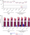
Figure 4.
Enrichment of the WNVF and WNVD groups in auto-Ab-positive individuals relative to the general population. (A) OR for the presence of auto-Abs in WNVIC relative to the general population, with adjustment for age and sex by Firth’s bias-corrected logistic regression. Firth’s correction can be used to estimate non-zero ORs and finite CIs despite the absence of auto-Ab carriers for some IFN combinations. The horizontal bars indicate the upper and lower limits of the 95% CI. (B) OR for the presence of auto-Abs in individuals with WNVF or WNVD relative to the general population, with adjustment for age and sex by logistic regression. The horizontal bars indicate the upper and lower limits of the 95% CI. (C) OR for the presence of auto-Abs in individuals with WNVD relative to the general population by age group, with adjustment for age and sex by logistic regression. ORs were calculated separately for patients with WNVD aged ≤65 and >65 yr. The horizontal bars indicate the upper and lower limits of the 95% CI. IFN-α, auto-Abs neutralizing IFN-α2 (regardless of their effects on other IFNs); IFN-ω, auto-Abs neutralizing IFN-ω (regardless of their effects on other IFNs); IFN-α ± ω ± β, auto-Abs neutralizing IFN-α2 and/or IFN-ω and/or IFN-β; IFN-α + ω, auto-Abs neutralizing both IFN-α2 and IFN-ω. ns: non-significant; ****P < 10−4.
Figure S5.
Figure S5.
Enrichment of the WNVD group and the two WNVD subgroups (with and without neuroinvasive-disease) in auto-Ab-positive individuals relative to the general population. (A) OR for the presence of auto-Abs in the WNVD group and the two WNVD subgroups (WNVD without evidence of neuroinvasive disease, and WNV neuroinvasive disease; WNND) relative to the general population, with adjustment for age and sex by Firth’s bias-corrected logistic regression. Firth’s correction can be used to estimate non-zero ORs and finite confidence intervals despite the absence of auto-Ab carriers for some IFN combinations. The horizontal bars indicate the upper and lower limits of the 95% CI. (B) Prevalence of auto-Abs against type I IFNs in the WNVIC, WNVF, and WNVD groups and the two WNVD subgroups (WNVD without evidence of neuroinvasive disease, and WNND) relative to the general population. IFN-α, auto-Abs neutralizing IFN-α2 (regardless of their effects on other IFNs); IFN-ω, auto-Abs neutralizing IFN-ω (regardless of their effects on other IFNs); IFN-α ± ω ± β, auto-Abs neutralizing IFN-α2 and/or IFN-ω and/or IFN-β; IFN-α + ω, auto-Abs neutralizing both IFN-α2 and IFN-ω. ns: non-significant. ****P < 10−4.
Figure 5.
Figure 5.
Testing of auto-Abs against type I IFNs in longitudinal samples. (A–C) Auto-Abs neutralizing IFN-α2 (A), IFN-ω (B), and IFN-β (C), as determined with the luciferase-based neutralization assay, at different time points after infection. Each sample was tested once with an IFN concentration of 10 ng/ml. The different symbols correspond to different patients testing negative or positive for auto-Abs neutralizing type I IFNs. d: day(s); w: week(s); m: month(s).
Figure 6.
Figure 6.
Auto-Abs in the CSF of patients with WNV neuroinvasive disease. (A–F) Luciferase-based neutralization assay to detect auto-Abs neutralizing IFN-α2 (A and B for the neutralization of 10 ng/ml and 100 pg/ml, respectively), IFN-β (C and D for the neutralization of 10 ng/ml and 100 pg/ml, respectively), and IFN-ω (E and F for the neutralization of 10 ng/ml and 100 pg/ml, respectively) in the CSF of patients with WNV neuroinvasive disease (y axis). The neutralizing activity of the patient’s serum or plasma is reported on the x axis. CSF samples were diluted 1:10. HEK293T cells were transfected with (1) a plasmid containing the firefly luciferase gene under the control of an ISRE-containing promotor and (2) a plasmid containing the Renilla luciferase gene. The cells were then treated with type I IFNs, and relative luciferase activity (RLA) was calculated by normalizing firefly luciferase activity against Renilla luciferase activity. An RLA <15% of the value for the mock treatment was considered to correspond to the neutralizing activity of the CSF (dotted line). Each sample was tested once.
Figure 7.
Figure 7.
WNV infection and IFN treatment in Vero-E6 cells. (A) WNV infection rate, as assessed based on the virus-induced CPE 48 h after infection, in Vero cells treated with various concentrations of IFN-α2 in the presence of plasma from patients with WNVD and neutralizing auto-Abs against IFN-α2 (n = 14), or plasma from patients with WNVD without auto-Abs neutralizing type I IFNs (n = 5). All the experiments involving the infection of Vero cells with WNV and all the WNV titration experiments were performed in triplicate. (B) Enhanced WNV replication, despite the presence of IFN-α2, in the presence of plasma from patients with auto-Abs against IFN-α2. WNV replication, assessed 48 h after infection, in Vero cells treated with IFN-α2 in the presence of plasma from patients with WNVD and neutralizing auto-Abs against IFN-α2 (n = 5) or plasma from patients with WNVD without auto-Abs neutralizing type I IFNs (n = 2) at different dilutions, or a commercial anti-IFN-α2 monoclonal antibody. All the experiments involving the infection of Vero cells with WNV and WNV titration experiments were performed in triplicate.

References

    1. Alotaibi, F., Alharbi N.K., Rosen L.B., Asiri A.Y., Assiri A.M., Balkhy H.H., Al Jeraisy M., Mandourah Y., AlJohani S., Al Harbi S., et al. . 2023. Type I interferon autoantibodies in hospitalized patients with Middle East respiratory syndrome and association with outcomes and treatment effect of interferon beta-1b in MIRACLE clinical trial. Influenza Other Respir. Viruses. 17:e13116. 10.1111/irv.13116 - DOI - PMC - PubMed
    1. Barzon, L., Pacenti M., Montarsi F., Fornasiero D., Gobbo F., Quaranta E., Monne I., Fusaro A., Volpe A., Sinigaglia A., et al. . 2022. Rapid spread of a new West Nile virus lineage 1 associated with increased risk of neuroinvasive disease during a large outbreak in northern Italy, 2022: One Health analysis. J. Trav. Med. taac125. 10.1093/jtm/taac125 - DOI - PMC - PubMed
    1. Bastard, P., Gervais A., Le Voyer T., Rosain J., Philippot Q., Manry J., Michailidis E., Hoffmann H.-H., Eto S., Garcia-Prat M., et al. . 2021a. Autoantibodies neutralizing type I IFNs are present in ∼4% of uninfected individuals over 70 years old and account for ∼20% of COVID-19 deaths. Sci. Immunol. 6:eabl4340. 10.1126/sciimmunol.abl4340 - DOI - PMC - PubMed
    1. Bastard, P., Michailidis E., Hoffmann H.-H., Chbihi M., Le Voyer T., Rosain J., Philippot Q., Seeleuthner Y., Gervais A., Materna M., et al. . 2021b. Auto-antibodies to type I IFNs can underlie adverse reactions to yellow fever live attenuated vaccine. J. Exp. Med. 218:e20202486. 10.1084/jem.20202486 - DOI - PMC - PubMed
    1. Bastard, P., Rosen L.B., Zhang Q., Michailidis E., Hoffmann H.-H., Zhang Y., Dorgham K., Philippot Q., Rosain J., Béziat V., et al. . 2020. Autoantibodies against type I IFNs in patients with life-threatening COVID-19. Science. 370:eabd4585. 10.1126/science.abd4585 - DOI - PMC - PubMed

Publication types