

Pleural Empyema Caused by Streptococcus intermedius and Fusobacterium nucleatum: A Distinct Entity of Pleural Infections
- PMID: 37348872
- PMCID: PMC10654859
- DOI: 10.1093/cid/ciad378
Pleural Empyema Caused by Streptococcus intermedius and Fusobacterium nucleatum: A Distinct Entity of Pleural Infections
Abstract
Background: Many community-acquired pleural infections are caused by facultative and anaerobic bacteria from the human oral microbiota. The epidemiology, clinical characteristics, pathogenesis, and etiology of such infections are little studied. The aim of the present prospective multicenter cohort study was to provide a thorough microbiological and clinical characterization of such oral-type pleural infections and to improve our understanding of the underlying etiology and associated risk factors.
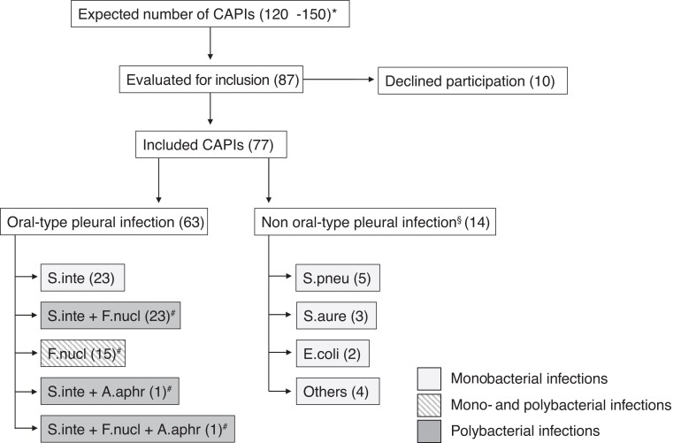
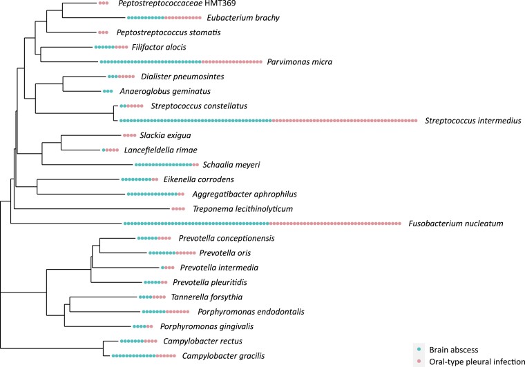
Methods: Over a 2-year period, we included 77 patients with community-acquired pleural infection, whereof 63 (82%) represented oral-type pleural infections. Clinical and anamnestic data were systematically collected, and patients were offered a dental assessment by an oral surgeon. Microbial characterizations were done using next-generation sequencing. Obtained bacterial profiles were compared with microbiology data from previous investigations on odontogenic infections, bacteremia after extraction of infected teeth, and community-acquired brain abscesses.
Results: From the oral-type pleural infections, we made 267 bacterial identifications representing 89 different species. Streptococcus intermedius and/or Fusobacterium nucleatum were identified as a dominant component in all infections. We found a high prevalence of dental infections among patients with oral-type pleural infection and demonstrate substantial similarities between the microbiology of such pleural infections and that of odontogenic infections, odontogenic bacteremia, and community-acquired brain abscesses.
Conclusions: Oral-type pleural infection is the most common type of community-acquired pleural infection. Current evidence supports hematogenous seeding of bacteria from a dental focus as the most important underlying etiology. Streptococcus intermedius and Fusobacterium nucleatum most likely represent key pathogens necessary for establishing the infection.
Keywords: Fusobacterium nucleatum; Streptococcus intermedius; 16S rRNA; next-generation sequencing; pleural infection.
© The Author(s) 2023. Published by Oxford University Press on behalf of Infectious Diseases Society of America.
Conflict of interest statement
Potential conflicts of interest. T. M. E. reports grants from GlaxoSmithKline and payment/honoraria from Boehringer Ingelheim, AstraZeneca, and SOS International. T. M. E. also reports participation on a Data and Safety Monitoring Board (DSMB) for a nutrition study, Helse Sør-Øst, Lillehammer. T. M. E. is leader of the Committee for the Education of Specialists in Pulmonary Medicine, Norway. Ø. K. is a co-founder and shareholder of Pathogenomix. T. M. L. is a member of the Professional Advisory Board, GenMark Diagnostics (ePlex), and has received honoraria for time spent. T. M. L. is also president of the UEMS (European Union of Medical Specialists) Section of Medical Microbiology and a member of the Professional Affairs Subcommittee, ESCMID (European Society of Clinical Microbiology and Infectious Disease) (both unpaid). S. L. reports payment/honoraria from the Norwegian Dental Association and the Norwegian Encyclopedia. S. L. also reports payment for expert testimony from the National Office for Health Service Appeals, the Norwegian System of Patient Injury Compensation and the Norwegian Courts of Justice. B. B. report personal payment for an idiopathic pulmonary fibrosis presentation from Boehringer Ingelheim. All other authors report no potential conflicts. All authors have submitted the ICMJE Form for Disclosure of Potential Conflicts of Interest. Conflicts that the editors consider relevant to the content of the manuscript have been disclosed.
Figures
References
-
- Corcoran JP, Wrightson JM, Belcher E, DeCamp MM, Feller-Kopman D, Rahman NM. Pleural infection: past, present, and future directions. Lancet Respir Med 2015; 3:563–77. - PubMed
-
- Dyrhovden R, Nygaard RM, Patel R, Ulvestad E, Kommedal O. The bacterial aetiology of pleural empyema. A descriptive and comparative metagenomic study. Clin Microbiol Infect 2019; 25:981–6. - PubMed
-
- Hassan M, Cargill T, Harriss E, et al. The microbiology of pleural infection in adults: a systematic review. Eur Respir J 2019; 54:1900542. - PubMed
-
- Maskell NA, Batt S, Hedley EL, Davies CW, Gillespie SH, Davies RJ. The bacteriology of pleural infection by genetic and standard methods and its mortality significance. Am J Resp Crit Care 2006; 174:817–23. - PubMed
Publication types
MeSH terms
LinkOut - more resources
Full Text Sources
Medical
