. 2023 Jul;8(7):1267-1279.
doi: 10.1038/s41564-023-01401-2. Epub 2023 Jun 22.

Bdellovibrio predation cycle characterized at nanometre-scale resolution with cryo-electron tomography

Affiliations

Bdellovibrio predation cycle characterized at nanometre-scale resolution with cryo-electron tomography

Mohammed Kaplan et al. Nat Microbiol. 2023 Jul.

Abstract

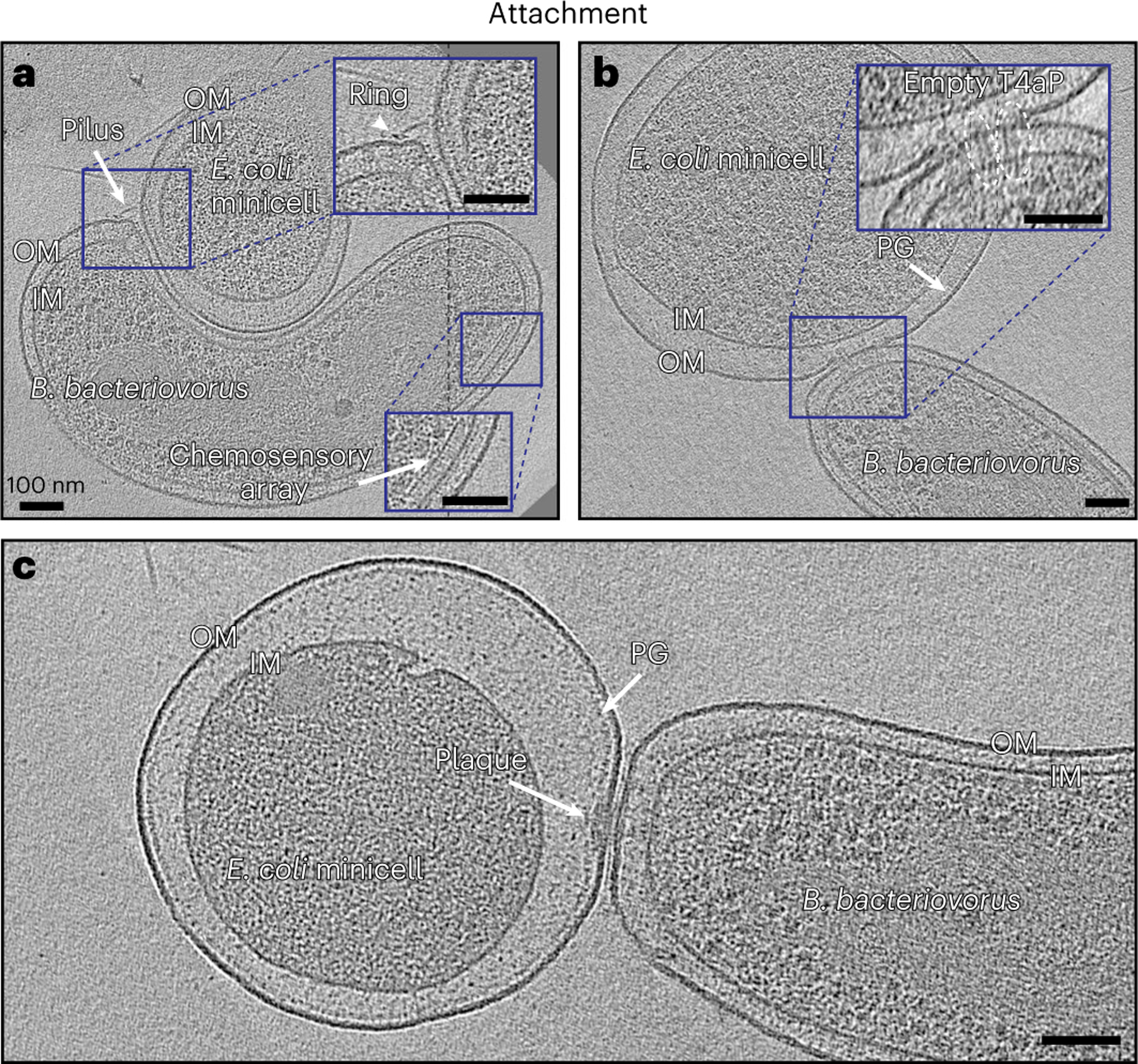
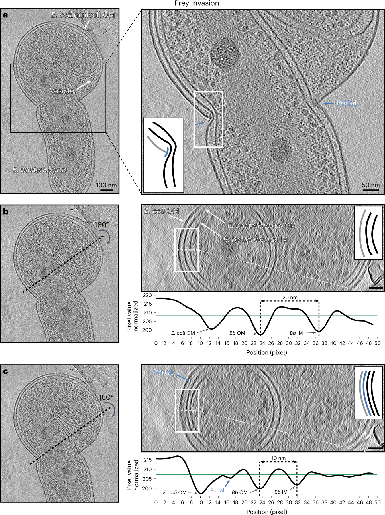
Bdellovibrio bacteriovorus is a microbial predator that offers promise as a living antibiotic for its ability to kill Gram-negative bacteria, including human pathogens. Even after six decades of study, fundamental details of its predation cycle remain mysterious. Here we used cryo-electron tomography to comprehensively image the lifecycle of B. bacteriovorus at nanometre-scale resolution. With high-resolution images of predation in a native (hydrated, unstained) state, we discover several surprising features of the process, including macromolecular complexes involved in prey attachment/invasion and a flexible portal structure lining a hole in the prey peptidoglycan that tightly seals the prey outer membrane around the predator during entry. Unexpectedly, we find that B. bacteriovorus does not shed its flagellum during invasion, but rather resorbs it into its periplasm for degradation. Finally, following growth and division in the bdelloplast, we observe a transient and extensive ribosomal lattice on the condensed B. bacteriovorus nucleoid.


Conflict of interest statement

Competing interests

The authors declare no competing interests.

Figures

Fig. 1 |
Fig. 1 |. Anatomy of attack-phase B. bacteriovorus.
a, A slice through an electron cryo-tomogram of an attack-phase B. bacteriovorus cell, with enlarged views of the flagellated (red) and biting (blue) poles. White rectangles highlight the flagellar motor (top) and non-piliated T4aP basal body (bottom), and the white ellipse highlights a rose-like complex. Question mark points to a cross-section through a periplasmic filamentous structure, and white arrow to a type IVa pilus. Scale bars, 50 nm. bd, Central slices through subtomogram averages of the B. bacteriovorus flagellar motor (b, 79 particles), rose-like complex (c, 132 particles) and non-piliated T4aP basal body (d, 335 particles). White arrows in d point to the extracellular ring of the T4aP basal body. Scale bars, 20 nm. e,f, Central slices through subtomogram averages of non-piliated T4aP basal bodies with (left, white arrow) and without (right) the lower periplasmic ring. These particles were classified using the built-in PCA and k-means clustering functions of the PEET package (for more details, see the legend of Supplementary Fig. 6). Dashed black lines indicate a composite of slices through the tomogram at different z-heights (a), and a composite of subtomogram averages aligned on the outer and inner membrane, respectively (c). IM, inner membrane.
Fig. 2 |
Fig. 2 |. Attachment of B. bacteriovorus to prey.
a, A slice through an electron cryo-tomogram showing a B. bacteriovorus cell attached to a prey (E. coli minicell) via T4aP. IM, inner membrane. b, A slice through an electron cryo-tomogram of B. bacteriovorus attached to prey (E. coli minicell) showing non-piliated T4aP basal bodies (white ellipses) penetrating to the prey’s PG layer. c, A slice through an electron cryo-tomogram of B. bacteriovorus attached to prey with a polar attachment plaque. Scale bars, 100 nm.
Fig. 3 |
Fig. 3 |. B. bacteriovorus flagellar absorption.
a, A slice through an electron cryo-tomogram showing a B. bacteriovorus cell attached to a prey via an attachment plaque with its flagellum resorbing into the periplasm. Enlargements in the red-boxed areas highlight different parts of the absorbed flagellum. Scale bars, 50 nm (main panel) and 20 nm (enlargements). b, A 3D segmentation of a and an enlarged view illustrating different parts of the absorbed flagellum.
Fig. 4 |
Fig. 4 |. B. bacteriovorus prey invasion.
a, A slice through an electron cryo-tomogram (left) and enlarged view (right) showing a non-productive invasion by a B. bacteriovorus of an E. coli minicell. Blue arrows and inset schematic highlight the portal. b, Right (top): cross-section through the yz plane of the tomogram shown in a along the black dotted line indicated on the left. Note that this slice does not include the portal. Right (bottom): average density profile taken along the white dashed line (inside the white rectangle) in the top panel. The distance between the predator’s inner membrane (IM) and OM is indicated (20 nm). c, Similar to b but for a yz slice where the portal is visible. The distance between the predator’s IM and OM is indicated (10 nm). The schematics in the right panels of ac represent the white-boxed areas in the corresponding slices, with the portal shown in blue, the prey OM in grey and the predator IM and OM in black. Scale bars, 100 nm (left panel of a) and 50 nm (other panels).
Fig. 5 |
Fig. 5 |. Anatomy of the bdelloplast.
ac, Slices (at different z-levels) through an electron cryo-tomogram of a V. cholerae bdelloplast containing two B. bacteriovorus after predator division. d, A 3D segmentation of the bdelloplast shown in ac. e, Enlargement (left) and 3D segmentation (right) of the white-boxed area in a, highlighting the features of the seal. f, Enlargement (left) and 3D segmentation (right) of the white-boxed area in c, highlighting the prey flagellar relic. Scale bars, 100 nm (ac) and 50 nm (e and f).
Fig. 6 |
Fig. 6 |. Ribosomal nucleoid lattice in the end-stage bdelloplast.
a,b, Slices (at different z-levels) through an electron cryo-tomogram of an end-stage E. coli minicell bdelloplast containing two B. bacteriovorus cells after predator division, highlighting the hexagonal arrangement of ribosomes around the nucleoids. c,d, Rotated views of a 3D segmentation of the nucleoids and ribosomes of the cryo-tomogram shown in a and b. Scale bars, 50 nm. e, Distances of individual ribosomes from the nucleoid surface measured in the 3D segmentations of the cryo-tomogram shown in a and b (‘experiment’, solid line), compared with a simulation of randomly distributed 20-nm-wide spheres packed in the same segmented volume (‘random’, dashed line). See also Supplementary Movie 21.

References

    1. Pérez J, Moraleda-Muñoz A, Marcos-Torres FJ & Muñoz-Dorado J Bacterial predation: 75 years and counting! Environ. Microbiol. 18, 766–779 (2016). - PubMed
    1. Stolp H & Starr MP Bdellovibrio bacteriovorus gen. et sp. n., a predatory, ectoparasitic, and bacteriolytic microorganism. Antonie van Leeuwenhoek 29, 217–248 (1963). - PubMed
    1. Stolp H & Petzold H Untersuchungen über einen obligat parasitischen Mikroorganismus mit lytischer Aktivität für Pseudomonas-Bakterien. J. Phytopathol. 45, 364–390 (1962).
    1. Sockett RE Predatory lifestyle of Bdellovibrio bacteriovorus. Annu. Rev. Microbiol. 63, 523–539 (2009). - PubMed
    1. Laloux G Shedding light on the cell biology of the predatory bacterium Bdellovibrio bacteriovorus. Front. Microbiol. 10, 3136 (2020). - PMC - PubMed

Publication types