. 2023 Jun 7:14:1042176.
doi: 10.3389/fendo.2023.1042176. eCollection 2023.

Mating modifies the expression of crucial oxidative-reductive transcripts in the pig oviductal sperm reservoir: is the female ensuring sperm survival?

Affiliations

Mating modifies the expression of crucial oxidative-reductive transcripts in the pig oviductal sperm reservoir: is the female ensuring sperm survival?

Manuel Álvarez-Rodríguez et al. Front Endocrinol (Lausanne). .

Abstract

Background: Mating induces large changes in the female genital tract, warranting female homeostasis and immune preparation for pregnancy, including the preservation of crucial oxidative status among its pathways. Being highly susceptible to oxidative stress, sperm survival and preserved function depend on the seminal plasma, a protection that is removed during sperm handling but also after mating when spermatozoa enter the oviduct. Therefore, it is pertinent to consider that the female sperm reservoir takes up this protection, providing a suitable environment for sperm viability. These aspects have not been explored despite the increasing strategies in modulating the female status through diet control and nutritional supplementation.

Aims: To test the hypothesis that mating modifies the expression of crucial oxidative-reductive transcripts across the entire pig female genital tract (cervix to infundibulum) and, particularly in the sperm reservoir at the utero-tubal junction, before ovulation, a period dominated by estrogen stimulation of ovarian as well as of seminal origin.

Methods: The differential expression of estrogen (ER) and progesterone (PR) receptors and of 59 oxidative-reductive transcripts were studied using a species-specific microarray platform, in specific segments of the peri-ovulatory sow reproductive tract in response to mating.

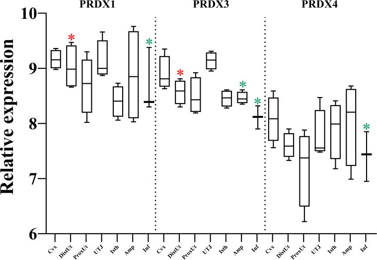
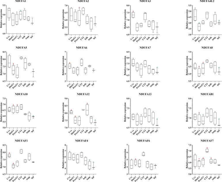
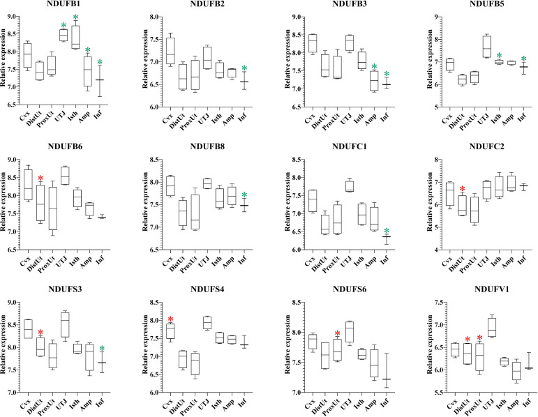
Results: Mating induced changes along the entire tract, with a conspicuous downregulation of both ER and PR and an upregulation of superoxide dismutase 1 (SOD1), glutaredoxin (GLRX3), and peroxiredoxin 1 and 3 (PRDX1, PRDX3), among other NADH Dehydrogenase Ubiquinone Flavoproteins, in the distal uterus segment. These changes perhaps helped prevent oxidative stress in the area adjacent to the sperm reservoir at the utero-tubal junction. Concomitantly, there were a downregulation of catalase (CAT) and NADH dehydrogenase (ubiquinone) oxidoreductases 1 beta subcomplex, subunit 1 (NDUFB1) in the utero-tubal junction alongside an overall downregulation of CAT, SOD1, and PRDX3 in the ampullar and infundibulum segments.

Conclusions: Natural mating is an inducer of changes in the expression of female genes commanding antioxidant enzymes relevant for sperm survival during sperm transport, under predominant estrogen influence through the bloodstream and semen. The findings could contribute to the design of new therapeutics for the female to improve oxidative-reductive balance.

Keywords: ROS; antioxidant; mating; periovulatory; porcine.


Conflict of interest statement

The authors declare that the research was conducted in the absence of any commercial or financial relationships that could be construed as a potential conflict of interest.

Figures

Figure 1
Figure 1
Summary of significant differential expression of progesterone and estrogen receptors. Box-plot representation of the log2 signal intensity of the selected transcripts by tissue, Cvx, cervix; DistUt, distal uterus; ProxUt, proximal uterus; UTJ, utero-tubal junction; Isth, isthmus; Amp, ampulla; and Inf, infundibulum. The green color “*” represents a relative decrease relative to the negative control (p < 0.05). PR, progesterone receptor; and ER, estrogen receptor.
Figure 2
Figure 2
Volcano plot depicting a summary of the differential expression analyses of transcripts in the reproductive internal genital tract segments (cervix, distal uterus, proximal uterus, utero-tubal junction, isthmus, ampulla, and infundibulum), 24 h after natural mating vs unmated, Sham (infusion of BTS extender) controls. The x-axis shows the log2 fold-changes in expression and the y-axis the statistical significance (-log10 p-value). This figure depicts p < 0.05 and p < 0.01 relative to the control.
Figure 3
Figure 3
PANTHER classification showing differential gene expression (DEG) of the 59 oxidative-reductive (OR) genes. The bar plots represent different percentages of DEGs classified according to (A) molecular function, (B) protein class, (C) cellular component, (D) biological process, and (E) pathway.
Figure 4
Figure 4
Summary of significant differential expression of peroxiredoxins (PRDX). Box-plot representation of the log2 signal intensity of the selected transcripts by tissue, Cvx, cervix; DistUt, distal uterus; ProxUt, proximal uterus; UTJ, utero-tubal junction; Isth, isthmus; Amp, ampulla; and Inf, infundibulum. The red color “*” represents a relative increase relative to the negative control (p < 0.05), whereas the green color “*” represents a relative decrease relative to the negative control (p < 0.05). PRDX1, peroxiredoxin 1; PRDX1, peroxiredoxin 3, and PRDX4, peroxiredoxin 4.
Figure 5
Figure 5
Summary of significant differential expression of superoxide dismutase (SOD); glutaredoxins (GLRX); and catalase (CAT). Box-plot representation of the log2 signal intensity of the selected transcripts by tissue, Cvx, cervix; DistUt, distal uterus; ProxUt, proximal uterus; UTJ, utero-tubal junction; Isth, isthmus; Amp, ampulla; and Inf, infundibulum. (A) SOD1, superoxide dismutase 1; cytoplasm; and SOD2, mitochondrial. (B) CAT, catalase. (C) GLRX1, glutaredoxin 1; GLRX2, glutaredoxin 2; and GLRX3, glutaredoxin 3. (D) ATOX1, antioxidant protein 1. The red color “*” represents a relative increase relative to the negative control (p < 0.05); whereas the green color “*” represents a relative decrease relative to the negative control (p < 0.05).
Figure 6
Figure 6
Summary of significant differential expression of caspases (CASP). Box-plot representation of the log2 signal intensity of the selected transcripts by tissue, Cvx, cervix; DistUt, distal uterus; ProxUt, proximal uterus; UTJ, utero-tubal junction; Isth, isthmus; Amp, ampulla; and Inf, infundibulum. The red color “*” represents a relative increase relative to the negative control (p < 0.05), whereas the green color “*” represents a relative decrease relative to the negative control (p < 0.05). CASP1, caspase 1; CASP2, caspase 2; CASP3, caspase 3; CASP8AP2, caspase 8 associated protein 2; CASP9, caspase 9; and CASP14, caspase 14.
Figure 7
Figure 7
Summary of significant differential expression of NADH dehydrogenase (ubiquinone) oxidoreductases (NDUF) Part 2. Box-plot representation of the log2 signal intensity of the selected transcripts by tissue, Cvx, cervix; DistUt, distal uterus; ProxUt, proximal uterus; UTJ, utero-tubal junction; Isth, isthmus; Amp, ampulla; and Inf, infundibulum. The red color “*” represents a relative increase relative to the negative control (p < 0.05), whereas the green color “*” represents a relative decrease relative to the negative control (p < 0.05). NDUFA1, NDUF 1 alpha subcomplex, subunit 1; NDUFA2, NDUF 1 alpha subcomplex, ubunit 2; NDUFA3, NDUF 1 alpha subcomplex, subunit 3; NDUFA4L2, 1 alpha subcomplex, 4-like 2; NDUFA5, NDUF 1 alpha subcomplex, subunit 5; NDUFA6, NDUF 1 alpha subcomplex, subunit 6; NDUFA7, NDUF 1 alpha subcomplex, subunit 7; NDUFA8, NDUF 1 alpha subcomplex, subunit 8; NDUFA10, NDUF 1 alpha subcomplex, subunit 10; NDUFA12, NDUF 1 alpha subcomplex, subunit 12; NDUFA13, NDUF 1 alpha subcomplex, subunit 13; NDUFAB1, NDUF 1 alpha subcomplex, subunit 5; NDUFAF1, NDUF 1 alpha subcomplex assembly factor 1; NDUFAF4, NDUF complex assembly factor 4; NDUFAF6, NDUF complex assembly factor 6; and NDUFAF7, NDUF complex assembly factor 7.
Figure 8
Figure 8
Summary of significant differential expression of NADH dehydrogenase (ubiquinone) oxidoreductases (NDUF) Part 2. Box-plot representation of the log2 signal intensity of the selected transcripts by tissue, Cvx, cervix; DistUt, distal uterus; ProxUt, proximal uterus; UTJ, utero-tubal junction; Isth, isthmus; Amp, ampulla; and Inf, infundibulum. The red color “*” represents a relative increase relative to the negative control (p < 0.05), whereas the green color “*” represents a relative decrease relative to the negative control (p < 0.05). NDUFB1, NDUF 1 beta subcomplex, subunit 1; NDUFB2, NDUF 1 beta subcomplex, ubunit 2; NDUFB3, NDUF 1 beta subcomplex, subunit 3; NDUFB5, 1 beta subcomplex, subunit 5; NDUFB6, NDUF 1 beta subcomplex, subunit 6; NDUBA8, NDUF 1 beta subcomplex, subunit 8; NDUFC1, NDUF 1 subunit C1; NDUFC2, NDUF 1 subunit C2; NDUFS3, NDUF iron-sulfur protein 3; NDUFS4, NDUF iron-sulfur protein 4; NDUFS6, NDUF iron-sulfur protein 6; and NDUFV1, NDUF flavoprotein 1.

Similar articles

Cited by

References

    1. Lu J, Huang J, Zhao S, Xu W, Chen Y, Li Y, et al. . FOXO1 is a critical switch molecule for autophagy and apoptosis of sow endometrial epithelial cells caused by oxidative stress. Oxid Med Cell Longev (2021) 2021:24. doi: 10.1155/2021/1172273 - DOI - PMC - PubMed
    1. Claus R. Physiological role of seminal components in the reproductivetract of the female pig. Biosci Proc (2020) 13:117–31. doi: 10.1530/biosciprocs.13.009 - DOI - PubMed
    1. Rodriguez-Martinez H, Petroni A, Einarsson S, Kindahl H. Concentrations of prostaglandin F2α in the pig oviductal fluid. Prostaglandins (1983) 25:413–24. doi: 10.1016/0090-6980(83)90045-X - DOI - PubMed
    1. Rodriguez-Martinez H, Nicander L, Viring S, Einarsson S, Larsson K. Ultrastructure of the uterotubal junction in preovulatory pigs. Anat Histol Embryol (1990) 19:16–36. doi: 10.1111/j.1439-0264.1990.tb00875.x - DOI - PubMed
    1. Alvarez-Rodriguez M, Martinez CA, Wright D, Rodriguez-Martinez H. Does the act of copulation per se, without considering seminal deposition, change the expression of genes in the porcine female genital tract? Int J Mol Sci (2020) 21:1–16. doi: 10.3390/ijms21155477 - DOI - PMC - PubMed

Publication types