. 2023 Aug;146(2):211-226.
doi: 10.1007/s00401-023-02598-6. Epub 2023 Jun 23.

Mutation ∆K281 in MAPT causes Pick's disease

Affiliations

Mutation ∆K281 in MAPT causes Pick's disease

Manuel Schweighauser et al. Acta Neuropathol. 2023 Aug.

Abstract

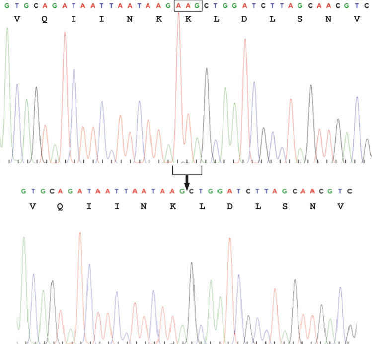
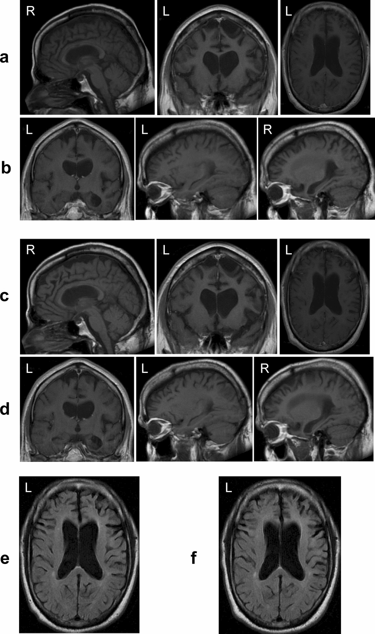
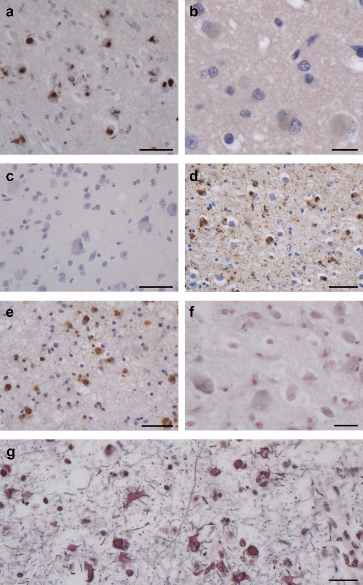
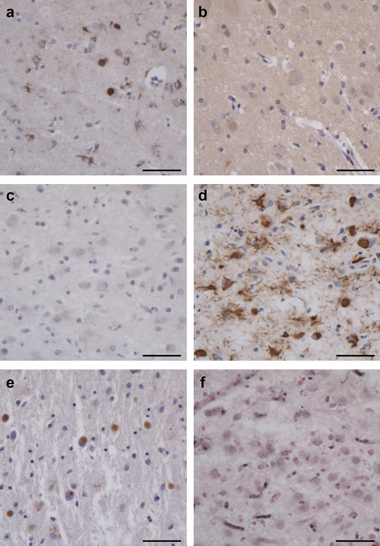
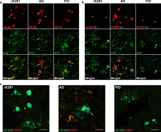
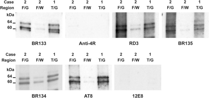
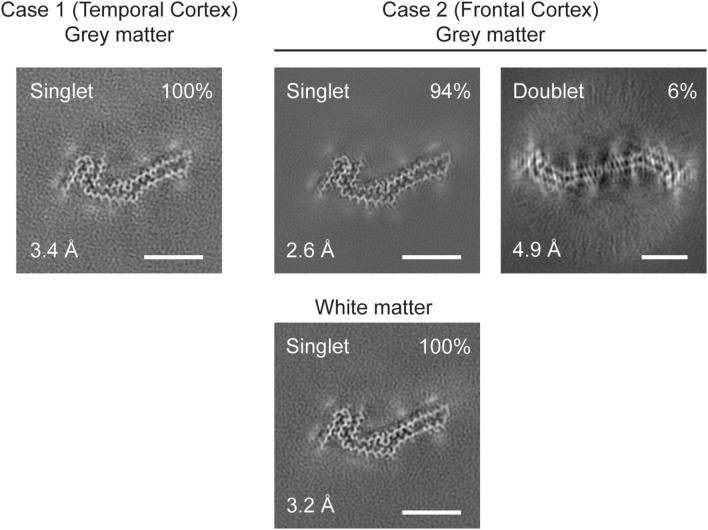
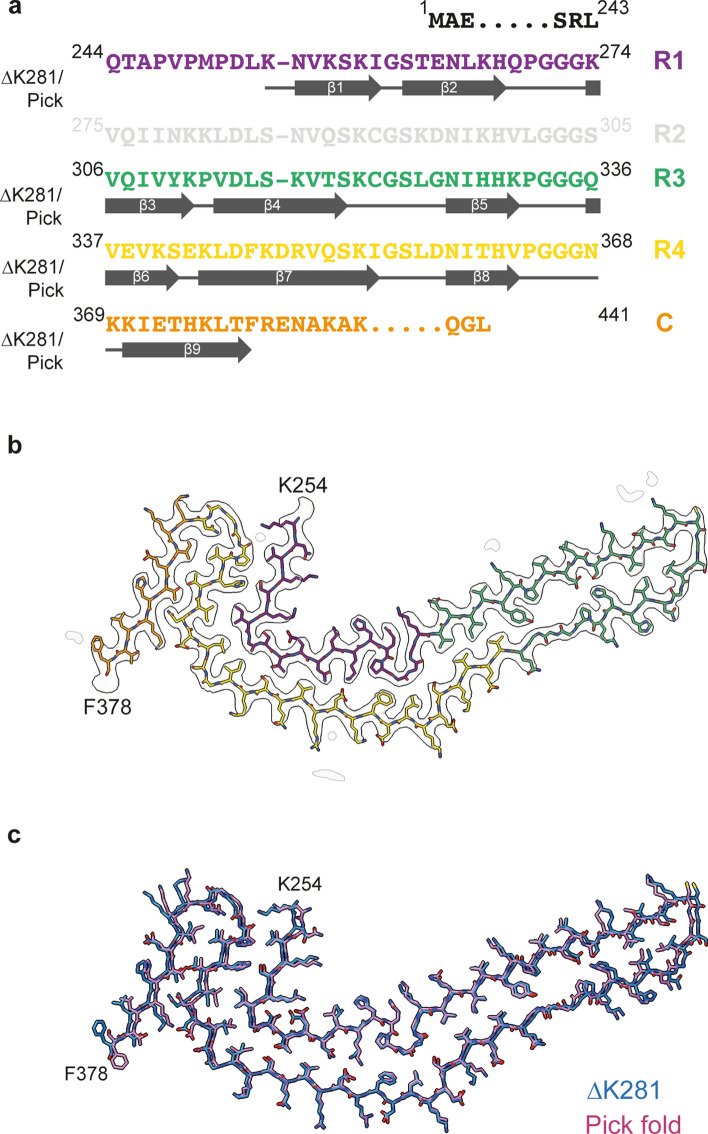
Two siblings with deletion mutation ∆K281 in MAPT developed frontotemporal dementia. At autopsy, numerous inclusions of hyperphosphorylated 3R Tau were present in neurons and glial cells of neocortex and some subcortical regions, including hippocampus, caudate/putamen and globus pallidus. The inclusions were argyrophilic with Bodian silver, but not with Gallyas-Braak silver. They were not labelled by an antibody specific for tau phosphorylated at S262 and/or S356. The inclusions were stained by luminescent conjugated oligothiophene HS-84, but not by bTVBT4. Electron cryo-microscopy revealed that the core of tau filaments was made of residues K254-F378 of 3R Tau and was indistinguishable from that of Pick's disease. We conclude that MAPT mutation ∆K281 causes Pick's disease.

Keywords: Electron cryo-microscopy; FTDP-17T; Luminescent conjugated oligothiophenes; MAPT mutation ∆K281; Pick’s disease; Silver staining; Tau.


Conflict of interest statement

The authors declare that they have no conflicts of interest.

Figures

Fig. 1
Fig. 1
Mutation ∆K281 in MAPT. A three-base deletion was present by DNA sequencing following PCR amplification of exon 10 of MAPT from case 1. Cloning of the MAPT exon 10 amplicon into pCR2.1, followed by sequencing, gave the wild-type sequence in one allele (top) and the deletion mutation in the other (bottom). Identical findings were obtained for case 2. In the wild-type sequence, two consecutive AAG codons encode K280 and K281. Deletion of one AAG codon does not allow one to decide which residue has been deleted. Current convention dictates to name the mutation after the most C-terminal amino acid. We therefore talk of ∆K281
Fig. 2
Fig. 2
Comparison of two T1-weighted MRI scans of the brain of case 1 with MAPT mutation ∆K281. (a, b, e), Scan obtained during the first visit, when case 1 was 50 years old; (c, d, f), Scan obtained 5 months later. (a, c, e), Sagittal, coronal and axial MR images show atrophy of the frontal lobe with knife-blade atrophy of the gyri [middle panel in (a) and (c)], atrophy of the corpus callosum and severe enlargement of the lateral ventricles. (b, d, f), Coronal and parasagittal MR images show atrophy of the frontal, parietal and temporal lobes, as well as severe enlargement of the lateral ventricles. The MR images from the second visit reveal progression of the atrophy; differences in the dimensions of the lateral ventricles between first and second scans are particularly noticeable
Fig. 3
Fig. 3
Staining of the frontal cortex from case 1 with MAPT mutation ∆K281. (a, d, e), Tau-positive inclusions in nerve cells and glial cells with antibodies RD3 (a), AT8 (d) and AT100 (e). (b, c), Tau-negative inclusions with antibodies anti-4R (b) and 12E8 (c). (f, g), Staining with Gallyas–Braak silver (f) and Bodian silver (g). Inclusions are not stained by Gallyas–Braak silver, but neuronal and glial inclusions are Bodian silver-positive. Scale bars: 50 µm (a, c, d, e), 20 µm (b) and 25 µm (f, g)
Fig. 4
Fig. 4
Staining of the frontal cortex from case 2 with MAPT mutation ∆K281. (a, d, e), Tau-positive inclusions in nerve cells and glial cells with antibodies RD3 (a), AT8 (d) and AT100 (e). (b, c), Tau-negative inclusions with antibodies anti-4R (b) and 12E8 (c). (f), Inclusions are not stained by Gallyas–Braak silver. Scale bars: 50 µm (af)
Fig. 5
Fig. 5
Labelling of Tau amyloid in frontal cortex from case 1 with MAPT mutation ∆K281 by HS-84, but not bTVBT4. Comparison with Alzheimer’s and Pick’s diseases. (a), Fluorescence images of frontal cortex sections from case 1 with MAPT mutation ∆K281, a case of Alzheimer’s disease (AD) and a case of Pick’s disease (PiD) labelled by HS-84 (red) and anti-tau antibody AT8 (green). HS-84 showed co-localisation with AT8 in neuronal (white arrowheads) and glial inclusions (yellow arrowheads) in case 1 (left panel) and in Pick’s disease (right panel). HS-84 showed co-localisation with AT8 in neurofibrillary tangles (white arrowhead) and neuropil threads (yellow arrowhead) in AD (middle panel). HS-84 also identified neuritic Aβ plaques in AD (white arrow). Scale bars, 20 µm. (b), Fluorescence images of frontal cortex sections from case 1 with MAPT mutation ∆K281, a case of AD and a case of Pick’s disease labelled by bTVBT4 (red) and anti-tau antibody AT8 (green). bTVBT4 did not label neuronal (white arrowhead) or glial tau inclusions (yellow arrowhead) in case 1 (left panel) or Pick’s disease (right panel). bTVBT4 labelled immunopositive neurofibrillary tangles (white arrowhead) and neuropil threads (yellow arrowhead) in AD (middle panel). Blue structures represent autofluorescent lipofuscin (LF). Scale bars, 20 µm. (c), Spectral images of frontal cortex sections from case 1 with MAPT mutation ∆K281 (left panel), a case of AD (middle panel) and a case of Pick’s disease (right panel) stained with HS-84 (green) and bTVBT4 (red). HS-84, but not bTVBT4, labelled inclusions (arrowheads) in case 1 (left panel) and Pick’s disease (right panel). Both HS-84 and bTVBT4 labelled Tau inclusions (arrowhead) in AD (middle panel). Only HS-84 identified neuritic Aβ plaques (arrow) in AD. Scale bars, 20 µm
Fig. 6
Fig. 6
Western blotting of sarkosyl-insoluble fractions from the temproal (T) and frontal (F) cortex of cases 1 and 2 with MAPT mutation ∆K281. Two major bands of 60 and 64 kDa and a minor band of 68 kDa were present in grey matter (G). Weaker bands of the same sizes were present in white matter from frontal cortex of case 2 (W). These bands were labelled by anti-Tau antibodies BR133, RD3, BR135, BR134 and AT8. They were not labelled by anti-4R or 12E8
Fig. 7
Fig. 7
Cryo-EM cross sections of Tau filaments from grey and white matter of frontal and temporal cortex of cases with MAPT mutation ∆K281. Cross-sections through the cryo-EM reconstructions, perpendicular to the helical axis and with a projected thickness of approximately one rung, are shown for grey matter of temporal cortex from case 1 and grey and white matter of frontal cortex from case 2 with MAPT mutation ∆K281. The resolution of the reconstructions and the percentages of each filament type are indicated. Singlet filaments predominated, with a small percentage of doublets in case 2. Scale bars, 5 nm
Fig. 8
Fig. 8
Tau filament fold from cases with MAPT mutation ∆K281. a Sequence alignment of the microtubule-binding repeats (R1-R4) of Tau with the observed 9 β-strand regions (arrows). b Sharpened high-resolution cryo-EM map of the ∆K281 singlet from grey matter of frontal cortex from case 2 with the atomic model overlaid. c Superposition of the backbone structures of the Tau filament fold from case 1 with mutation ∆K281 (blue) and the Pick fold (red)

References

    1. Åslund A, Sigurdson CJ, Klingstedt T, Grathwohl S, Bolmont T, Dickstein DL, et al. Novel pentameric thiophene derivatives for in vitro and in vivo optical imaging of a plethora of protein aggregates in cerebral amyloidoses. ACS Chem Biol. 2009;4:673–684. doi: 10.1021/cb900112v. - DOI - PMC - PubMed
    1. Barghorn S, Zheng-Fischhöfer Q, Ackmann M, Biernat J, von Bergen M, Mandelkow EM, et al. Structure, microtubule interactions, and paired helical filament aggregation by tau mutants of frontotemporal dementias. Biochemistry. 2000;39:11714–11721. doi: 10.1021/bi000850r. - DOI - PubMed
    1. Boeve BF, Tremont-Lukats IW, Waclawik AJ, Murrell JR, Hermann B, Jack CR, et al. Longitudinal characterization of two siblings with frontotemporal dementia and parkinsonism linked to chromosome 17 associated with the S305N tau mutation. Brain. 2005;128:752–772. doi: 10.1093/brain/awh356. - DOI - PubMed
    1. Braak H, Braak E. Cortical and subcortical argyrophilic grains characterize a disease associated with adult-onset dementia. Neuropathol Appl Neurobiol. 1989;15:13–26. doi: 10.1111/j.1365-2990.1989.tb01146.x. - DOI - PubMed
    1. Bronner IF, ter Meulen BC, Azmani A, Severijnen LA, Willemsen R, Kamphorst W, et al. Hereditary Pick’s disease with the G272V tau mutation shows predominant three-repeat tau pathology. Brain. 2005;128:2645–2653. doi: 10.1093/brain/awh591. - DOI - PubMed

Publication types

LinkOut - more resources