. 2023 Jun 23;14(1):107.
doi: 10.1007/s12672-023-00715-1.

HSF1 is a novel prognostic biomarker in high-risk prostate cancer that correlates with ferroptosis

Affiliations

HSF1 is a novel prognostic biomarker in high-risk prostate cancer that correlates with ferroptosis

GaoZhen Jia et al. Discov Oncol. .

Abstract

Background: Prostate cancer (PC) is the most common cancer in older men in Europe and the United States and has the second highest death rate among male cancers. The transcription of heat shock proteins by Heat shock factor 1 (HSF1) is known to regulate cell growth and stress. Nevertheless, the impact of HSF1 on ferroptosis in PC through heat shock protein 10 (HSPE1) remains unexplored.

Methods: This study employed a range of analytical techniques, including proteomics sequencing, LC-MS/MS, CHIP-qPCR, Western blotting, immunohisto -chemistry, JC-1, CKK-8, MDA, and ROS assays. Bioinformatics analysis was performed using the UALCAN,GEPIA, PCaDB and Metascape platforms.

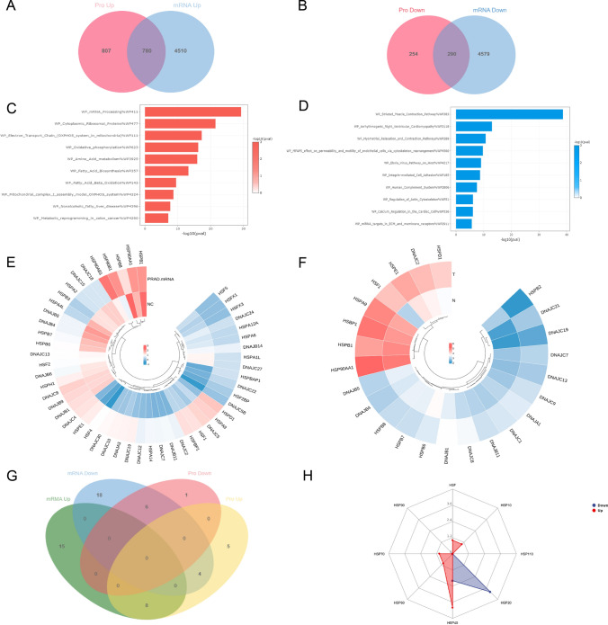
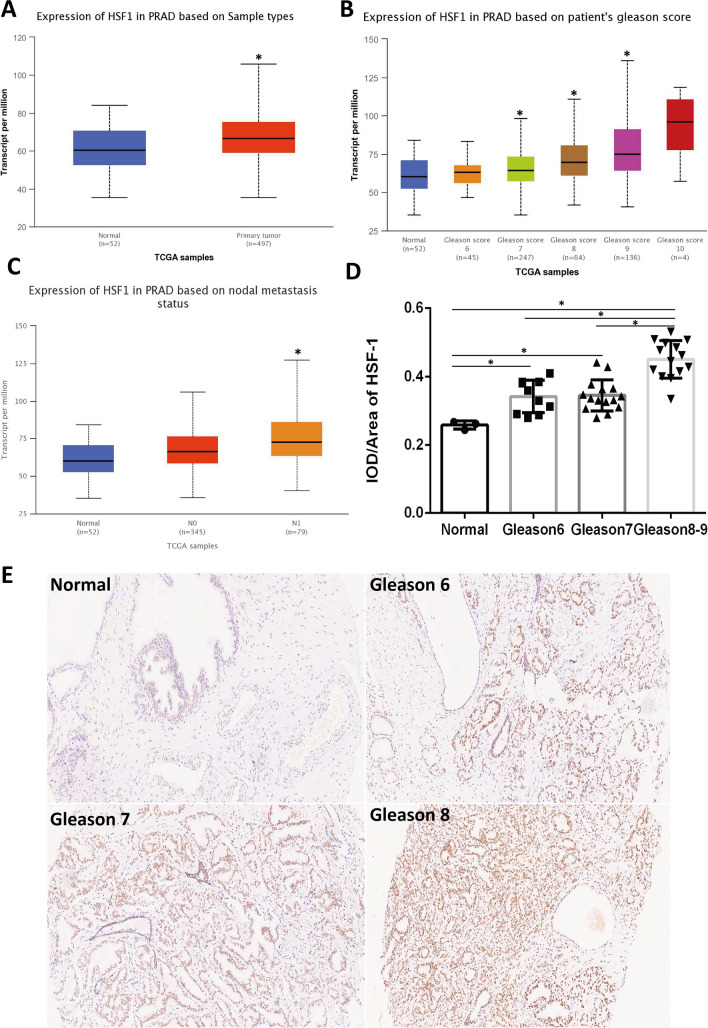
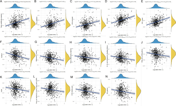
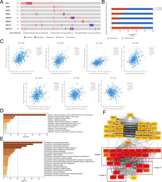
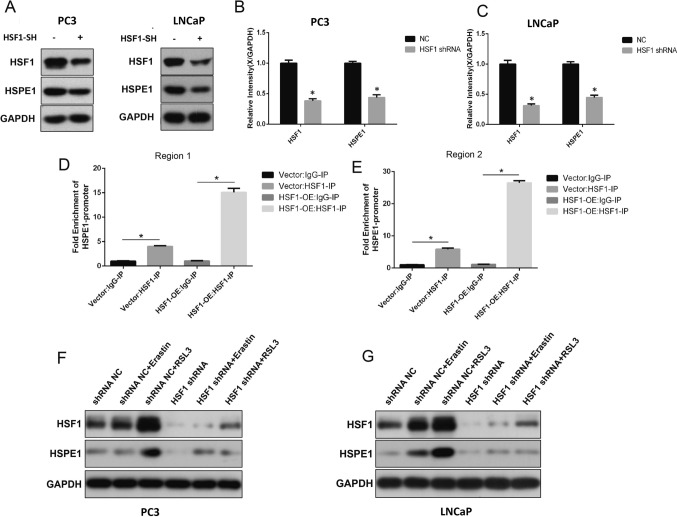
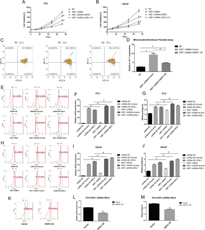
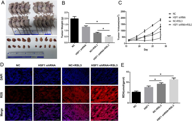
Results: Compared with levels observed in tumor-adjacent tissue, the levels of proteins associated with fatty acids, amino acids and the oxidative phosphorylation metabolic pathway were significantly upregulated in high-risk PC tissue (Gleason score ≥ 8). HSF1 mRNA and protein levels in high-risk PC tissues were significantly higher than those observed in medium-risk PC (Gleason score = 7) and low-risk PC (Gleason score ≤ 6) tissues. ssGSEA showed that HSF1 was involved in the proliferation and anti-apoptotic processes of PC. Further bioinformatics analysis showed that HSF1 potentially affects the mitochondrial oxidative phosphorylation (OXPHOS) system by targeting HSPE1. In addition, HSF1 alleviates ROS and MDA levels to enhance the resistance of prostate cancer cells to ferroptosis by regulating HSPE1 in vitro, and HSF1 knockout promotes the susceptibility of PC to RSL3 treatment by increasing ferroptosis in vivo.

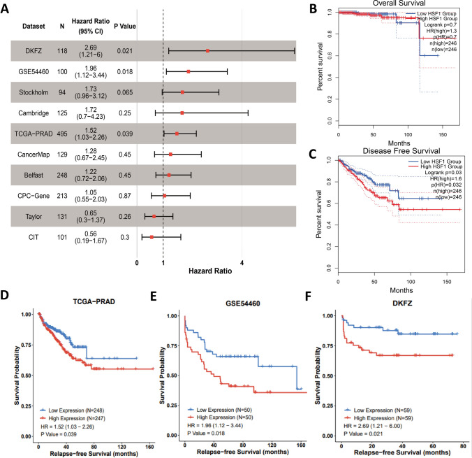
Conclusion: Collectively, our findings suggest that HSF1 exerts a significant influence on PC. HSF1 may represent a promising biomarker for identifying high-risk PC, and the elimination of HSF1 could potentially enhance the therapeutic effectiveness of RSL3.

Keywords: Ferroptosis; HSF1; HSPE1; High-risk prostate cancer; RSL3.


Conflict of interest statement

The authors declare no competing financial interests.

Figures

Fig. 1
Fig. 1
Proteomic analysis of high-risk prostate cancer. A, B Volcano map and heat map of differentially expressed protein. C, D Enrichment analysis of up-regulated and down-regulated protein WIKI pathways. E The heatmaps depicting the differential expression of Heat Shock Proteins (HSPs) at the mRNA level were obtained from the TCGA-PRAD database. F The heatmaps depicting the differential expression of Heat Shock Proteins (HSPs) at the protein level were obtained from our proteomic data. G The Venn graph shows intersection of differentially expressed HSPs in mRNA level and protein level. H Distribution of differentially expressed HSPs (According to molecular mass)
Fig. 2
Fig. 2
The expression of HSF1 in prostate cancer. AC mRNA expression of HSF1 in TCGA (*P < 0.05). D Immunohistochemical semi-quantitative analysis (*P < 0.05). E Representative immunohistochemistry images of HSF1 in different prostate cancer tissues
Fig. 3
Fig. 3
Survival analysis of HSF1. A Forest plot of the univariate Cox regression analysis in ten sets. B, C Kaplan–Meier survival curves in TCGA (OS, overall survival; DFS, disease free survival). DF Kaplan–Meier survival curves in TCGA, GSE54460, and DKFZ (RFS, relapse free survival)
Fig. 4
Fig. 4
ssGSEA analysis of HSF1 and pathways, and.the correlations between individual gene and pathway score was analysed with Spearman. AE The pathways that exhibit a positive correlation with HSF1 are presented. FN The pathways that exhibit a negative correlation with HSF1 are presented
Fig. 5
Fig. 5
Mutation and downstream pathway of HSF1. A Mutation analysis in Cbioportal database. B Correlation between HSF1 mutation proportion and Gleason Score. C Correlation between HSF1 and HSPs expression levels. D, E Enrichment analysis of the top 300 HSPE1-positively correlated genes. F PPI analysis of HSPE1-positively correlated genes
Fig. 6
Fig. 6
HSF1 can directly regulate HSPE1 expression in prostate cancer. AC Expression levels of HSF1 and HSPE1 after HSF1 shRNA knockdown were tested by WB (*P < 0.05). D, E ChIP-qPCR assay of HSF1 binding to the HSPE1 promoter (*P < 0.05). F, G Expression levels of HSF1 and HSPE1 after treated with Erastin or RSL3 were tested by WB
Fig. 7
Fig. 7
HSF1 affects oxidative stress and lipid peroxidation levels by regulating HSPE1 in vitro. A, B CKK-8 assay after HSF1 knockout (*P < 0.05). C, D Mitochondrial membrane potential assay in PC3 cell line via JC-1 staining and flow cytometry (*P < 0.05). EG ROS and MDA assays in PC3 cell lines after HSF1 knockout (*P < 0.05). HJ ROS and MDA assays in LNCaP cell lines after HSF1 knockout. K, M ROS and MDA assays in PC3 cell lines after HSPE1 overexpression (*P < 0.05)
Fig. 8
Fig. 8
Experiments in vivo. AC Tumor weight(g) and tumor growth curves (mm3) of mice subcutaneously inoculated with PC3 cells (* P < 0.05). D, E ROS staining and MDA assay in tumor tissues were performed after 4 weeks (*P < 0.05)

Similar articles

Cited by

References

    1. Parkin DM, Bray F, Ferlay J, et al. Global cancer statistics, 2002. CA Cancer J Clin. 2005;55(2):74–108. doi: 10.3322/canjclin.55.2.74. - DOI - PubMed
    1. Cooperberg MR, Cowan J, Broering JM, Carroll PR. High-risk prostate cancer in the United States, 1990–2007. World J Urol. 2008;26(3):211–218. doi: 10.1007/s00345-008-0250-7. - DOI - PMC - PubMed
    1. D'Amico AV, Whittington R, Malkowicz SB, et al. Biochemical outcome after radical prostatectomy, external beam radiation therapy, or interstitial radiation therapy forclinically localized prostate cancer. JAMA. 1998;280(11):969–974. doi: 10.1001/jama.280.11.969. - DOI - PubMed
    1. Chung BH. The role of radical prostatectomy in high-risk prostate cancer. Prostate Int. 2013;1(3):95–101. doi: 10.12954/PI.13018. - DOI - PMC - PubMed
    1. Mossanen M, Krasnow RE, Nguyen PL, Trinh QD, Preston M, Kibel AS. Approach to the patient with high-risk prostate cancer. Urol Clin N Am. 2017;44(4):635. doi: 10.1016/j.ucl.2017.07.009. - DOI - PubMed

LinkOut - more resources