. 2023 Jul 10;33(13):2702-2716.e3.
doi: 10.1016/j.cub.2023.05.054. Epub 2023 Jun 22.

Modulation and neural correlates of postmating sleep plasticity in Drosophila females

Affiliations

Modulation and neural correlates of postmating sleep plasticity in Drosophila females

José M Duhart et al. Curr Biol. .

Abstract

Sleep is essential, but animals may forgo sleep to engage in other critical behaviors, such as feeding and reproduction. Previous studies have shown that female flies exhibit decreased sleep after mating, but our understanding of the process is limited. Here, we report that postmating nighttime sleep loss is modulated by diet and sleep deprivation, demonstrating a complex interaction among sleep, reproduction, and diet. We also find that female-specific pC1 neurons and sleep-promoting dorsal fan-shaped body (dFB) neurons are required for postmating sleep plasticity. Activating pC1 neurons leads to sleep suppression on standard fly culture media but has little sleep effect on sucrose-only food. Published connectome data suggest indirect, inhibitory connections among pC1 subtypes. Using calcium imaging, we show that activating the pC1e subtype inhibits dFB neurons. We propose that pC1 and dFB neurons integrate the mating status, food context, and sleep drive to modulate postmating sleep plasticity.

Keywords: Drosophila; diet; dorsal fan-shaped body; egg laying; mating; motivation; nutrition; pC1 neurons; reproduction; sleep.


Conflict of interest statement

Declaration of interests The authors declare no competing interests.

Figures

Figure 1.
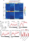
Figure 1.. Food type modulates postmating nighttime sleep loss.
(A) Experimental design. (B and C) Sleep profiles of virgin and mated females on normal food (B) or 5% sucrose (C), measured using multi-beam monitors. N = 38–40. (D and E) Daytime (D) and nighttime (E) sleep for flies shown in (B) and (C). (F and G) Sleep profiles of virgin and mated females on normal food (F) or 5% sucrose (G), measured using single-beam monitors. N = 88–91. (H and I) Daytime (H) and nighttime (I) sleep for flies shown in (F) and (G). Wild-type (Iso31) flies were used in all panels. The white and black bars in the experimental design and below the x-axis in sleep profiles indicate 12-h light and 12-h dark periods, respectively. In this and subsequent figures, bar graphs present mean ± SEM and individual measurements; *p<0.05, **<0.01, ***p<0.001, ****p<0.0001, and ns: not significant. Two-way ANOVA followed by Sidak’s post hoc test (D, E, H, and I). See also Figure S1.
Figure 2.
Figure 2.. Food composition modulates postmating changes in nighttime sleep and egg laying.
(A) Position heatmaps indicating the duration spent at different tube positions per 30-min bin computed from the multi-beam data shown in Figures 1B and 1C. The top of each heatmap (Food) is closest to the food. (B) Number of eggs laid per female on normal food or 5% sucrose. N = 16 vials (4 females/vial). (C-F) Sleep profiles of virgin and mated females on normal food (C), 7.75% sucrose (D), 5% sucrose + 2.75% yeast (E), or 5% sucrose + 2.75% tryptone (F). N = 47–72. (G) Nighttime sleep for flies in (C–F). Wild-type (Iso31) flies were used in all panels. Two-way ANOVA followed by Sidak’s post hoc test (B and G). See also Figure S2.
Figure 3.
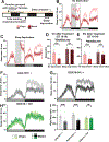
Figure 3.. Sleep deprivation and the inhibition of dFB-projecting neurons attenuate the impact of mating on nighttime sleep.
(A) Experimental design for panels (B–E). (BC) Sleep profiles of Iso31 virgin and mated females on normal food, without (B) or with (C) mechanical sleep deprivation from ZT 12 to ZT 18 (gray rectangle). Dotted rectangles represent the 1-h and 6-h period after deprivation (ZT 18–19 and ZT 18–24). N = 55–56. (D-E) Sleep amounts during the 1-h (D) or 6-h (E) period after sleep deprivation for flies in (B) and (C). (F-H) Sleep profiles of virgin and mated females with impaired neurotransmitter release from dFB-projecting neurons (R23E10 > TNT, H) and their parental controls (F and G) on normal food. N = 41–48. (I) Nighttime sleep for flies in (F–H). Two-way ANOVA followed by Sidak’s post hoc test (D, E, and I). See also Figure S3.
Figure 4.
Figure 4.. pC1 neurons are required for postmating sleep suppression.
(A) Neural circuit for increased egg laying after mating (adapted from ). (BF) Sleep profiles of virgin and mated females with impaired neurotransmitter release from pC1a,c,e subtypes (pC1-SS1(a,c,e) > TNT, D) or all pC1 subtypes (pC1-SS2(a-e) > TNT, F) and their parental controls (B, C, and E) on normal food. pC1-SS1 and pC1-SS2 target pC1a,c,e, and pC1a-e, respectively. N = 41–95. (G) Nighttime sleep for flies in (B–F). (H) Experimental design for (I–R). (I-L) Sleep profiles of virgin (I and K) or mated (J and L) females expressing the warmth-activated TrpA1 channel in pC1 (pC1-SS1(a,c,e) > TrpA1 or pC1-SS2(a-e) > TrpA1) neurons and their parental controls on normal food. TrpA1 was activated by raising the temperature from 22°C to 27°C. N = 42–64. (M) Nighttime sleep change (sleep at 27°C – baseline sleep at 22°C) for flies in (I–L). (N–Q) Sleep profiles of virgin (N and P) or mated (O and Q) females of the same genotypes as (I-L) on 5% sucrose. N = 30–32. (R) Nighttime sleep change for flies in (N–Q). Two-way ANOVA followed by Sidak’s (G) or Tukey’s (M, and R) post hoc test. See also Figures S4 and S5.
Figure 5.
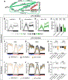
Figure 5.. Activation of the pC1e subtype suppresses sleep.
(A) Simplified diagram of the connections among pC1 and OviIN neurons based on an analysis of the hemibrain connectome dataset using the neuPrint tool. Percentage of input to the postsynaptic target and synapse number (in parentheses) for each connection are presented over the connecting arrows. Only connections that represent more than 0.4% of the input to the postsynaptic target are shown. (B–D) Sleep profiles of virgin and mated females with impaired neurotransmitter release from pC1e (pC1e > TNT, D) and their parental controls (B and C) on normal food. N = 62–150. (E) Nighttime sleep for flies in (B-D). (F–G) Sleep profiles of virgins (F) and mated (G) females expressing the warmth-activated TrpA1 channel in pC1e (pC1e > TrpA1) and their parental controls on normal food. TrpA1 was activated by raising the temperature from 22°C to 29°C. N = 42–73. (H) Nighttime sleep change (sleep at 29°C – baseline sleep at 22°C) for flies in (F) and (G). (I–J) Sleep profiles of virgins (I) and mated (J) females of the same genotypes as (F) and (G) on 5% sucrose food. TrpA1 was activated by raising the temperature from 22°C to 29°C. N = 21–49. (K) Nighttime sleep change for flies in (I) and (J). Two-way ANOVA followed by Sidak’s (E) or Dunnett’s (H and K) post hoc test. See also Figure S6.
Figure 6.
Figure 6.. dFB-projecting neurons are inhibited by the activation of pC1a,c,e or pC1e neurons.
(A) GCaMP7b signal in dFB-projecting neurons (R23E10 > GCaMP7b) before and shortly after ATP perfusion in a dissected female brain expressing the ATP-sensitive P2X2 receptor in pC1a,c,e neurons. The “fire” lookup table is used. Scale bar: 30 μm. (B) Fluorescence traces of normalized GCaMP7b signal (ΔF/F) in the cell bodies of dFB neurons in response to ATP perfusion (gray area) in females expressing P2X2 in pC1a,c,e neurons (pC1a,c,e > P2X2). Flies carrying R23E10-LexA, LexAop-GCaMP7b, and UAS-P2X2, but not pC1-SS1(a,c,e), served as negative controls. N = 20–21 brains. (C) Minimum (Min) ΔF/F responses for brains in (B). (D) GCaMP7b signal in dFB-projecting neurons (R23E10 > GCaMP7b) before and shortly after ATP perfusion in a dissected female brain expressing P2X2 in pC1e neurons. The “fire” lookup table is used. Scale bar: 30 μm. (E) Fluorescence traces of normalized GCaMP7b signal (ΔF/F) in the cell bodies of dFB neurons in response to ATP perfusion in females expressing P2X2 in pC1e neurons (pC1e > P2X2). Flies lacking pC1e-splitG4 served as negative controls. N = 15 brains. (F) Minimum (Min) ΔF/F responses for brains in (E). (G) Model for postmating sleep plasticity. Sex Peptide deposited by males during copulation leads to the inhibition of SPSN, SAG, pC1a, pC1d, and OviIN. The inhibition of GABAergic OviIN neurons results in the disinhibition (activation) of pC1c,e subtypes. Activating pC1c,e neurons in mated females leads to the inhibition of the sleep-promoting dFB neurons through indirect connections (dotted lines). OviIN receives information about the sugar content. Information about additional nutrients may be integrated at the level of pC1c,e or their downstream neurons. Unpaired Student’s t test (C, F).

Similar articles

Cited by

References

    1. Duhart JM, Inami S, and Koh K (2021). Many faces of sleep regulation: beyond the time of day and prior wake time. FEBS J 10.1111/febs.16320. - DOI - PMC - PubMed
    1. Beckwith EJ, and French AS (2019). Sleep in Drosophila and Its Context. Front Physiol 10. 10.3389/fphys.2019.01167. - DOI - PMC - PubMed
    1. Machado DR, Afonso DJ, Kenny AR, Öztürk-Çolak, A., Moscato, E.H., Mainwaring, B., Kayser, M., and Koh, K. (2017). Identification of octopaminergic neurons that modulate sleep suppression by male sex drive. Elife 6. 10.7554/eLife.23130. - DOI - PMC - PubMed
    1. Beckwith EJ, Geissmann Q, French AS, and Gilestro GF (2017). Regulation of sleep homeostasis by sexual arousal. Elife 6. 10.7554/eLife.27445. - DOI - PMC - PubMed
    1. Chen D, Sitaraman D, Chen N, Jin X, Han C, Chen J, Sun M, Baker BS, Nitabach MN, and Pan Y (2017). Genetic and neuronal mechanisms governing the sex-specific interaction between sleep and sexual behaviors in Drosophila. Nat Commun 8, 1–14. 10.1038/s41467-017-00087-5. - DOI - PMC - PubMed

Publication types

Substances

LinkOut - more resources