. 2023 Jul 6;110(7):1046-1067.
doi: 10.1016/j.ajhg.2023.06.002. Epub 2023 Jun 22.

Using the ACMG/AMP framework to capture evidence related to predicted and observed impact on splicing: Recommendations from the ClinGen SVI Splicing Subgroup

Collaborators, Affiliations

Using the ACMG/AMP framework to capture evidence related to predicted and observed impact on splicing: Recommendations from the ClinGen SVI Splicing Subgroup

Logan C Walker et al. Am J Hum Genet. .

Abstract

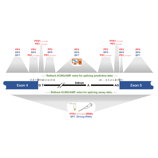
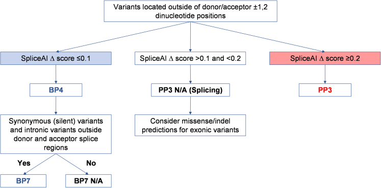
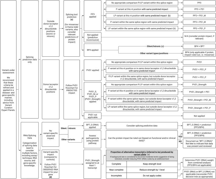
The American College of Medical Genetics and Genomics (ACMG)/Association for Molecular Pathology (AMP) framework for classifying variants uses six evidence categories related to the splicing potential of variants: PVS1, PS3, PP3, BS3, BP4, and BP7. However, the lack of guidance on how to apply such codes has contributed to variation in the specifications developed by different Clinical Genome Resource (ClinGen) Variant Curation Expert Panels. The ClinGen Sequence Variant Interpretation Splicing Subgroup was established to refine recommendations for applying ACMG/AMP codes relating to splicing data and computational predictions. We utilized empirically derived splicing evidence to (1) determine the evidence weighting of splicing-related data and appropriate criteria code selection for general use, (2) outline a process for integrating splicing-related considerations when developing a gene-specific PVS1 decision tree, and (3) exemplify methodology to calibrate splice prediction tools. We propose repurposing the PVS1_Strength code to capture splicing assay data that provide experimental evidence for variants resulting in RNA transcript(s) with loss of function. Conversely, BP7 may be used to capture RNA results demonstrating no splicing impact for intronic and synonymous variants. We propose that the PS3/BS3 codes are applied only for well-established assays that measure functional impact not directly captured by RNA-splicing assays. We recommend the application of PS1 based on similarity of predicted RNA-splicing effects for a variant under assessment in comparison with a known pathogenic variant. The recommendations and approaches for consideration and evaluation of RNA-assay evidence described aim to help standardize variant pathogenicity classification processes when interpreting splicing-based evidence.

Keywords: ACMG/AMP codes; BP4; BP7; ClinGen; PP3; PS1; PVS1; RNA splicing; variant classification.


Conflict of interest statement

Declaration of interests A.L., L.M.V., S.H., H.Z., R.K., D.B.-B., A.C., A.T., and T.P. are employed by fee-for-service laboratories performing clinical sequencing services.

Figures

None
Graphical abstract
Figure 1
Figure 1
Schematic showing sequence motifs at intron-exon junctions and terminology applied in this report Pre-mRNA splicing, the process of intron removal from nascent pre-mRNA followed by exon ligation, is principally reliant on the following cis-acting regulatory elements: the splice donor +1,2 dinucleotide flanking the 5′ end of the intron; the splice acceptor −1,2 dinucleotide flanking the 3′ end of the intron; the polypyrimidine tract and branch point site (usually at positions −18 to −4458) upstream of the 3′ splice acceptor. So-called consensus sequences for different splicing regulatory elements have been defined by comparing position weight matrices for surrounding sequence. The vast majority of introns (>98%) are recognized by highly conserved dinucleotides at the 5′ boundary (GT) and 3′ boundary (AG)., Intron categories may variously be designated by the boundary dinucleotide sequence, spliceosome (likely) excising the intron (U2-type spliceosomes for most GT-AG introns, U12-type spliceosomes for most AT-AC introns, with some exceptions), and/or by comparing position weight matrices for surrounding sequence., Exon-intron boundaries for consensus donor and acceptor site motifs (including the polypyrimidine tract) defined by Burge et al. are as follows: 11 bases for the donor splice site motif (from the last 3 exonic to the first 8 intronic bases) and 14 bases for the acceptor splice site motif (from the last 12 intronic to the first 2 exonic bases). More detailed examination of the position weight matrix plot for the most prevalent U2-type intron reveals degeneracy at some positions in these motifs; thus, impact on splicing is most likely for variants located in the donor splice region (the last 3 bases of the exon and 3–6 nucleotides of intronic sequence adjacent to the exon) and the acceptor splice region (the first base of the exon and from 3 to 20 nucleotides upstream from the exon boundary). For secondary variant analysis, a minimal splice region was defined as above for the donor splice region, with the acceptor splice region designated as only the first base of the exon and the third nucleotide upstream from the exon boundary. Exonic and intronic splicing enhancer and silencer elements also play an important role in exon definition (not shown here).
Figure 2
Figure 2
Schematic demonstrating assignment of gene-specific codes to splice donor/acceptor ±1,2 dinucleotide variants based on a modified version of the original ClinGen SVI PVS1 framework Original framework refers to recommendations as published. It is important to note that each PVS1 assigned weight may be reduced if there is evidence of potential rescue mechanisms. For example, skipping of either exon 4 or 7 may lead to a protein that retains partial function. Annotating gene-specific lists of naturally occurring splicing events can provide greater evidence of potential “rescue” isoforms. Also see Box S1.
Figure 3
Figure 3
Model for optimizing thresholds for prediction algorithms of alternative splicing (A) Schematic demonstrating how collation of 3 variant datasets (in vitro splicing data, splicing prediction scores, and clinical classification data) enable calibration of splicing prediction algorithms for pathogenicity. While clinically classified variant data is preferable, splicing assay data can be used as an imperfect surrogate for pathogenicity. More extensive annotation of alternative splicing events and level of aberration will lead to an improved correlation of splicing events with variant pathogenicity. The distribution of hypothetical computationally predicted splice scores is illustrated, showing significant overlap of non-spliceogenic/spliceogenic datasets (left side) and benign/pathogenic datasets (right side). The low, intermediate, and high prediction score used to assign ACMG/AMP code weighting can be determined by calculating likelihood ratios for different score categories and obtaining consensus on the score thresholds to be applied. (B) Process for calibrating splicing prediction score thresholds for computational tools. A worked example of a likelihood ratio calculation is shown in Table S5. Note: truth datasets were filtered to exclude splice donor/acceptor ±1,2 dinucleotide variants, which are captured by the PVS1 decision tree process.
Figure 4
Figure 4
An exemplar PP3, BP4, and BP7 decision tree for maximum SpliceAI splicing prediction scores and calibrated cutoff scores The analytical process is shown in Figure 3B and data shown in Table 1. BP7 should not be applied for donor/acceptor splice regions given their higher prior for harboring spliceogenic variants. This may be defined as the splice region (a conservative application already implemented by several VCEPs) or the minimal splice region. PP3 may still be applied for missense or insertion-deletion variants that show computational evidence for a deleterious effect for change in protein sequence.
Figure 5
Figure 5
Decision tree for application of bioinformatic codes and RNA-splicing assay results for variant interpretation (A) Alternative prediction tools/thresholds may be appropriate for variants that impact sites other than GT-AT donor-acceptor motifs. (B) LP variants at the canonical positions should only be used as evidence if additional supporting clinical evidence is present. (C) Silent (excluding last 3 nucleotides of exon and first nucleotide of exon) and intronic variants at or beyond the +7 and −21 positions (conservative designation for donor/acceptor splice region) or otherwise at or beyond the +7 and −4 positions (less conservative designation for the minimal donor/acceptor splice region). (D) If multiple impacts are observed from a splicing assay, use flowchart for the most conservative application of PVS1 based on experimental data. (E) We recommend that these thresholds be refined and applied in a disease- and gene-specific manner, including advice from VCEPs. Categorization as complete or near complete needs to consider multiple factors, including assay/technique, RNA source, and validation of assay weights using established controls. Examples of laboratory-specific approaches and suggested operational thresholds have been reported previously.,,,, See Table S9 for additional considerations for interpretation of mRNA assay data.

Update of

Similar articles

Cited by

References

    1. Richards S., Aziz N., Bale S., Bick D., Das S., Gastier-Foster J., Grody W.W., Hegde M., Lyon E., Spector E., et al. Standards and guidelines for the interpretation of sequence variants: a joint consensus recommendation of the American College of Medical Genetics and Genomics and the Association for Molecular Pathology. Genet. Med. 2015;17:405–424. doi: 10.1038/gim.2015.30. - DOI - PMC - PubMed
    1. Abou Tayoun A.N., Pesaran T., DiStefano M.T., Oza A., Rehm H.L., Biesecker L.G., Harrison S.M., ClinGen Sequence Variant Interpretation Working Group ClinGen SVI Recommendations for interpreting the loss of function PVS1 ACMG/AMP variant criterion. Hum. Mutat. 2018;39:1517–1524. doi: 10.1002/humu.23626. - DOI - PMC - PubMed
    1. Brnich S.E., Abou Tayoun A.N., Couch F.J., Cutting G.R., Greenblatt M.S., Heinen C.D., Kanavy D.M., Luo X., McNulty S.M., Starita L.M., et al. Recommendations for application of the functional evidence PS3/BS3 criterion using the ACMG/AMP sequence variant interpretation framework. Genome Med. 2019;12:3. doi: 10.1186/s13073-019-0690-2. - DOI - PMC - PubMed
    1. Ghosh R., Harrison S.M., Rehm H.L., Plon S.E., Biesecker L.G., ClinGen Sequence Variant Interpretation Working Group Updated recommendation for the benign stand-alone ACMG/AMP criterion. Hum. Mutat. 2018;39:1525–1530. doi: 10.1002/humu.23642. - DOI - PMC - PubMed
    1. Spurdle A.B., Greville-Heygate S., Antoniou A.C., Brown M., Burke L., de la Hoya M., Domchek S., Dörk T., Firth H.V., Monteiro A.N., et al. Towards controlled terminology for reporting germline cancer susceptibility variants: an ENIGMA report. J. Med. Genet. 2019;56:347–357. doi: 10.1136/jmedgenet-2018-105872. - DOI - PubMed

Publication types

LinkOut - more resources