. 2023 Jul 6;110(7):1068-1085.
doi: 10.1016/j.ajhg.2023.06.001. Epub 2023 Jun 22.

Null and missense mutations of ERI1 cause a recessive phenotypic dichotomy in humans

Affiliations

Null and missense mutations of ERI1 cause a recessive phenotypic dichotomy in humans

Long Guo et al. Am J Hum Genet. .

Abstract

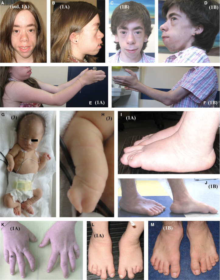
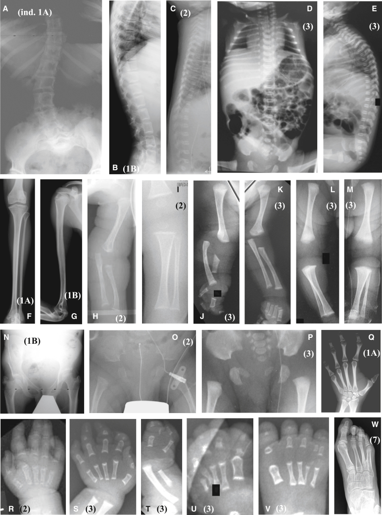
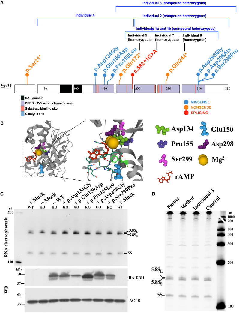
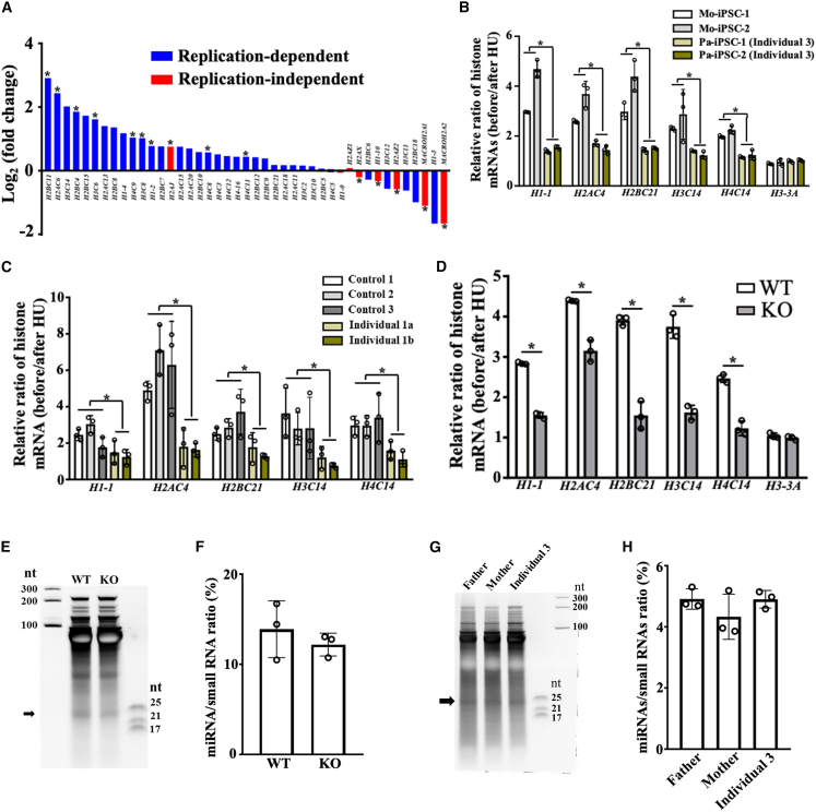
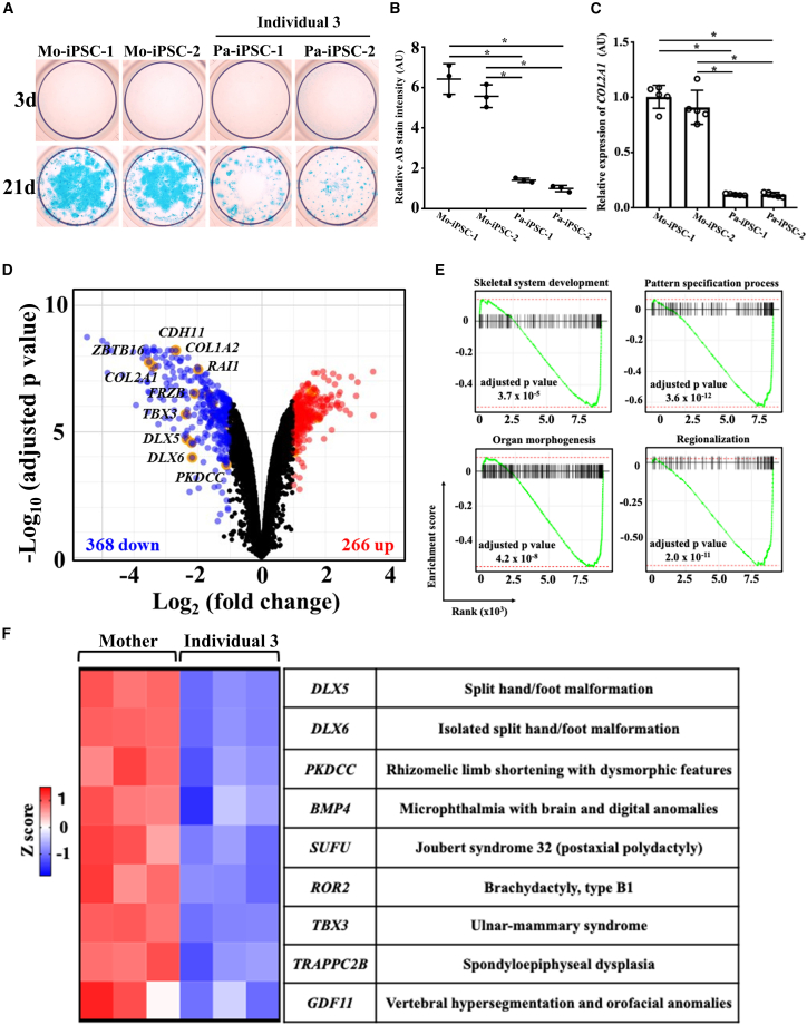
ERI1 is a 3'-to-5' exoribonuclease involved in RNA metabolic pathways including 5.8S rRNA processing and turnover of histone mRNAs. Its biological and medical significance remain unclear. Here, we uncover a phenotypic dichotomy associated with bi-allelic ERI1 variants by reporting eight affected individuals from seven unrelated families. A severe spondyloepimetaphyseal dysplasia (SEMD) was identified in five affected individuals with missense variants but not in those with bi-allelic null variants, who showed mild intellectual disability and digital anomalies. The ERI1 missense variants cause a loss of the exoribonuclease activity, leading to defective trimming of the 5.8S rRNA 3' end and a decreased degradation of replication-dependent histone mRNAs. Affected-individual-derived induced pluripotent stem cells (iPSCs) showed impaired in vitro chondrogenesis with downregulation of genes regulating skeletal patterning. Our study establishes an entity previously unreported in OMIM and provides a model showing a more severe effect of missense alleles than null alleles within recessive genotypes, suggesting a key role of ERI1-mediated RNA metabolism in human skeletal patterning and chondrogenesis.

Keywords: ERI1; exoribonuclease; ribosomopathy; short stature; skeletal dysplasia; spondyloepimetaphyseal dysplasia.


Conflict of interest statement

Declaration of interests The Department of Molecular and Human Genetics at Baylor College of Medicine receives revenue from clinical genetic testing completed at Baylor Genetics Laboratories.

Figures

Figure 1
Figure 1
Photographs of affected individuals (A and B) Individual 1A. Note zygomatic hypoplasia, prominent alveolar processes of the maxilla and mandible, and small and low-set ears. (C and D) Individual 1B. Note zygomatic hypoplasia and prominent alveolar processes of the maxilla and mandible. (E and F) Maximal elbow extension for individuals 1A and 1B. Note distal camptodactyly in individual 1B. (G and H) Individual 3, born at full term. Short stature with short limbs (G) and oligo-syndactyly of the right hand (H) and the right foot (G). The black box in (G) is used to increase anonymity. (I and J) Lateral view of feet of individuals 1A (I) and 1B (J). Note short feet. (K) Hands of individual 1A. Clinodactyly of the proximal interphalangeal joint of second right digit. (L and M) Dorsal view of feet of individuals 1A (L) and 1B (M). Note short feet and 4-5 syndactyly.
Figure 2
Figure 2
Radiographs of affected individuals (A) Lower spine of individual 1A with lumbar scoliosis. (B) Lateral spine of individual 1B showing flattened vertebral bodies and irregular plates. (C–E) Platyspondyly in individuals 2 (C) and 3 (D and E). (F–M) Long tubular bones showing epi-metaphyseal dysplasia characterized by irregular epiphyses and frayed metaphyses in individuals 1A (F), 1B (G), 2 (H and I), and 3 (J–M). The right upper limb of individual 3 shows ulnar hypoplasia and radial bowing with elbow dislocation (J). (N–P) Hypoplasia of pelvis and epi-metaphyseal dysplasia of femurs in individuals 1B (N), 2 (O), and 3 (P). Note right coxa vara and left coxa valga (N), iliac hypoplasia and delayed pubic ossification (O and P). (Q) Right hand of individual 1A. Note single thumb phalanx, ulnar deviation of the second finger at proximal interphalangeal joint, and narrow metacarpal bones. (R) Left hand of individual 2 showing brachy-syndactyly of the third and fourth fingers. (S–V) Hands and feet of individual 3. The left hand (S) shows brachydactyly with hypoplasia of all middle phalanges, the first proximal and distal phalanges, and the second and third distal phalanges as well as ulnar clinodactyly of the second and third fingers and radial clinodactyly of the fourth and fifth fingers. The proximal end of the proximal phalanges and distal ends of the metacarpals are conspicuously cupped. The right hand shows oligo-syndactyly (split hand) (T). Both feet show postaxial syndactyly and the third and fourth metatarsal bases may be fused (U and V). The right fourth metatarsal is hypoplastic (V). (W) Right foot of individual 7. Note absence of the fifth ray, proximal fusion of the third and fourth metatarsal bones, and widened fourth metatarsus. The black boxes in (J), (L), and (U) mask arrows previously used in clinical discussions.
Figure 3
Figure 3
Functional analysis of ERI1 pathogenic variants (A) Schematic overview of ERI1 showing the position of the variants identified in the individuals. ERI1 includes two functional domains: amino-terminal SAP (SAF-A/B, Acinus, and PIAS) domain and carboxy-terminal 3′ exonuclease domain. (B) 3D structure of ERI1 with annotated residues Asp134, Glu150, Pro155, Asp298, and Ser299 generated via UniProKB (https://www.uniprot.org/uniprot/Q8IV48). PDB: 1ZBU. rAMP, riboadenine-5′-monophosphate. (C) The upper panel shows ethidium bromide-stained RNA from the wild type (WT) and ERI1 knockout (KO) HeLa cells. Nt, nucleotide. WT, p.Asp134Gly, p.Glu150Asp, p.Pro155Leu, p.Asp298Gly, and p.Ser299Pro ERI1 were produced in the KO cells to rescue the 3′-to-5′ exoribonuclease activity of ERI1 in catalyzing 5.8S ribosomal RNA (rRNA) processing. The lower panel shows western blot of whole-cell lysates. (D) Ethidium bromide-stained RNA from the lymphoblastoid cell lines of the affected individual 3, his parents, and a healthy control. 5.8SL, long form of 5.8S rRNA; 5.8SS, short form of 5.8S rRNA.
Figure 4
Figure 4
Effect of ERI1 deficiency on histone mRNAs and miRNAs (A) Histone mRNA levels of the family 3-derived iPSCs in the process of chondrogenic differentiation (day 8). Both replication-dependent and replication-independent histone genes are ranked according to the fold change (FC) of the differentially expressed genes in Mo-iPSC-1 versus Pa-iPSC-1. p < 0.05. n = 3 biologically independent samples. Statistical significance was assessed via two-sided t test. p value was adjusted by false discovery rate (FDR). (B–D) qPCR for histone mRNAs before and after hydroxyurea (HU) treatment for 45 min. All markers are replication-dependent histone genes except H3-3A. (B) Family 3-derived iPSCs. (C) Fibroblasts from three healthy control individuals and the two affected individuals in family 1. (D) Wild type (WT) and knock-out (KO) HeLa cells. The data in (B)–(D) indicate mean ± SD. n = 3 biological replicates. Statistical test was assessed via multiple t tests. p < 0.05. (E–H) SYBR Gold-stained small RNAs from the wild type (WT) and ERI1 knockout (KO) HeLa cells (E) and family 3-derived lymphoblastoid cells (G). nt, nucleotide. The arrows show single-strand RNAs between 21 and 25 nt, corresponding to the abundance of miRNAs. Quantification of the miRNA to total small RNA (<300 nt) ratio in HeLa cells (F) and lymphoblastoid cells (H). The data indicate mean ± SD. p = 0.44 and 0.33. n = 3 biological replicates. Statistical significance was assessed via two-sided t test and one-way ANOVA.
Figure 5
Figure 5
Chondrogenic differentiation of the affected-individual-derived induced pluripotent stem cells (iPSCs) (A) Alcian blue (AB) staining for the chondrocytic nodules induced from the healthy mother iPSCs (Mo-iPSC-1 and -2) and affected-individual-derived iPSCs (Pa-iPSC-1 and -2) in family 3. (B) Quantification of AB staining; n = 3 biologically independent samples. p < 0.05. Error bars represent SD. (C) qPCR for COL2A1; n = 5 biologically independent samples. p < 0.05. AU, arbitrary unit. The data in (B) and (C) indicate mean ± SD. Statistical test was assessed via one-way ANOVA. (D) Volcano plot of RNA-seq data for the differentiating iPSCs at day 8 showing differentially expressed genes (DEGs) in Mo-iPSC-1 versus Pa-iPSC-1 (n = 3 biologically independent samples) with respect to fold change (FC) and significance (adjusted p value). Each dot represents an individual gene. Blue dots represent significant downregulated DEGs (FC < 0.5) and red dots represent significant upregulated DEGs (FC > 2). Statistical significance was assessed via moderated t test in limma. p value is adjusted by false discovery rate. (E) Gene set enrichment analysis for RNA-seq data in (D). The genes belonging to the corresponding GO terms are enriched at the bottom of the entire ranked list of genes. (F) Heatmap of DEGs associated causally with human skeletal dysplasia.
Figure 6
Figure 6
Skeletal dysplasia of Eri1 knockout (KO) mice (A) X-ray images of paws of the wild type (WT) and Eri1 KO mice at 16 weeks. Scale bar, 1 mm. (B) Quantitative analysis of the paw length shown in (A) (n = 9 mice). p < 0.05. The above quantitative data indicate mean ± SD and statistical significance was assessed via two-sided t test. Error bars represent SD. (C) X-ray images of the WT and Eri1 KO mice at 16 weeks. The areas highlighted by the dotted box in the upper panels are magnified in the lower panels. Scale bar, 1 cm. (D) Measurement of the height of the caudal vertebral bodies shown in (C) (n = 9 mice). p < 0.05.
Figure 7
Figure 7
Hypothesis of the pathological mechanism underlying the phenotypic dichotomy between null and missense variants of ERI1 Both null and missense mutations of ERI1 cause loss of 3′-to-5′ exoribonuclease activity of ERI1, leading to defective processing of 5.8S rRNA and impaired degradation of replication-dependent histone mRNA in humans, which interrupt diverse cellular processes including ribosome biogenesis and coordination of histone gene expression with cell cycle progression. These defects singly or jointly alter gene expression profiling that regulates acral patterning, thus inducing digit abnormalities. ERI1 missense mutations also produce exonuclease-dead proteins, which may compete with other exonucleases having functional redundancy with ERI1, thus aggravating the dysfunction of ERI1-mediated RNA metabolism. The aggravated dysfunction or additional unknown effects lead to impaired chondrogenesis that causes spondyloepimetaphyseal dysplasia, finally contributing to the phenotypic dichotomy between null and missense variants of ERI1. Solid arrow, causal connection; dashed arrow, hypothetical connection; red line, additional effects of exonuclease-dead ERI1 to the loss of the exoribonuclease activity.

References

    1. Zuo Y., Deutscher M.P. Exoribonuclease superfamilies: Structural analysis and phylogenetic distribution. Nucleic Acids Res. 2001;29:1017–1026. - PMC - PubMed
    1. Simmer F., Tijsterman M., Parrish S., Koushika S.P., Nonet M.L., Fire A., Ahringer J., Plasterk R.H.A. Loss of the putative RNA-directed RNA polymerase RRF-3 makes C. Elegans hypersensitive to RNAi. Curr. Biol. 2002;12:1317–1319. - PubMed
    1. Gu W., Shirayama M., Conte D., Vasale J., Batista P.J., Claycomb J.M., Moresco J.J., Youngman E.M., Keys J., Stoltz M.J., et al. Distinct Argonaute-Mediated 22G-RNA Pathways Direct Genome Surveillance in the C. elegans Germline. Mol. Cell. 2009;36:231–244. - PMC - PubMed
    1. Han T., Manoharan A.P., Harkins T.T., Bouffard P., Fitzpatrick C., Chu D.S., Thierry-Mieg D., Thierry-Mieg J., Kim J.K. 26G endo-siRNAs regulate spermatogenic and zygotic gene expression in Caenorhabditis elegans. Proc.Natl.Acad.Sci.USA. 2009;106:18674–18679. - PMC - PubMed
    1. Bühler M., Verdel A., Moazed D. Tethering RITS to a Nascent Transcript Initiates RNAi- and Heterochromatin-Dependent Gene Silencing. Cell. 2006;125:873–886. - PubMed