

Longitudinal Gray Matter Trajectories in Pediatric Mild Traumatic Brain Injury
- PMID: 37353339
- PMCID: PMC10437012
- DOI: 10.1212/WNL.0000000000207508
Longitudinal Gray Matter Trajectories in Pediatric Mild Traumatic Brain Injury
Abstract
Background and objectives: This prospective, longitudinal cohort study examined trajectories of brain gray matter macrostructure after pediatric mild traumatic brain injury (mTBI).
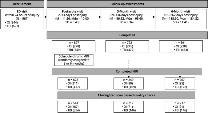
Methods: Children aged 8-16.99 years with mTBI or mild orthopedic injury (OI) were recruited from 5 pediatric emergency departments. Reliable change between preinjury and 1 month postinjury symptom ratings was used to classify mTBI with or without persistent symptoms. Children completed postacute (2-33 days) and/or chronic (3 or 6 months) postinjury T1-weighted MRI, from which macrostructural metrics were derived using automated segmentation. Linear mixed-effects models were used, with multiple comparisons correction.
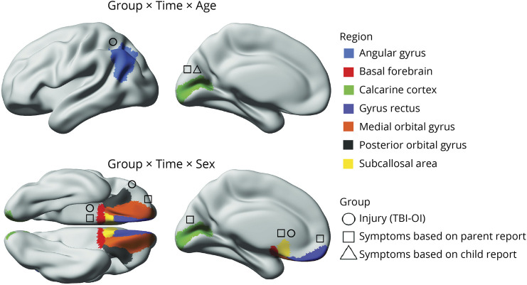
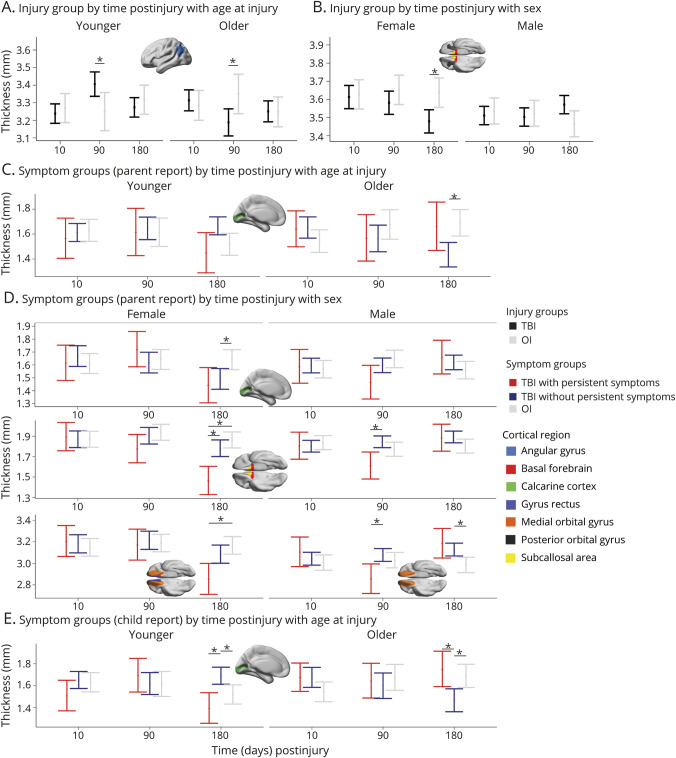
Results: Groups (N = 623; 407 mTBI/216 OI; 59% male; age mean = 12.03, SD = 2.38 years) did not differ in total brain, white, or gray matter volumes or regional subcortical gray matter volumes. However, time postinjury, age at injury, and biological sex-moderated differences among symptom groups in cortical thickness of the angular gyrus, basal forebrain, calcarine cortex, gyrus rectus, medial and posterior orbital gyrus, and the subcallosal area all corrected p < 0.05. Gray matter macrostructural metrics did not differ between groups postacutely. However, cortical thinning emerged chronically after mTBI relative to OI in the angular gyrus in older children (d [95% confidence interval] = -0.61 [-1.15 to -0.08]); and in the basal forebrain (-0.47 [-0.94 to -0.01]), subcallosal area (-0.55 [-1.01 to -0.08]), and the posterior orbital gyrus (-0.55 [-1.02 to -0.08]) in females. Cortical thinning was demonstrated for frontal and occipital regions 3 months postinjury in males with mTBI with persistent symptoms vs without persistent symptoms (-0.80 [-1.55 to -0.05] to -0.83 [-1.56 to -0.10]) and 6 months postinjury in females and younger children with mTBI with persistent symptoms relative to mTBI without persistent symptoms and OI (-1.42 [-2.29 to -0.45] to -0.91 [-1.81 to -0.01]).
Discussion: These findings signal little diagnostic and prognostic utility of postacute gray matter macrostructure in pediatric mTBI. However, mTBI altered the typical course of cortical gray matter thinning up to 6 months postinjury, even after symptoms typically abate in most children. Collapsing across symptom status obscured the neurobiological heterogeneity of discrete clinical outcomes after pediatric mTBI. The results illustrate the need to examine neurobiology in relation to clinical outcomes and within a neurodevelopmental framework.
© 2023 American Academy of Neurology.
Conflict of interest statement
A. L. Ware, A. Onicas, C. Lebel, N. Abdeen, M. H. Beauchamp, C. Beaulieu, B. Bjornson, W. Craig, M. Dehaes, Q. Doan, S. Deschenes, S. B. Freedman, B. G. Goodyear, J. Gravel and A.-A. Ledoux report no disclosures relevant to the manuscript; R. Zemek has received competitively funded research grants from Canadian Institutes of Health Research (CIHR), Ontario Neurotrauma Foundation (ONF), Physician Services Incorporated (PSI) Foundation, CHEO Foundation, Ontario Brain Institute (OBI), and Ontario SPOR Support Unit (OSSU), and the National Football League (NFL) Scientific Advisory Board. R. Zemek holds a Clinical Research Chair in Pediatric Concussion from University of Ottawa, and is the co-founder, Scientific Director, and a minority shareholder in 360 Concussion Care, an interdisciplinary concussion clinic; K.O. Yeates reports no disclosures relevant to the manuscript. Go to
Figures
References
-
- Gilchrist J, Thomas K, Xu L, McGuire LC, Coronado VG. Nonfatal traumatic brain injuries related to sports and recreation activities among persons aged ≤19 years—United States, 2001–2009. MMWR Morb Mortal Wkly Rep. 2011;60:1337-1342. - PubMed
Publication types
MeSH terms
Grants and funding
LinkOut - more resources
Full Text Sources
Medical